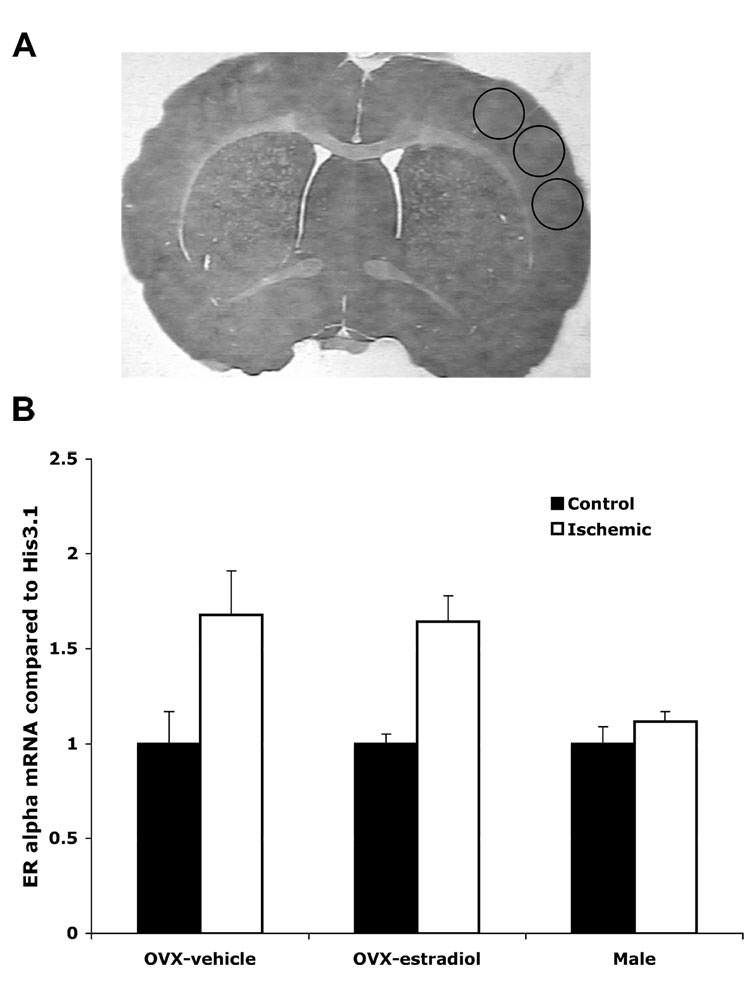
Fig. 2.
ERα mRNA expression is increased on the ischemic cortex of vehicle and estradiol-treated OVX female, but not male rats. Real-time PCR using ERα specific primers was performed on RNA isolated from the cortex of OVX females given oil or estradiol and male rats 24 hours after MCAO. Data was normalized to Histone 3.1 and compared to the control side of the cortex. Asterisks on the graph indicate significant differences from control side of the cortex (p< 0.05, n=3). The three circles in A represent areas where micropunches were taken from the 300µm sections.

