Abstract
Deliberate ingestion of foreign bodies is common amongst prison inmates. The motives behind the ingestion are variable. As the only designated hospital in Northern Ireland treating acute surgical pathologies in the prison population, we reviewed our experience of foreign body ingestion between March 1998 and June 2007. Types of foreign objects, symptomatology, haematological analyses, radiological findings, operative intervention and complications were retrieved from case notes. A literature search was performed using Medline to correlate this clinical data with published evidence to produce therapeutic guidelines to assist the surgical multi-disciplinary team.
Eleven prisoners presented with foreign body ingestion over the study period (M=8 and F=3, mean age: 28.1 years, range 21–48). Mean follow-up was 597 days (range 335–3325 days). Although the literature states that most foreign bodies usually pass spontaneously without the need for intervention, this study demonstrates a higher intervention rate of 36% within the Northern Irish prison population in comparison with other prisoners.
Keywords: Gastrointestinal, Foreign Body, Ingestion, Prison
INTRODUCTION
Ingestion of foreign bodies is a common clinical problem. Difficulties can often occur in both diagnostic and management protocols. Approximately 80% of cases occur in the paediatric population with ingestion frequently occurring accidentally1. In adults, the unintentional swallowing of objects occurs mainly in the elderly population and those patients with learning disabilities and alcohol dependence, whereas intentional episodes occur commonly in psychiatric patients and prisoners2. In the latter group razor blades, batteries and other sharp metallic items are most commonly encountered1.
In the general population, 80–90% of foreign bodies will pass spontaneously3. However, endoscopic intervention is required in 10–20% of patients with less than 1% of patients requiring surgery3. The prison population is a unique environment with different emotional and physical constraints where both the nature and motives behind ingestion are ambiguous with further difficulties encountered in the diagnosis and management of such patients.
OBJECTIVE
To perform a clinical epidemiological review of all patients from the prison population in Belfast who presented to the Belfast City Hospital for management of ingested foreign bodies and to correlate these clinical data with published evidence to produce therapeutic guidelines to assist the surgical multi-disciplinary team.
METHODS
A nine-year retrospective review of all prisoners presenting with foreign body ingestion to the Belfast City Hospital Surgical Unit was completed. Clinical records were then reviewed for data regarding patient demographics, type of foreign body ingested, intention of ingestion, clinical presentation, past medical and social history, haematological analyses, radiological findings, management details including the need for operative intervention and complications. A further assessment of any previous admissions with similar complaints was completed from review of the hospital patient administration system. Follow up was completed for all patients by the prison medical teams with specialist review in the Belfast City Hospital as required.
RESULTS
Demographics
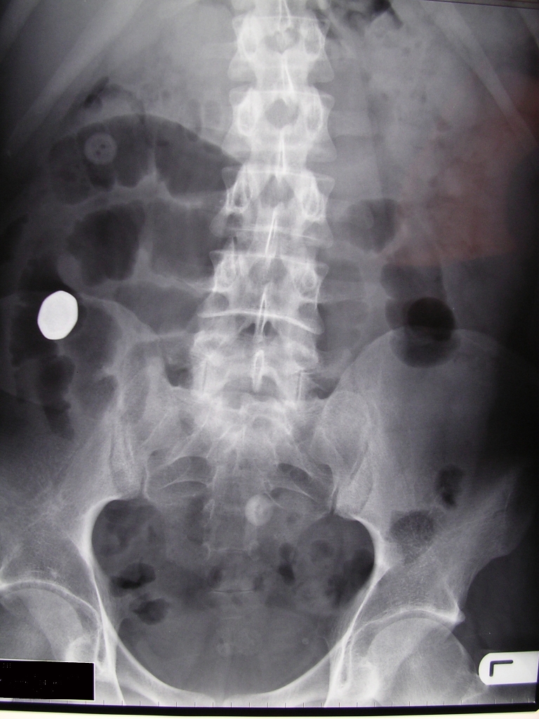
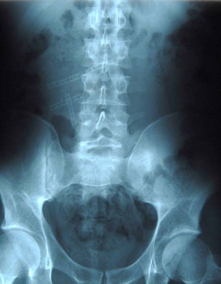
Eleven prisoners (8 males, 3 females) presented with gastrointestinal foreign bodies over the study period. Mean age was 28.1 years (range 21 – 48). Mean admission duration was 2.3 days. Ingested foreign bodies identified included razor blades (n=6), batteries (n = 3), a 20-pence coin (n = 1) and a wrist watch (n = 1), (Figs 1 – 3). Presenting symptoms included reduced appetite (n = 4), vomiting (n = 8), abdominal pain (n = 8) and constipation (n = 5). None of the patients had a significant gastrointestinal past medical history. Most smoked tobacco (n = 8) and consumed alcohol (n = 6) on a regular basis. See Table I for clinical data for all 11 patients.
Fig 1.
Razor blades in the small bowel.
Fig 3.
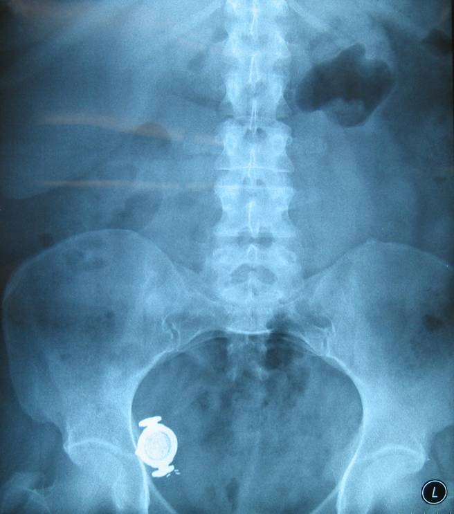
Wrist-watch in right iliac fossa. Failure to progress beyond terminal ileum.
Table I.
Clinical data for all 11 patients (RIF=Right iliac fossa, RUQ=Right upper quadrant, SB=Small bowel, SBO=Small bowel obstruction).
| No | Age | Sex | Type of FOREIGN BODY | Intentional | Repeated ingestion | AXR | CT Scan | Endoscopy: attempted retrieval | Surgery | Hospital Stay (days) | Outcome |
|---|---|---|---|---|---|---|---|---|---|---|---|
| 1 | 29 | M | Razors | Yes | 3 Razors in SB | No | No | 1 | Alive | ||
| 2 | 23 | M | Razors | Yes | 1 Razor in SB | No | No | 1 | Alive | ||
| 3 | 31 | M | 20p coin | Yes | 20p coin in RIF, SBO | Complete SBO | Yes – colonoscopy: failed | Laparotomy: Resection of terminal ileum and Right hemicolectomy | 6 | Alive : diagnosed with Crohn's disease | |
| 4 | 25 | F | Razors | Yes | Razor in RUQ | No | No | 1 | Alive | ||
| 5 | 26 | M | Razors | Yes | 1 Razor in SB | No | No | 1 | Alive | ||
| 6 | 21 | M | Batteries | Yes | Yes – Razors | 3 Batteries in SB | No | No | 1 | Alive | |
| 7 | 23 | M | Razors | Yes | 5 Razors in SB, 1 in rectum | No | No | 1 | Alive | ||
| 8 | 24 | M | Razors | Yes | Yes – Razors and Metallic Rod | 2 Razors in SB and rectum | No | No | 1 | Alive | |
| 9 | 37 | F | Batteries | Yes | Yes – Batteries | 6 Batteries in SB, SBO | No | Laparotomy: enterotomy | 6 | Alive | |
| 10 | 22 | M | Batteries | Yes | Yes – Batteries | 2 Batteries in SB, SBO | No | Laparotomy: enterotomy | 8 | Alive | |
| 11 | 48 | F | Watch | Yes | Watch in RIF | Yes – successful extraction | No | 4 | Alive | ||
Fig 2.
20-pence coin in the right iliac fossa. Dilated loops of small bowel proximal to coin. Reproduced with permission from the Irish Journal of Medical Science - Reference 4
Investigations
Haematological analyses were normal on admission except for a mean raised C-reactive protein of 47.2 mg/l (range 2 – 221). A plain abdominal X-ray confirmed the presence of a metallic foreign body in all patients. An erect chest X-ray was normal in all patients. Computerised Tomography (CT) imaging was required in patient 3, which confirmed small bowel obstruction secondary to an impacted 20-pence coin at the terminal ileum.
Management
Seven patients were managed conservatively. Patients 3 and 11 underwent attempted endoscopic retrieval of the foreign body. Patient 3 admitted to swallowing a 20-pence coin. The coin had impacted in the terminal ileum and attempts at endoscopic retrieval were unsuccessful. He proceeded to laparotomy where the terminal ileum and right colon were resected with a primary anastomosis performed. Histopathological analysis revealed Crohn's Disease with stricturing distal to the impacted 20-pence coin4. Patient 11 was admitted after intentionally swallowing a wrist-watch 6-weeks prior to presentation. Endoscopic retrieval from the ileo-caecal junction was successful (Figs 3, 4). Two further patients (9 & 10) required operative intervention for continuing obstructive symptomatology. An enterotomy was performed with extraction of the metallic foreign body followed by primary closure in both instances.
Fig 4.
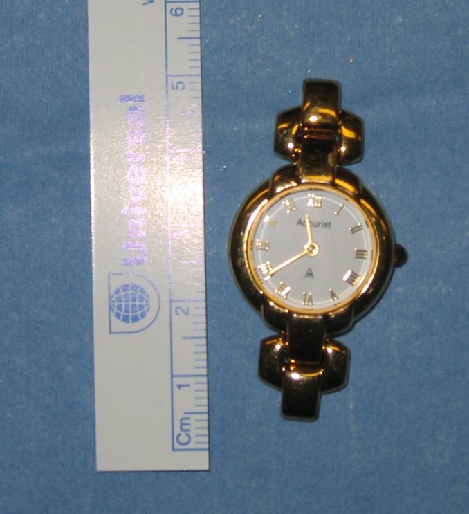
Endoscopic retrieval of wrist-watch was successful. The watch was still functioning.
Outcome
Mean follow-up was 597 days (range 335–3325 days) and was complete for all patients. There were no significant post-operative or long-term complications. All (n = 11) cases of foreign body ingestion were intentional. Almost half of the patients (n = 4) repeated the ingestion of foreign objects (Fig 5).
Fig 5.
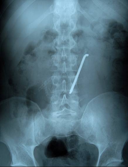
Re-ingestion of metallic rod combined with previous ingestion of razor blades as shown in Figure 1.
DISCUSSION
Ingestion of metallic foreign bodies remains a common problem amongst prison inmates. Swallowing multiple objects at once or repeating the ingestion is a frequent occurrence2. The motives behind the ingestions vary. Underlying psychiatric conditions such as schizophrenia, depression, self-mutilation, masochism and suicidal tendencies, attempts to escape incarceration by transfer to a hospital or psychiatric unit, genuine accidental ingestions and attempts at drug trafficking are common motives1,3,5–6. Blaho et al demonstrated a very high incidence of foreign body ingestion from 2 different prison populations during a 5-week study period where 14 ingestions were noted in 8 male prisoners6.
The foreign bodies ingested and the methods of ingestion were similar. Communication between the inmates of both prisons was well established6. This study suggested imitative behaviour as an underlying cause.
There is no published epidemiological data describing the true prevalence of foreign body ingestion in the prison population. From review of published case series, patients affected range from 22 to167 cases in the United States series to 261 cases from larger European studies3,7–8.
The majority of foreign bodies that reach the gastrointestinal tract will pass spontaneously. Foreign body impaction may occur at areas of anatomical narrowing (the cricopharyngeus, the lower oesophageal sphincter, the pylorus, the ileocaecal valve and the anus), physiological angulation (the curvature of the duodenum) or areas of pathological strictures (terminal ileum in Crohn's disease)1,2,4,6,9. The most common sites for perforation are the lower oesophagus and terminal ileum2,9. Bleeding occurs when injury to the gastrointestinal mucosa occurs.5 Generally, foreign bodies greater than 2–2.5 cm in size will not enter the pyloric canal and those exceeding 6–10 cm in length will not progress through the curvature of the duodenum1–2,10.
Treatment depends on the patient's age and symptomatology, nature and type of ingested foreign body and the anatomical location especially if impacted. Foreign bodies may be managed conservatively or therapeutically with endoscopic, laparoscopic or open surgical methodologies. Blunt objects such as coins can impact in the oesophagus resulting in partial or complete obstruction. Endoscopic retrieval should be attempted in all instances, as prolonged lodgment can lead to pressure necrosis, perforation or fistula formation1–2. A prospective in vivo study by Feigel et al demonstrated that a retrieval net was superior to the basket or forceps technique11. If the blunt object has passed in to the stomach and is less than 2cm in diameter, a conservative outpatient management protocol with weekly radiographs should be adopted. If the blunt object remains in the stomach, it has been recommended to delay endoscopic retrieval for 1–2 months to facilitate any opportunity for spontaneous passage2,5,11,12. Blaho et al also recommend a 3–4 week waiting period to allow passage before attempts at endoscopic retrieval are made5. Zuloaga et al suggest a two month waiting period12. One of the patients in our study presented with a five week history of colicky right iliac fossa pain. The abdominal X-ray on presentation revealed a wrist-watch in the right iliac fossa suggestive of probable impaction at the terminal ileum (Fig 3). Repeat X-rays showed failure of progression of the object. Colonoscopic retrieval of the wrist-watch was successful on day-3 post-admission (Fig 4).
Endoscopic retrieval of sharp objects, such a razor blades, straightened paperclips and needles that are lodged in the oesophagus should be performed urgently1,2. If the object progresses into the stomach or duodenum, immediate attempts at endoscopic retrieval should be undertaken, as the risk of perforation at the ileocaecal valve is approximately 35%1,2,9. If the sharp object has passed beyond the duodenum, the patient should be monitored with daily radiographs and remain under strict observation. Surgical intervention may be required if a sharp object fails to progress radiologically after 72-hours. Emergency laparotomy is required if the patient develops acute clinical signs1,2. All six patients that swallowed razor blades in our study presented after the blades had passed into the small bowel. None of them developed symptoms of obstruction or perforation and consequently were successfully managed conservatively.
Batteries (disc or button) require urgent endoscopic retrieval if lodged in the oesophagus due to the possible risk of chemical burns, electrical discharge and liquefaction necrosis which can lead to subsequent perforation1,2. Once the battery has passed into the stomach, retrieval is only indicated if it remains in the stomach beyond 48 hours, or if it is a larger battery measuring more than 2cm in diameter2. Once beyond the duodenojejunal flexure, 85.4% are passed within 72-hours13. A follow-up radiograph twice per week is sufficient2. Laxatives and anti acids have no proven benefit in management, however gastric lavage has been described to facilitate removal14. Our study showed that 67% of patients that swallowed batteries developed small bowel obstruction requiring a laparotomy with enterotomy. Murshid et al also describe the successful laparoscopic extraction of a sewing needle from the terminal ileum of a 17-year-old female after failure of the endoscopic approach9. Packages of narcotics should not be removed endoscopically as the risk of rupture and leak of the toxic substance is high1,2. Surgical intervention is reserved for those cases where signs of obstruction or leakage of substance occurs2. A management protocol is outlined in Table II.
Table II.
Recommended management protocol for ingested foreign bodies.
| Type of Object | Site of Object | Management Protocol |
|---|---|---|
| Batteries | Oesophagus | Urgent endoscopic retrieval |
| Stomach and Duodenum | If > 48 hrs → endoscopic retrieval | |
| If > 2 cm → endoscopic retrieval | ||
| DJ Flexure | Twice weekly X-rays | |
| If signs of Obstruction/Bleeding/Perforation → urgent endoscopic retrieval +/− laparotomy | ||
| Sharp Metallic Objects | Oesophagus | Urgent endoscopic retrieval |
| Stomach and Duodenum | Urgent endoscopic retrieval | |
| DJ Flexure | Daily x-rays/strict observation | |
| If fails to progress > 72 hrs → laparotomy | ||
| If signs of Obstruction/Bleeding/Perforation → laparotomy | ||
| Blunt Metallic Objects | Oesophagus | Endoscopic retrieval |
| Stomach and Duodenum | If < 2cm → weekly X-rays/conservative management | |
| If > 2 cm → observe with weekly X-rays for 1–2 months. If failure to progress → endoscopic retrieval | ||
| DJ Flexure | Weekly X-rays/conservative management | |
| If signs of Obstruction/Bleeding/Perforation → urgent endoscopic retrieval +/− laparotomy | ||
Many prisoners who deliberately swallow foreign bodies ingest multiple objects or repeat the ingestion in a further episode5,7. Weiland et al demonstrated that amongst 22 male prisoners there were 75 separate hospitalisations, with a total of 256 objects swallowed7. In our study 36% of prisoners repeated the ingestion. Patient 8 was managed conservatively following ingestion of razor blades. He represented a few weeks later with ingestion of further razor blades and a metallic rod (Fig 5).
CONCLUSION
Although the literature states that most foreign bodies in the general population usually pass spontaneously without the need for intervention, this study demonstrates a major difference within the prison population where surgical or endoscopic intervention was required in 36% of these patients (18% required a laparotomy, 9% required endoscopic intervention, and a further 9% required both endoscopic and open removal), in contrast to 1% in the general population1,2. Weiland et al conducted a 10 year study (Wisconsin, USA) on 22 male prisoners with a total of 256 ingested foreign bodies; 40% of the objects passed spontaneously, 30% were managed endoscopically whereas 30% required surgery7. Similarly Barros et al conducted a 6-year study (Madrid, Spain) where 167 patients (including 70 prisoners) were reviewed. Surgery was required in 30% of the patients3.
Identification of prisoners who have a pre-incarceration tendency to swallow foreign objects or those who repeatedly ingest objects, and subjecting them to psychiatric and behaviour modification therapy may prove efficacious. However, this was not assessed in this study.
The authors have no conflict of interest.
Footnotes
Winner of the best clinical paper, Ulster Society for Gastroenterology Spring Meeting, Belfast April 2007.
Presented at the Association of Surgeons of Great Britain and Ireland - Annual Meeting, Bournemouth, UK - 14th to 16th May 2008.
REFERENCES
- 1.Pavlidis TE, Marakis GN, Triantafyllou A, Psarras K, Kontoulis TM, Sakantamis AK. Management of ingested foreign bodies: how justifiable is a waiting policy? Internet J Surg. 2007;9(1) doi: 10.1097/SLE.0b013e31816b78f5. [DOI] [PubMed] [Google Scholar]
- 2.Eisen GM, Baron TH, Dominitz JA, Feigel DO, Goldstein JL, Johanson JF, et al. American Society for Gastrointestinal Endoscopy. Guideline for the management of ingested foreign bodies. Gastrointest Endosc. 2002;55(7):802–6. doi: 10.1016/s0016-5107(02)70407-0. [DOI] [PubMed] [Google Scholar]
- 3.Barros JL, Caballero A, Jr, Rueda JC, Monturiol JM. Foreign body ingestion: management of 167 cases. World J Surgery. 1991;15(6):783–8. doi: 10.1007/BF01665320. [DOI] [PubMed] [Google Scholar]
- 4.O'Donnell ME, Gibson N, Sharif MA, Spence RA, Lee J. Crohn's disease of the terminal ileum: a cheap diagnosis. Ir J Med Sci. doi: 10.1007/s11845-007-0080-4. Epub 2007 Aug 18. [DOI] [PubMed] [Google Scholar]
- 5.Blaho KE, Merigian KS, Winbery SL, Park LJ, Cockrell M. Foreign body ingestion in the Emergency Department: case reports and review of treatment. J Emerg Med. 1998;16(1):21–6. doi: 10.1016/s0736-4679(97)00229-1. [DOI] [PubMed] [Google Scholar]
- 6.Abraham B, Alao AO. An unusual foreign body ingestion in a schizophrenic patient: case report. Int J Psychiatry Med. 2005;35(3):313–8. doi: 10.2190/7AE8-3AV0-W3UA-TKV4. [DOI] [PubMed] [Google Scholar]
- 7.Weiland ST, Schurr MJ. Conservative management of ingested foreign bodies. J Gastrointest Surg. 2002;6:496–500. doi: 10.1016/s1091-255x(01)00027-0. [DOI] [PubMed] [Google Scholar]
- 8.Magdalena Castineira C, Magdalena Lopez C. [Foreign body ingestion among prisoners] Spanish. Rev Esp Enferm Dig. 1994;85(4):254–6. [PubMed] [Google Scholar]
- 9.Murshid KR, Khairy GE. Laparoscopic removal of a foreign body from the intestine. J R Coll Surg Edinb. 1998;43(2):109–11. [PubMed] [Google Scholar]
- 10.Sinnatamby CS, editor. Last's Anatomy: Regional and Applied. 10th ed. London: Churchill Livingstone; 2003. pp. 241–56. [Google Scholar]
- 11.Faigel DO, Stotland BR, Kochman ML, Hoops T, Judge T, Kroser J, et al. Device choice and experience level in endoscopic foreign object retrieval: an in vivo study. Gastrointest Endosc. 1997;45(6):490–2. doi: 10.1016/s0016-5107(97)70179-2. [DOI] [PubMed] [Google Scholar]
- 12.Zuloaga J, Olivera J, Candia A, Arias V, De La Fuente A. [Ingestion of foreign bodies in a prison population; surgical treatment] Spanish. Rev Esp Enferm Apar Dig. 1987;72:617–21. [PubMed] [Google Scholar]
- 13.Litovitz TL. Battery ingestions: product accessibility and clinical course. Pediatrics. 1985;75(3):469–76. [PubMed] [Google Scholar]
- 14.Namasivayam S. Button battery ingestion: a solution to a management dilemma. Pediatr Surg Int. 1999;15(5):383–4. doi: 10.1007/s003830050605. [DOI] [PubMed] [Google Scholar]


