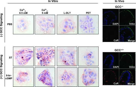
Fig. 4.
Ligand-dependent GCC signaling upregulates surface CaR receptors in normal and transformed intestinal epithelial cells. Membranes of non-permeabilized T84 cells (in vitro) were stained (brown) with specific rabbit polyclonal anti-CaR in the presence of low (0.5 mM; left column) or high (5 mM; right three columns) Ca2+o (magnification ×40). In blue, counterstaining of nuclei with hematoxylin. Tumor cells were treated for 24 h with phosphate-buffered saline (PBS) (vehicle control; upper row), ST (1 μM; middle row) or 8-br-cGMP (5 mM; lower row) in the absence (left two columns) or presence (right two columns) of the indicated CNG inhibitors (see Figure 1 legend for keys). Also, epithelial cells in villus tips from the jejunum of GCC+/+ and GCC−/− mice were stained with 4′,6-diamidino-2-phenylindole (DAPI) (blue, nuclei) and specific antibodies against villin (green, enterocyte brush-border membranes) and CaR (red) and subjected to confocal microscopy (magnification ×63).

