Abstract
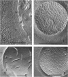
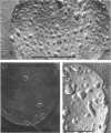
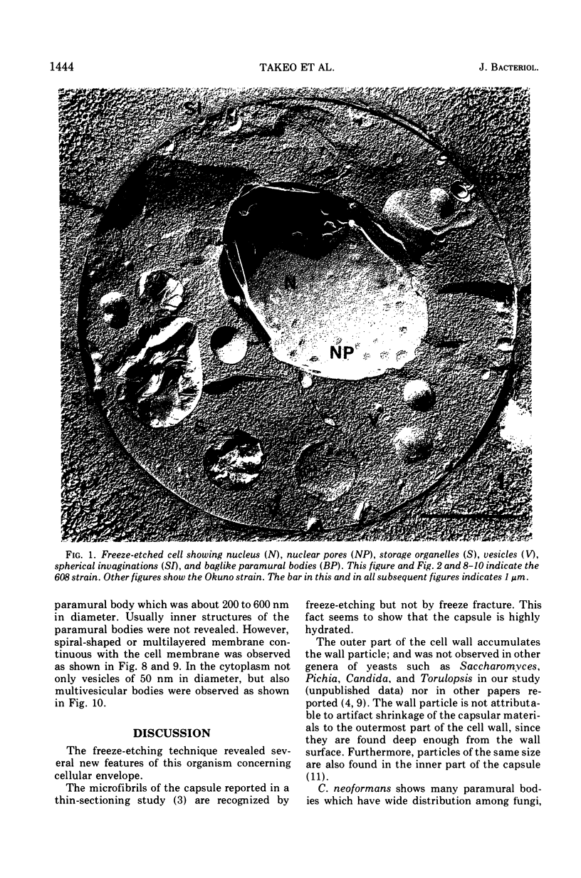
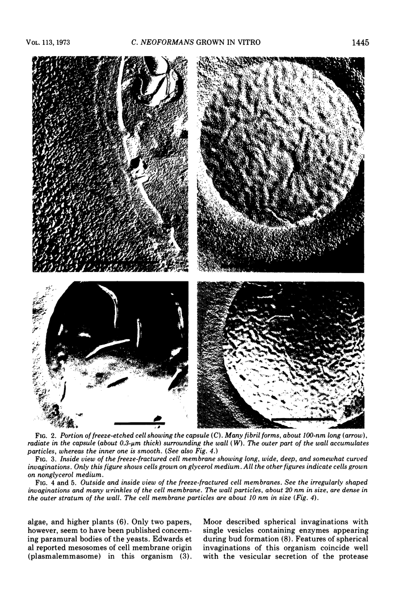
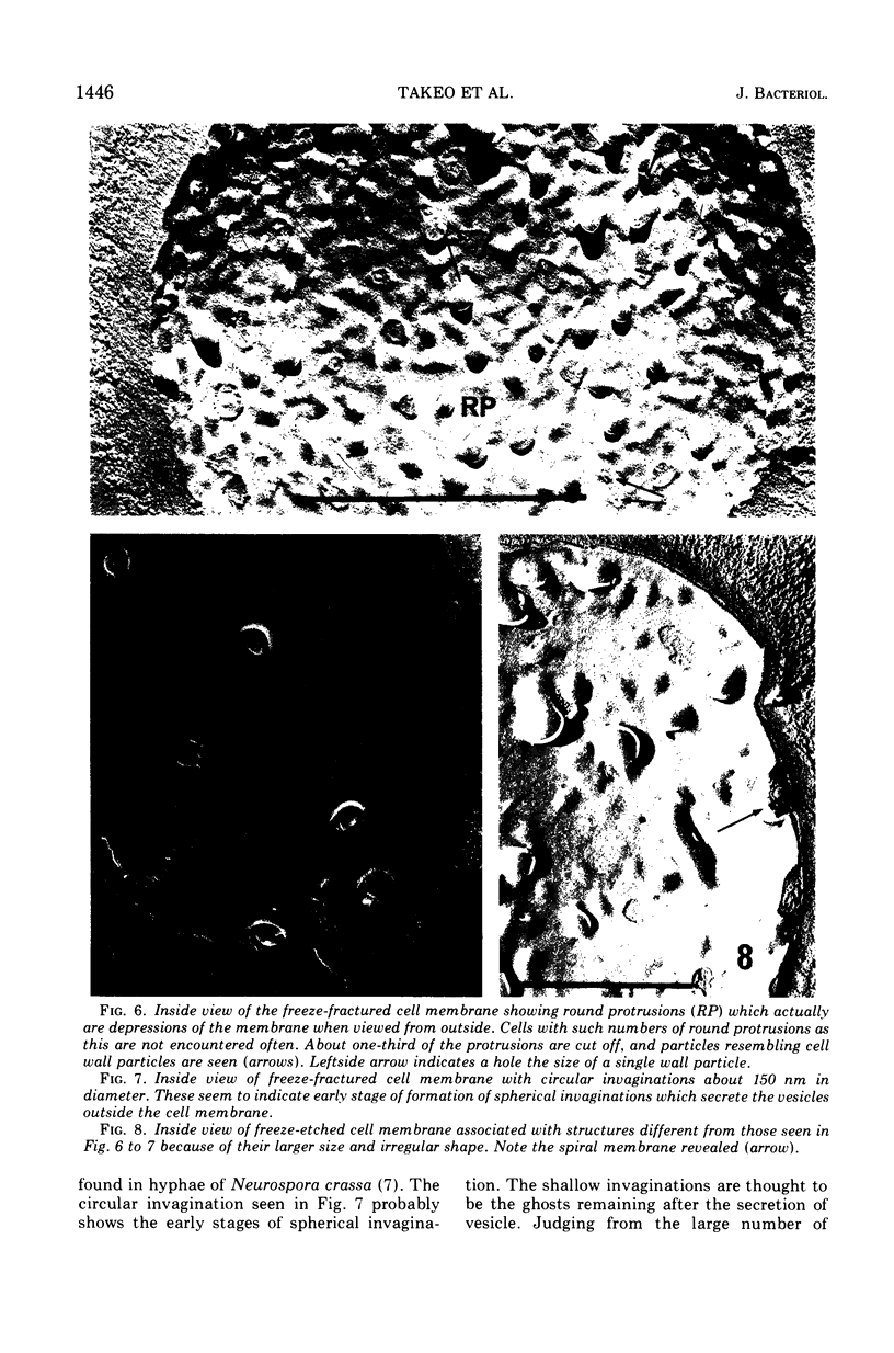
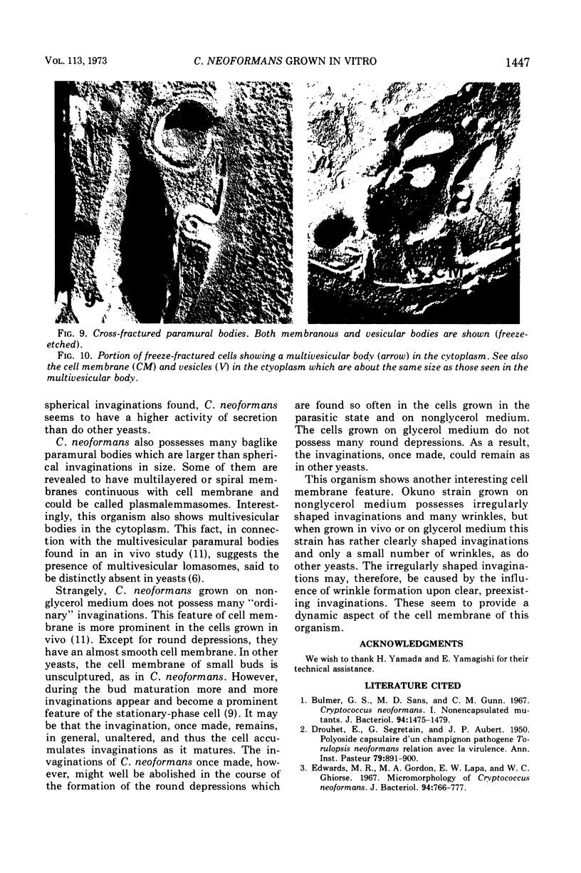
Cryptococcus neoformans grown on culture media was observed by the freeze-etching technique. In the capsule, short fibrils were seen when freezeetched. This organism was unique in the appearance of the cell wall, which showed two strata. The outer one was dense with particles of about 20 nm in diameter, whereas the inner one was sparse in particles. The appearance of the cell membrane of this organism differed distinctly depending on the culture media. When grown on glycerol medium, the cell membrane possessed, as do other yeasts, clear but somewhat longer and curved invaginations. The membrane of cells grown on nonglycerol medium exhibited, however, only a few invaginations of irregular shape. Instead, characteristically of this organism, the cell membrane showed round depressions of 40 to 200 nm in diameter which were the surface view of the paramural bodies. In cross-fractured cells, both types of paramural bodies were found. Some of them contained a single vesicle of about 50 nm in diameter. These seem to play a role in secreting the cytoplasmic vesicles. Data suggesting the existence of multivesicular bodies in the cytoplasm and multivesicular lomasomes were also obtained. Some of the baglike paramural bodies showed multilayered membrane. These are thought to be plasmalemmasomes. This organism was similar to other yeasts reported in other respects.
Full text
PDF
Images in this article
Selected References
These references are in PubMed. This may not be the complete list of references from this article.
- Bulmer G. S., Sans M. D., Gunn C. M. Cryptococcus neoformans. I. Nonencapsulated mutants. J Bacteriol. 1967 Nov;94(5):1475–1479. doi: 10.1128/jb.94.5.1475-1479.1967. [DOI] [PMC free article] [PubMed] [Google Scholar]
- DROUHET E., SEGRETAIN G., AUBERT J. P. Polyoside capsulaire d'un champignon pathogene Torulopsis neoformans; relation avec la virulence. Ann Inst Pasteur (Paris) 1950 Dec;79(6):891–900. [PubMed] [Google Scholar]
- Edwards M. R., Gordon M. A., Lapa E. W., Ghiorse W. C. Micromorphology of Cryptococcus neoformans. J Bacteriol. 1967 Sep;94(3):766–777. doi: 10.1128/jb.94.3.766-777.1967. [DOI] [PMC free article] [PubMed] [Google Scholar]
- Guth E., Hashimoto T., Conti S. F. Morphogenesis of ascospores in Saccharomyces cerevisiae. J Bacteriol. 1972 Feb;109(2):869–880. doi: 10.1128/jb.109.2.869-880.1972. [DOI] [PMC free article] [PubMed] [Google Scholar]
- Matile P., Jost M., Moor H. Intrazelluläre Lokalisation proteolytischer Enzyme von Neurospora crassa. Z Zellforsch Mikrosk Anat. 1965 Oct 12;68(2):205–216. [PubMed] [Google Scholar]
- Moor H. Endoplasmic reticulum as the initiator of bud formation in yeast (S. cerevisiae). Arch Mikrobiol. 1967 Jun 6;57(2):135–146. doi: 10.1007/BF00408697. [DOI] [PubMed] [Google Scholar]
- Takeo K., Uesaka I., Uehira K., Nishiura M. Fine structure of Cryptococcus neoformans grown in vivo as observed by freeze-etching. J Bacteriol. 1973 Mar;113(3):1449–1454. doi: 10.1128/jb.113.3.1449-1454.1973. [DOI] [PMC free article] [PubMed] [Google Scholar]


