

Abstract
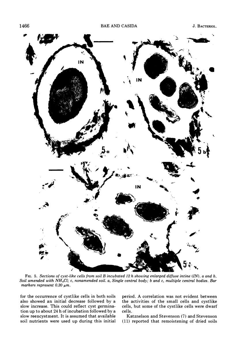
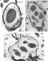
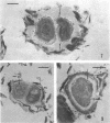
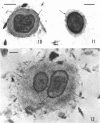
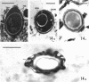
Air-dried soils were adjusted to 50% moisture-holding capacity and incubated for 2 weeks at 30 C. Samples were removed at intervals, and their total microbial populations were physically separated and concentrated from the soil debris for sectioning and ultrastructure examination. Although the total numbers of cell sections in these preparations remained relatively constant during the soil incubations, the percentages of dwarf cells (≤0.3 μm in diameter), minute cells (≤0.2 μm in diameter), and cells with a cystlike structure decreased with time followed by a slow increase. During this period, a corresponding increase and decrease occurred in the percentages of cells in the 0.3- to 0.5-μm diameter range, but dividing cells were rarely observed. The percentages of spores and of cells with electron-transparent areas also increased and then decreased during incubation. When nutrients were added to these soils, the initial increases in cell size occurred at what appeared to be a faster rate. But this probably was related to a corresponding increase in total cell numbers which also occurred. The responses of the spores, cystlike cells, and cells with electron-transparent areas to nutrient additions were not consistent although all conditions of incubation, regardless of nutrient addition, increased the occurrence of an enlarged diffuse intine-like layer for the cystlike cells. In addition to the above, incubated soils contained cells, which were mainly in the 0.3- to 0.5-μm cell diameter range, that had an internal membrane surrounding the general area of the nuclear material. Changes in additional fine structure features of the microbial populations are described.
Full text
PDF


Images in this article


Selected References
These references are in PubMed. This may not be the complete list of references from this article.
- Bae H. C., Cota-Robles E. H., Casida L. E. Microflora of soil as viewed by transmission electron microscopy. Appl Microbiol. 1972 Mar;23(3):637–648. doi: 10.1128/am.23.3.637-648.1972. [DOI] [PMC free article] [PubMed] [Google Scholar]
- Freese E. B., Cole R. M., Klofat W., Freese E. Growth, sporulation, and enzyme defects of glucosamine mutants of Bacillus subtilis. J Bacteriol. 1970 Mar;101(3):1046–1062. doi: 10.1128/jb.101.3.1046-1062.1970. [DOI] [PMC free article] [PubMed] [Google Scholar]
- Hitchins V. M., Sadoff H. L. Morphogenesis of cysts in Azotobacter vinelandii. J Bacteriol. 1970 Oct;104(1):492–498. doi: 10.1128/jb.104.1.492-498.1970. [DOI] [PMC free article] [PubMed] [Google Scholar]
- Hoeniger J. F., Stuart P. F., Holt S. C. Cytology of spore formation in Clostridium perfringens. J Bacteriol. 1968 Nov;96(5):1818–1834. doi: 10.1128/jb.96.5.1818-1834.1968. [DOI] [PMC free article] [PubMed] [Google Scholar]
- KATZNELSON H., STEVENSON I. L. Observations on the metabolic activity of the soil microflora. Can J Microbiol. 1956 Oct;2(6):611–622. doi: 10.1139/m56-074. [DOI] [PubMed] [Google Scholar]
- KELLENBERGER E., RYTER A., SECHAUD J. Electron microscope study of DNA-containing plasms. II. Vegetative and mature phage DNA as compared with normal bacterial nucleoids in different physiological states. J Biophys Biochem Cytol. 1958 Nov 25;4(6):671–678. doi: 10.1083/jcb.4.6.671. [DOI] [PMC free article] [PubMed] [Google Scholar]
- Korch C. T., Doi R. H. Electron microscopy of the altered spore morphology of a ribonucleic acid polymerase mutant of Bacillus subtilis. J Bacteriol. 1971 Mar;105(3):1110–1118. doi: 10.1128/jb.105.3.1110-1118.1971. [DOI] [PMC free article] [PubMed] [Google Scholar]
- Spurr A. R. A low-viscosity epoxy resin embedding medium for electron microscopy. J Ultrastruct Res. 1969 Jan;26(1):31–43. doi: 10.1016/s0022-5320(69)90033-1. [DOI] [PubMed] [Google Scholar]
- Vela G. R., Cagle G. D., Holmgren P. R. Ultrastructure of Azotobacter vinelandii. J Bacteriol. 1970 Nov;104(2):933–939. doi: 10.1128/jb.104.2.933-939.1970. [DOI] [PMC free article] [PubMed] [Google Scholar]
- WYSS O., NEUMNN M. G., SOCOLOFSKY M. D. Development and germination of the Azotobacter cyst. J Biophys Biochem Cytol. 1961 Aug;10:555–565. doi: 10.1083/jcb.10.4.555. [DOI] [PMC free article] [PubMed] [Google Scholar]