Abstract
OBJECTIVE—Hepatic triglyceride is closely associated with hepatic insulin resistance and is known to be decreased by thiazolididinediones. We studied the effect of pioglitazone on hepatic triglyceride content and the consequent effect on postprandial endogenous glucose production (EGP) in type 2 diabetes.
RESEARCH DESIGN AND METHODS—Ten subjects with type 2 diabetes on sulfonylurea therapy were treated with pioglitazone (30 mg daily) for 16 weeks. EGP was measured using a dynamic isotopic methodology after a standard liquid test meal both before and after pioglitazone treatment. Liver and muscle triglyceride levels were measured by 1H magnetic resonance spectroscopy, and intra-abdominal fat content was measured by magnetic resonance imaging.
RESULTS—Pioglitazone treatment reduced mean plasma fasting glucose and mean peak postprandial glucose levels. Fasting EGP decreased after pioglitazone treatment (16.6 ± 1.0 vs. 12.2 ± 0.7 μmol · kg−1 · min−1, P = 0.005). Between 80 and 260 min postprandially, EGP was twofold lower on pioglitazone (2.58 ± 0.25 vs. 1.26 ± 0.30 μmol · kg−1 · min−1, P < 0.001). Hepatic triglyceride content decreased by ∼50% (P = 0.03), and muscle (anterior tibialis) triglyceride content decreased by ∼55% (P = 0.02). Hepatic triglyceride content was directly correlated with fasting EGP (r = 0.64, P = 0.01) and inversely correlated to percentage suppression of EGP (time 150 min, r = −0.63, P = 0.02). Muscle triglyceride, subcutaneous fat, and visceral fat content were not related to EGP.
CONCLUSIONS—Reduction in hepatic triglyceride by pioglitazone is very closely related to improvement in fasting and postprandial EGP in type 2 diabetes.
Hepatic triglyceride has been shown to be strongly associated with hepatic insulin resistance in type 2 diabetes (1–3). The exact mechanism by which hepatic triglyceride induces hepatic insulin resistance is unknown but is thought to relate to accumulation of intracellular fatty acid metabolites and consequent activation of a serine kinase cascade and induction of cellular insulin resistance (4). Reduction in hepatic triglyceride content by moderate weight reduction normalizes rates of basal endogenous glucose production (EGP) in patients with type 2 diabetes (2). Hepatic insulin resistance is also associated with impaired postprandial suppression of EGP in type 2 diabetes, but the effect of reduction of hepatic triglyceride content on postprandial suppression of EGP in type 2 diabetes is unknown.
Thiazolidinediones, such as pioglitazone, possess insulin-sensitizing properties and have been shown to decrease hepatic triglyceride content in type 2 diabetes (5). Because thiazolidinediones have been shown to reduce both fasting and postprandial glucose levels (6,7), we hypothesized that pioglitazone treatment in type 2 diabetes would reduce hepatic triglyceride content and consequently reduce basal and postprandial EGP. In addition, visceral fat has also been implicated in hepatic insulin resistance, and pioglitazone has been reported to decrease visceral fat content (8). The effect of this on EGP requires definition.
To test our hypothesis, we used noninvasive 1H magnetic resonance spectroscopy to assess hepatic triglyceride and intramyocellular triglyceride content and magnetic resonance imaging to quantify intra-abdominal fat content in 10 type 2 diabetic patients before and after treatment with pioglitazone. EGP was measured using a dynamic isotopic methodology after a standard liquid test meal.
RESEARCH DESIGN AND METHODS
We studied 10 suboptimally controlled healthy subjects with type 2 diabetes (6 men and 4 women; 9 Caucasians and 1 Asian; ≥2 years duration; mean age 52.1 ± 2.8 years; age range 38–64 years; A1C >7.5%; and no history of weight loss or ketonuria at diagnosis) on maximum-tolerated sulfonylurea treatment, who required additional antidiabetes medications. Apart from sulfonylurea treatment, no subject was taking any medications that would affect glucose or lipid metabolism, and subjects on statins were excluded. Subjects who had previous treatment with metformin, thiazolidinediones, or insulin and those with history of noncompliance with treatment were excluded. Two subjects were on tolbutamide, three were on glimiperide, and five were on gliclazide treatment.
After a 4-week run-in period to ensure metabolic stability, 30 mg pioglitazone once daily was given for 16 weeks. Two metabolic study days were undertaken: one before and one after pioglitazone treatment. Localized 1H magnetic resonance spectra of liver and skeletal muscle and abdominal fat distribution were obtained before and after completion of pioglitazone treatment. The study protocol was approved by the Newcastle and North Tyneside Local Research Ethics Committee. Full verbal and written explanation was given, and written consent was obtained before commencement of the studies.
Each subject was studied twice: once before pioglitazone treatment and once after 16 weeks of pioglitazone treatment. Subjects refrained from alcohol or vigorous exercise for 3 days before the study day and consumed their habitual weight-maintaining diet. After a 12-h overnight fast, all subjects arrived at 0700 h, and anthropometric measurements were recorded. Bioimpedance was performed using a Holtain BC Analyser (Holtain, Dyfed, U.K.), and the percentage of body fat was derived. An intravenous cannula for infusion was sited in an antecubital fossa vein and a second cannula in a distal forearm vein, this hand being placed in a heated box at 50°C to allow sampling of arterialized blood. Baseline blood samples were then taken. After patients were rested for 30 min, at time −120 min, a fasting plasma glucose–adjusted prime of 6,6-dideuterated glucose was given intravenously (9), and a continuous infusion of 6,6-dideuterated glucose (0.04 mg · ml−1 · min−1) was commenced. A period of 120 min was allowed for equilibrium of 6,6-dideuterated-glucose; the end of this period was taken to be time 0. At this time, a standard liquid test meal (100 g carbohydrate, 12.5 g fat, and 16 g protein) containing 2 g 2-deuterated glucose was given, and the subjects consumed this over a 10-min period. The rate of infusion of 6,6-dideuterated glucose was adjusted in a stepwise fashion to match the anticipated pattern of endogenous glucose release after the meal and was as follows: −120 to 0 min, 100% of basal infusion rate; 0–3 min, 100%; 3–8 min, 70%; 8–18 min, 55%; 18–28 min, 30%; 28–45 min, 15%; 45–70 min, 25%; 70–160 min, 35%; 160–270 min, 55%; and 270–360 min, 80%. The metabolic study was finished at 360 min. The rate of infusion was determined iteratively from eight pilot studies. Plasma enrichments of 6,6-dideuterated glucose were analyzed in each pilot study, and rate of infusion was modified in subsequent studies until the anticipated pattern of EGP was mimicked.
Frequent blood samples were taken for measuring plasma glucose, 2-deuterated glucose, 6,6-dideuterated glucose, triglycerides, free fatty acids (FFAs), insulin, C-peptide, and glucagon. Substrate oxidation rates were calculated from indirect calorimetry data derived from constant flow hood calorimeter (Delta Trac 17), using standard formulas (10). In one subject, the after-pioglitazone study was terminated at 260 min because of symptoms of hypoglycemia (blood glucose 3.7 mmol/l). The same subject had reported symptoms of hypoglycemia during the pioglitazone treatment period, and the sulfonylurea dose had been reduced. The study was stopped at 120 min after meal ingestion in one other subject on both study days because of vasovagal symptoms.
Metabolite and hormone assays.
Plasma glucose was measured on a Yellow Springs glucose analyzer (Yellow Springs Instruments, Yellow Springs, OH). FFA was measured on a Roche Cobas centrifugal analyzer using a Wako kit (Wako Chemicals, Neuss, Germany). Plasma triglycerides were measured on a Roche Cobas centrifugal analyzer using a colorimetric assay (ABX Diagnostics, Montpellier, France). Serum insulin and C-peptide assays were both measured using Dako ELISA kits (Dako, Ely, Cambridge, U.K.). Plasma glucagon was measured using the glucagon radioimmunoassay kit (Linco Research, St. Charles, MO), and tubes were counted using a Packard Cobra gamma counter. 2H2 percent enrichment in plasma glucose was determined by gas chromatography–mass spectroscopy using a Thermo Voyager single quadrupole mass spectrometer interfaced to a Thermo Trace GC, with automated injection via a Thermo AS2000 autosampler (Thermo Scientific, Waltham, MA). The coefficient of variation (CV) for the precision of plasma 2H2 atom percent excess (APE) measurement was 4.1%.
Calculations of EGP.
The profile of exogenous glucose concentration, i.e., the component of total glucose concentration due to exogenous glucose ingestion, was initially calculated from 2-deuterated glucose (11). We then calculated the time course of the endogenous glucose concentration, i.e., the component of total glucose due to EGP only, by subtracting the calculated exogenous component and the 6,6-dideuterated glucose concentration from the measured total glucose concentration. The steady-state values of plasma clearance rate and basal EGP (basal EGP = plasma clearance rate × basal glucose concentration) were estimated from the 6,6-dideuterated glucose decay curve after the prime dose of 6,6-dideuterated glucose given 2 h before the meal (12). Because 6,6-2H2-glucose had been infused mimicking the expected behavior of EGP, the ratio of 6,6-2H2-glucose to endogenously produced glucose was almost constant (tracer-to-tracee clamp technique), allowing reliable estimation of EGP (13). EGP was calculated using both the model of Steele et al. (14,15) and the two-compartment model of Radziuk et al. (16), with the tracer-to-tracee ratio derivative calculated after the signal was smoothed using an algorithm based on stochastic nonparametric deconvolution (17). Calculated EGP was similar in both models, and the results from the Steele model are presented. In three subjects, the EGP profiles could not be assessed because of modeling factors in the postprandial period, and their data were excluded from the EGP analysis. Relationships between EGP and its determinants were determined in all subjects with paired data available (seven subjects).
1H magnetic resonance spectroscopy.
Localized 1H nuclear magnetic resonance spectra of liver and muscle were obtained in a 1.5-Tesla magnetic resonance scanner with a 1H transmitter/receiver coil placed over the relevant tissue. Liver spectra were acquired by applying the breath hold–triggered stimulation echo acquisition sequence. Spectra were collected without water suppression, using a point-resolved spectroscopy sequence (18), with a 2- × 2- × 2-cm voxel in the soleus muscle and anterior tibialis muscle and a 3- × 3- × 3-cm voxel in the liver, using an echo time of 25 ms and repetition time of 5,000 ms with 32 acquisitions and a spectral resolution of 1 Hz.
Spectra were analyzed using an magnetic resonance user interface (19). Automatic phase correction was performed using the water peak in the spectra, and the water peak was assigned to 4.68 ppm. Lipid spectral peaks were assigned as described in Boesch et al. (20), and spectra peak amplitudes were estimated by fitting Lorentzian curves to the spectra. To calculate molar density of triglycerides from the amplitudes of water and intramyocellular lipid estimated in the spectral fitting, we used the formulas described by Szczepaniak et al. (21).
Abdominal fat distribution.
The volume of total and visceral fat compartments was determined as described previously (22). To determine the visceral fat content, a region encompassing the viscera was drawn manually by an operator using a computer mouse. The visceral fat volume was determined by multiplying the number of pixels brighter than the threshold in this region by the pixel size and slice thickness. Regions were also drawn manually around brightly appearing structures such as bone marrow to subtract their contribution from the total fat volume. The subcutaneous fat volume was quantified by subtracting the visceral fat volume from the total fat volume. The intrasubject CVs in measurement of subcutaneous and visceral fat were 4.9 and 3.8%, respectively.
Statistical analysis.
All data are expressed as means ± SE. Statistical analyses were performed using MINITAB software (Minitab, State College, PA). Comparisons were carried out using Student's two-tailed t test where appropriate, and Wilcoxon's signed-rank test was used to compare EGP. Relationships were tested using the linear correlation analysis. Stepwise regression analysis was used for determining the determinants of EGP. A P value of <0.05 was considered to indicate statistical significance.
RESULTS
Plasma glucose, A1C, and body weight.
Fasting mean plasma glucose (10.1 ± 0.5 vs. 7.8 ± 0.5 mmol/l, P = 0.02) and A1C (8.6 ± 0.3 vs. 6.7 ± 0.38% P < 0.001) were significantly lower after pioglitazone treatment (Table 1). After 16 weeks of pioglitazone treatment, both body weight and BMI increased significantly, and increases in both fat mass and fat-free mass contributed to the overall weight gain (Table 1).
TABLE 1.
Anthropometric and laboratory measurements before and after pioglitazone treatment for 16 weeks
| Before pioglitazone | After pioglitazone | P value | |
|---|---|---|---|
| Body weight (kg) | 87.4 ± 4.7 | 93.6 ± 4.7 | 0.001 |
| BMI (kg/m2) | 31.2 ± 1.2 | 33.0 ± 1.1 | 0.001 |
| Fat mass (kg) | 32.9 ± 3.5 | 35.6 ± 3.4 | 0.001 |
| Fat-free mass (kg) | 54.6 ± 3.5 | 58.0 ± 3.5 | <0.001 |
| A1C | 8.6 ± 0.3 | 6.7 ± 0.3 | <0.001 |
| Fasting plasma glucose (mmol/l) | 10.1 ± 0.5 | 7.8 ± 0.5 | 0.02 |
| Peak plasma glucose (mmol/l) | 18.7 ± 2.3 | 15.6 ± 3.8 | <0.05 |
| Fasting plasma insulin (pmol/l) | 102.2 ± 14.8 | 81.1 ± 14.8 | NS |
| Fasting plasma C-peptide (nmol/l) | 1.3 ± 0.1 | 1.0 ± 0.1 | 0.005 |
| Fasting plasma triglyceride (mmol/l) | 2.3 ± 0.4 | 1.9 ± 0.6 | NS |
| Fasting plasma FFA (μmol/l) | 0.73 ± 0.08 | 0.66 ± 0.05 | NS |
| Baseline glucagon (pg/ml) | 150.9 ± 24.2 | 112.4 ± 17.2 | 0.005 |
Date are means ± SE.
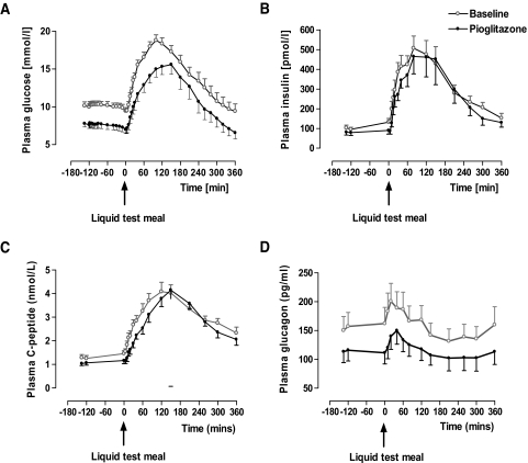
After meal ingestion, plasma glucose rose on both study days, but mean peak plasma glucose was significantly lower (18.7 ± 2.3 vs. 15.6 ± 3.8 mmol/l, P < 0.05) and delayed (100 vs. 150 min) after pioglitazone treatment. The postprandial plasma glucose remained significantly lower until the end of the study (360 min, 9.4 ± 1.0 vs. 6.5 ± 0.8 mmol/l, P = 0.04) (Fig. 1A).
FIG. 1.
Change in plasma glucose (A), plasma insulin (B), plasma C-peptide (C), and plasma glucagon (D) before and after the meal and before (○) and after (•) pioglitazone treatment. Data are means ± SE.
Plasma insulin and C-peptide.
Fasting insulin levels (102.2 ± 14.8 vs. 81.1 ± 14.8 pmol/l, NS) and after-meal mean peak insulin levels (506.2 ± 62.5 vs. 464.5 ± 87.5 pmol/l, NS) did not change significantly after pioglitazone treatment (Fig. 1B). However, fasting C-peptide levels were significantly reduced after pioglitazone treatment (1.3 ± 0.1 vs. 1.0 ± 0.1 nmol/l, P = 0.005) and remained significantly lower until measured 80 min postprandially (Fig. 1C).
Insulin secretion rates and hepatic insulin extraction.
Using C-peptide and plasma insulin levels, insulin secretion rate and hepatic insulin extraction were derived. Insulin secretion rates were similar in the postprandial period both at baseline and after pioglitazone treatment (peak insulin secretion rate 1,197 ± 125 vs. 1,205 ± 165 pmol/min; NS). Similarly, mean postprandial hepatic insulin extraction was unchanged after pioglitazone treatment (39.3 ± 0.01 vs. 40.2 ± 0.01%, NS).
Glucagon.
Fasting glucagon levels were significantly decreased on pioglitazone treatment (150.9 ± 24.2 vs. 112.4 ± 17.2 pg/ml, P = 0.005), and there was a lower and delayed postprandial peak (207.4 ± 24.1 pg/ml at 20 min vs. 150.1 ± 23.9 pg/ml at 40 min, P < 0.01). Postprandial glucagon levels reached a nadir at 210 min on both study days and then rose slightly on both study days during the postabsorptive period (Fig. 1D).
FFA and triglyceride.
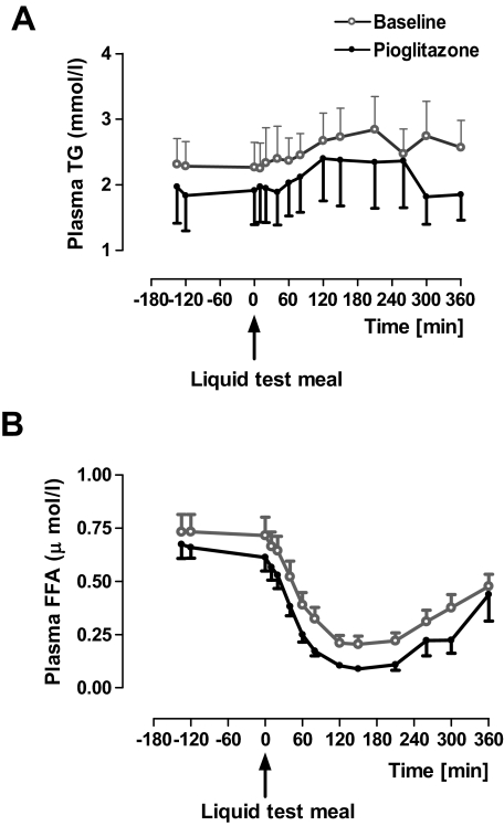
Fasting plasma FFA levels were slightly but not statistically significantly lower (0.73 ± 0.08 vs. 0.66 ± 0.05 μmol/l, NS). Postprandially, FFA concentrations were suppressed on both study days, more so after pioglitazone treatment (0.20 ± 0.04 vs. 0.08 ± 0.02 μmol/l at 150 min [nadir], P = 0.018). Levels remained significantly lower until 210 min (0.22 ± 0.04 vs. 0.10 ± 0.02 μmol/l, P = 0.03) (Fig. 2A). Fasting plasma triglyceride levels were slightly lower after pioglitazone treatment (2.3 ± 0.4 vs. 1.9 ± 0.6 mmol/l, NS) and remained lower throughout the postprandial period (Fig. 2B).
FIG. 2.
Change in plasma triglyceride (A) and plasma FFAs (B) before and after the meal and before (○) and after (•) pioglitazone treatment. Data are means ± SE.
EGP.
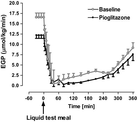
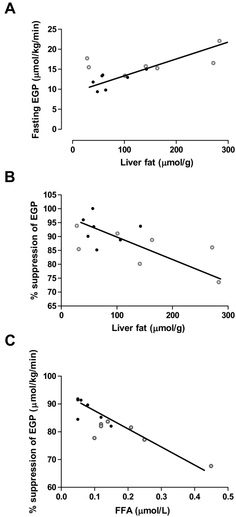
At the end of the first 120-min basal period, mean plasma APEs of 6,6-2H2-glucose were 1.48 ± 0.05 and 1.61 ± 0.09 before and after pioglitazone treatment, respectively. The consequent rate of fall of plasma 6,6-2H2-glucose APE on the change in infusion protocol was similar during the postprandial period on both study days (31 and 38% at 30 min and 47 and 51% at 60 min). At nadir, between 120 and 180 min, plasma APEs were 0.6 ± 0.01 and 0.7 ± 0.01 before and after pioglitazone treatment, respectively. Fasting EGP was significantly reduced after pioglitazone treatment (16.6 ± 1.0 vs. 12.2 ± 0.7 μmol · kg−1 · min−1, P = 0.005) (Fig. 3). Although postprandial suppression of EGP was rapid on both study days (at 40 min, 96% suppression [baseline study] and 93% suppression [pioglitazone study]), postprandial EGP was significantly lower at 150 min after pioglitazone treatment (2.50 ± 0.61 vs. 0.82 ± 0.25 μmol · kg−1 · min−1, P = 0.05). Between 80 and 260 min, mean EGP was twofold lower after pioglitazone treatment (2.58 ± 0.25 vs. 1.26 ± 0.30 μmol · kg−1 · min−1, P < 0.001) (Fig. 3). When expressed as percentage suppression from baseline, postprandial suppression was still greater after pioglitazone treatment (82 vs. 90%, between 180 and 210 min, P = 0.02). Fasting EGP was directly correlated with hepatic triglyceride content before and after pioglitazone treatment (r = 0.64, P = 0.01) (Fig. 4A). Likewise, hepatic triglyceride content correlated inversely with percentage suppression of EGP at 150 min (r = −0.63, P = 0.02) (Fig. 4B). There was a strong negative correlation between FFA levels at nadir (150 min) and percentage suppression of EGP at 210 min (r = −0.87, P < 0.001) (Fig. 4C). A stepwise regression analysis, with EGP (fasting and postprandial) as the dependent variable and hepatic triglyceride, muscle triglyceride, plasma insulin, plasma glucagon, visceral fat, plasma FFA, age, and BMI as independent variables showed that for fasting EGP, hepatic triglyceride was the most significant and independent variable (step 1, adjusted r2 = 53, P < 0.001) followed by fasting plasma glucagon (step 2, adjusted r2 = 84; P = 0.005). For postprandial percentage suppression of EGP at 150 min, hepatic triglyceride was the only significant and independent variable (step 1, adjusted r2 = 36; P = 0.03). There was no independent relationship between changes in plasma glucagon or molar insulin-to-glucagon ratio with EGP.
FIG. 3.
Change in EGP during the study period and before (○) and after (•) pioglitazone treatment. Data are means ± SE.
FIG. 4.
Correlation between fasting EGP and liver fat content (r = 0.64, P = 0.01) (A), postprandial EGP (percentage suppression at 150 min) and liver fat content (r = −0.63, P = 0.02) (B), as well as postprandial EGP (percentage suppression at 210 min) and FFA concentration (r = −0.87, P < 0.001), (C) before (○) and after (•) pioglitazone treatment.
1H magnetic resonance spectroscopy.
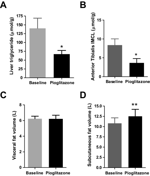
Despite increase in body weight, hepatic triglyceride content decreased by ∼50% after pioglitazone treatment (140.1 ± 28.1 vs. 67.0 ± 10.3 μmol/g, P = 0.03) (Fig. 5A), and tibialis anterior muscle triglyceride content decreased by ∼55% (8.37 ± 1.6 vs. 3.65 ± 1.14 μmol/g, P = 0.02) (Fig. 5B). Soleus triglyceride content was slightly lower after pioglitazone treatment (23.8 ± 3.3 vs. 20.8 ± 3.1 μmol/g, NS). Intramyocellular triglyceride content did not correlate with fasting (r = 0.27, P = 0.37) or mean postprandial EGP (80–260 min; r = 0.19, P = 0.52).
FIG. 5.
Liver triglyceride (A), muscle triglyceride (B), visceral fat (C), and subcutaneous fat (D) content at baseline and after pioglitazone treatment. Data are means ± SE. *P < 0.04; **P < 0.001.
Abdominal fat content.
Pioglitazone treatment was associated with a significant increase in subcutaneous fat content (10.8 ± 1.4 vs. 12.5 ± 1.7 l, P = 0.003) (Fig. 5D) and decreased visceral fat–to–subcutaneous fat ratio (0.68 ± 0.1 vs. 0.57 ± 0.1, P = 0.02). Total visceral fat content was unchanged (6.2 ± 0.3 vs. 6.2 ± 0.5 l, NS) (Fig. 5C). Visceral fat content did not correlate with fasting (r = −0.05, P = 0.88) or mean postprandial EGP (80–260 min) (r = 0.01, P = 0.76).
Substrate oxidation.
Fasting glucose oxidation was significantly lower (2.2 ± 0.2 vs. 1.5 ± 0.2 mg · kg−1 · min−1, P < 0.01) after pioglitazone treatment and accounted for 18 and 12% of fasting glucose disposal before and after pioglitzone treatment, respectively. Postprandial glucose oxidation remained lower after pioglitazone treatment until 120 min after the meal. Fasting (0.4 ± 0.1 vs. 0.2 ± 0.1 mg · kg−1 · min−1, NS) and postprandial lipid oxidation (0.2 ± 0.1 vs. 0.1 ± 0.1 mg · kg−1 · min−1, NS) were slightly but not significantly lower after pioglitazone treatment. Resting and postprandial energy expenditure was similar on both study days.
DISCUSSION
In keeping with our hypothesis, pioglitazone treatment in type 2 diabetes resulted in an ∼50% decrease in hepatic triglyceride and 55% reduction in muscle triglyceride, despite significant weight gain (∼6 kg). This was associated with a 26% decrease in fasting EGP and a twofold decrease in postprandial EGP. Hepatic triglyceride content was directly correlated with fasting EGP (r = 0.64, P = 0.01) and inversely correlated with percentage suppression of EGP (time 150 min, r = −0.63, P = 0.02) before and after pioglitazone treatment. By stepwise regression, hepatic triglyceride was found to be a significant independent predictor of fasting EGP, accounting for 53% of its variation (P < 0.001). Similarly, hepatic triglyceride content accounted for 36% of the variation seen in the percentage suppression of postprandial EGP observed at 150 min (P = 0.03). In contrast, although muscle triglyceride content decreased with pioglitazone treatment, it did not correlate with EGP. There was no relationship between visceral fat content with EGP.
In type 2 diabetes, hepatic insulin resistance is believed to underlie the elevated basal rates of EGP and subnormal postprandial suppression of EGP (23,24). In a recent study, elevated EGP was found to be a primary mechanism for postprandial hyperglycemia in type 2 diabetes (25). Hepatic triglyceride content is characteristically increased in type 2 diabetes and is an important predictor of hepatic insulin resistance (1,26), and this is true even independent of obesity (27). In addition, hepatic triglyceride content correlated negatively with postprandial net hepatic glycogen synthesis in type 2 diabetes (3). Although the exact cause of the excess accumulation of hepatic triglyceride in type 2 diabetes is unclear, we have previously shown this is likely to be at least partly secondary to increased postprandial uptake of dietary fatty acids by the liver (28). Other plausible mechanisms include increased FFA delivery from visceral depots and reduced intrahepatic FFA oxidation (29).
Hepatic steatosis induced by 3-day high-fat feeding resulted in reduced insulin activation of insulin receptor substrate (IRS)-1–and IRS-2–associated phosphatidylinositol 3-kinase activity, increased gluconeogenesis, and liver-specific insulin resistance (30). By reversal of this principle, a very–low-fat diet induced decrease in hepatic triglyceride in patients with type 2 diabetes was shown to markedly improve both EGP suppression during insulin infusion and hepatic insulin sensitivity (2). Similarly, we observed a ∼50% decrease in hepatic triglyceride content after pioglitazone treatment (Fig. 5A), and this was associated with significant improvement in fasting and postprandial EGP (Fig. 3).
Increased EGP in type 2 diabetes is contributed to by both increased gluconeogenesis and glycogenolysis (25), but the effect of gluconeogenesis may predominate (31,32). The results of this study suggest that pioglitazone-induced reduction in EGP is likely to reflect changes in either gluconeogenesis and/or glycogenolysis. In a recent study, pioglitazone treatment improved fasting and postprandial glycemia, principally via inhibition of gluconeogenesis (33). The effect of thiazolidinediones on glycogenolysis is yet to be determined. Pioglitazone has been shown to decrease gluconeogenesis (34) and to inhibit expression of key genes involved in gluconeogenesis (35). In addition, FFAs are potent stimulators of gluconeogenesis, and thiazolidinediones decrease FFA levels (36,37). Pioglitazone also markedly increases adiponectin levels (38,39), and adiponectin has been shown to decrease gluconeogenesis and EGP (40,41). Hyperglucagonemia is associated with increased basal EGP (42), and we observed a major decrease in fasting glucagon levels after pioglitazone treatment. Because fasting insulin levels were relatively unchanged after pioglitazone treatment, the effect of pioglitazone on fasting EGP is likely to be secondary to its effect, either directly or indirectly, on the liver.
We observed that the initial postprandial suppression of EGP both before and after pioglitazone was relatively normal. Using a similar dynamic tracer methodology, we have previously reported similar initial postprandial suppression of EGP in subjects with type 2 diabetes compared with matched controls (43). The conventional dual tracer approach with fixed priming and fixed-rate tracer infusion has been extensively used to measure EGP and documents subnormal suppression of EGP in type 2 diabetes (44,45). However, by infusing the intravenous tracer to mimic the expected pattern of EGP, the tracer-to-tracee ratio is maintained at an approximate constant, and the EGP estimate is model independent and is not overestimated (43). This has recently been quantitatively assessed (13). In contrast to our observations, a recent study reported a much slower rate of suppression of EGP after pioglitazone (33), and this is likely to be a consequence of using a fixed-rate tracer infusion for a dynamic meal study.
The rapid early suppression of EGP needs to be considered. Hepatic insulin action is relatively unimpaired at high insulin levels (46), and glucose effectiveness is also preserved in type 2 diabetes (47). In the immediate postprandial period, the change in plasma FFAs is fairly subtle, and glucagon levels actually rise. It has been suggested that FFA concentrations signal the suppression of EGP (portal hypothesis) (48). However, our data do not support this because there is robust suppression (∼75%) of EGP despite only an ∼15% decrease in FFAs by the 30-min postprandial measurement. It is hence most likely that the direct effect of rising hepatic sinusoidal insulin is the primary determinant of EGP in this period.
Impaired postprandial suppression of FFAs and glucagon is a characteristic feature of type 2 diabetes (44,49), and both FFAs and glucagon (25,50,51) have been implicated in the abnormal regulation of EGP. Recently, Woerle et al. (25) have demonstrated that postprandial EGP was significantly correlated with insulin-to-glucagon molar ratio, with postprandial decrements in both plasma glucagon and plasma FFAs. In the current study, between 80 and 260 min after the meal, pioglitazone treatment resulted in a twofold greater suppression of EGP compared with pretreatment. Unlike the immediate postprandial period, insulin levels fall during this period, and FFAs and glucagon may exert a more pronounced effect on EGP. Insulin levels and insulin secretion rates were relatively unchanged between the 2 study days, but postprandial FFA and glucagon levels were significantly lower after pioglitazone treatment. We observed a strong correlation between the nadir FFA levels and percentage suppression of EGP at 210 min (Fig. 4C). Hence, it is likely that pioglitazone-induced interruption of intrahepatic fatty acid supply may underlie the beneficial effects of EGP during this time.
Although pioglitazone treatment decreased muscle triglyceride content as seen in previous studies (52,53), we observed no correlation between muscle triglyceride and EGP. The mechanism of reduction of muscle triglyceride by pioglitazone is unclear. Pioglitazone is postulated to increase tissue fatty acid oxidation through its effect on increasing adiponectin and consequent activation of adenosine monophosphate–activated protein kinase (53), but muscle fatty acid oxidative enzyme levels were not observed to change after pioglitazone treatment in a recent study (52). In contrast to previous studies, we did not observe any significant change in visceral fat content after pioglitazone treatment. The fact that EGP improved independent of change in visceral fat content implies that visceral fat is unlikely to be a critical determinant of EGP, and this clearly refutes the portal hypothesis in the control of EGP (48).
Potential shortcomings of the study must be considered. First, the small study sample size is a potential weakness of the study, but the subject characteristics are typical for type 2 diabetes, and the main findings are clear-cut. The small sample size is also likely to explain the lack of statistical significance of lipid oxidation between the study days. Second, the study was a within-subject comparative study by design and did not have a placebo arm. Third, gluconeogenesis and glycogenolysis were not separately quantified; hence, the precise mechanism of reduction in EGP with pioglitazone could not been determined. Nonetheless, the study defines the precise effects of pioglitazone on postprandial glucose metabolism and the determinants of EGP in type 2 diabetes.
In summary, we have demonstrated that 16-week treatment with pioglitazone improves fasting and postprandial EGP suppression and decreases liver and muscle triglyceride content despite overall increase in body weight. Increased hepatic triglyceride content is associated with subnormal postprandial suppression of EGP and is an independent determinant of fasting and postprandial EGP. This study provides further insight into the mechanism of action of pioglitazone and suggests that hepatic triglyceride is a useful therapeutic target for management of type 2 diabetes.
Acknowledgments
This research was supported by an unrestricted educational grant from Takeda Pharmaceuticals, U.K. The company had no input into study design, data interpretation, or manuscript preparation.
We gratefully acknowledge the cooperation and time given by all our volunteers.
Published ahead of print at http://diabetes.diabetesjournals.org on 5 June 2008.
The costs of publication of this article were defrayed in part by the payment of page charges. This article must therefore be hereby marked “advertisement” in accordance with 18 U.S.C. Section 1734 solely to indicate this fact.
REFERENCES
- 1.Ryysy L, Hakkinen AM, Goto T, Vehkavaara S, Westerbacka J, Halavaara J, Yki-Jarvinen H: Hepatic fat content and insulin action on free fatty acids and glucose metabolism rather than insulin absorption are associated with insulin requirements during insulin therapy in type 2 diabetic patients. Diabetes 49 :749 –758,2000 [DOI] [PubMed] [Google Scholar]
- 2.Petersen KF, Dufour S, Befroy D, Lehrke M, Hendler RE, Shulman GI: Reversal of nonalcoholic hepatic steatosis, hepatic insulin resistance, and hyperglycemia by moderate weight reduction in patients with type 2 diabetes. Diabetes 54 :603 –608,2005 [DOI] [PMC free article] [PubMed] [Google Scholar]
- 3.Krssak M, Brehm A, Bernroider E, Anderwald C, Nowotny P, Dalla Man C, Cobelli C, Cline GW, Shulman GI, Waldhausl W, Roden M: Alterations in postprandial hepatic glycogen metabolism in type 2 diabetes. Diabetes 53 :3048 –3056,2004 [DOI] [PubMed] [Google Scholar]
- 4.Shulman GI: Cellular mechanisms of insulin resistance. J Clin Invest 106 :171 –176,2000 [DOI] [PMC free article] [PubMed] [Google Scholar]
- 5.Bajaj M, Suraamornkul S, Pratipanawatr T, Hardies LJ, Pratipanawatr W, Glass L, Cersosimo E, Miyazaki Y, DeFronzo RA: Pioglitazone reduces hepatic fat content and augments splanchnic glucose uptake in patients with type 2 diabetes. Diabetes 52 :1364 –1370,2003 [DOI] [PubMed] [Google Scholar]
- 6.Suter SL, Nolan JJ, Wallace P, Gumbiner B, Olefsky JM: Metabolic effects of new oral hypoglycemic agent CS-045 in NIDDM subjects. Diabetes Care 15 :193 –203,1992 [DOI] [PubMed] [Google Scholar]
- 7.Miyazaki Y, Mahankali A, Matsuda M, Glass L, Mahankali S, Ferrannini E, Cusi K, Mandarino LJ, DeFronzo RA: Improved glycemic control and enhanced insulin sensitivity in type 2 diabetic subjects treated with pioglitazone. Diabetes Care 24 :710 –719,2001 [DOI] [PubMed] [Google Scholar]
- 8.Miyazaki Y, Mahankali A, Matsuda M, Mahankali S, Hardies J, Cusi K, Mandarino LJ, DeFronzo RA: Effect of pioglitazone on abdominal fat distribution and insulin sensitivity in type 2 diabetic patients. J Clin Endocrinol Metab 87 :2784 –2791,2002 [DOI] [PubMed] [Google Scholar]
- 9.Hother-Nielsen O, Beck-Nielsen H: On the determination of basal glucose production rate in patients with type 2 (non-insulin-dependent) diabetes mellitus using primed-continuous 3-3H-glucose infusion. Diabetologia 33 :603 –610,1990 [DOI] [PubMed] [Google Scholar]
- 10.Frayn KN: Calculation of substrate oxidation rates in vivo from gaseous exchange. J Appl Physiol 55 :628 –634,1983 [DOI] [PubMed] [Google Scholar]
- 11.Cobelli C, Toffolo G: Constant specific activity input allows reconstruction of endogenous glucose concentration in non-steady state. Am J Physiol 258 :E1037 –E1040,1990 [DOI] [PubMed] [Google Scholar]
- 12.Cobelli C FD, Toffolo G: Tracer Kinetics in Biomedical Research: From Data to Model. New York, Kluwer Academic/Plenum,2000
- 13.Toffolo G, Basu R, Dalla Man C, Rizza R, Cobelli C: Assessment of postprandial glucose metabolism: conventional dual- vs. triple-tracer method. Am J Physiol Endocrinol Metab 291 :E800 –E806,2006 [DOI] [PubMed] [Google Scholar]
- 14.Steele R: Influences of glucose loading and of injected insulin on hepatic glucose output. Ann N Y Acad Sci 82 :420 –430,1959 [DOI] [PubMed] [Google Scholar]
- 15.Steele R, Wall JS, De Bodo RC, Altszuler N: Measurement of size and turnover rate of body glucose pool by the isotope dilution method. Am J Physiol 187 :15 –24,1956 [DOI] [PubMed] [Google Scholar]
- 16.Radziuk J, Norwich KH, Vranic M: Experimental validation of measurements of glucose turnover in nonsteady state. Am J Physiol 234 :E84 –E93,1978 [DOI] [PubMed] [Google Scholar]
- 17.Sparacino G, Pillonetto G, Capello M, De Nicolao G, Cobelli C: WINSTODEC: a stochastic deconvolution interactive program for physiological and pharmacokinetic systems. Comput Methods Programs Biomed 67 :67 –77,2002 [DOI] [PubMed] [Google Scholar]
- 18.Bottomley PA: Spatial localization in NMR spectroscopy in vivo. Ann N Y Acad Sci 508 :333 –348,1987 [DOI] [PubMed] [Google Scholar]
- 19.Naressi A, Couturier C, Castang I, de Beer R, Graveron-Demilly D: Java-based graphical user interface for MRUI, a software package for quantitation of in vivo/medical magnetic resonance spectroscopy signals. Comput Biol Med 31 :269 –286,2001 [DOI] [PubMed] [Google Scholar]
- 20.Boesch C, Slotboom J, Hoppeler H, Kreis R: In vivo determination of intra-myocellular lipids in human muscle by means of localized 1H-MR-spectroscopy. Magn Reson Med 37 :484 –493,1997 [DOI] [PubMed] [Google Scholar]
- 21.Szczepaniak LS, Babcock EE, Schick F, Dobbins RL, Garg A, Burns DK, McGarry JD, Stein DT: Measurement of intracellular triglyceride stores by H spectroscopy: validation in vivo. Am J Physiol 276 :E977 –E989,1999 [DOI] [PubMed] [Google Scholar]
- 22.Thomas EL, Saeed N, Hajnal JV, Brynes A, Goldstone AP, Frost G, Bell JD: Magnetic resonance imaging of total body fat. J Appl Physiol 85 :1778 –1785,1998 [DOI] [PubMed] [Google Scholar]
- 23.Gerich JE: Is muscle the major site of insulin resistance in type 2 (non-insulin-dependent) diabetes mellitus? Diabetologia 34 :607 –610,1991 [DOI] [PubMed] [Google Scholar]
- 24.DeFronzo RA: Pathogenesis of type 2 (non-insulin dependent) diabetes mellitus: a balanced overview. Diabetologia 35 :389 –397,1992 [DOI] [PubMed] [Google Scholar]
- 25.Woerle HJ, Szoke E, Meyer C, Dostou JM, Wittlin SD, Gosmanov NR, Welle SL, Gerich JE: Mechanisms for abnormal postprandial glucose metabolism in type 2 diabetes. Am J Physiol Endocrinol Metab 290 :E67 –E77,2006 [DOI] [PubMed] [Google Scholar]
- 26.Banerji MA, Buckley MC, Chaiken RL, Gordon D, Lebovitz HE, Kral JG: Liver fat, serum triglycerides and visceral adipose tissue in insulin-sensitive and insulin-resistant black men with NIDDM. Int J Obes Relat Metab Disord 19 :846 –850,1995 [PubMed] [Google Scholar]
- 27.Seppala-Lindroos A, Vehkavaara S, Hakkinen AM, Goto T, Westerbacka J, Sovijarvi A, Halavaara J, Yki-Jarvinen H: Fat accumulation in the liver is associated with defects in insulin suppression of glucose production and serum free fatty acids independent of obesity in normal men. J Clin Endocrinol Metab 87 :3023 –3028,2002 [DOI] [PubMed] [Google Scholar]
- 28.Ravikumar B, Carey PE, Snaar JE, Deelchand DK, Cook DB, Neely RD, English PT, Firbank MJ, Morris PG, Taylor R: Real-time assessment of postprandial fat storage in liver and skeletal muscle in health and type 2 diabetes. Am J Physiol Endocrinol Metab 288 :E789 –E797,2005 [DOI] [PubMed] [Google Scholar]
- 29.Yki-Jarvinen H: Fat in the liver and insulin resistance. Ann Med 37 :347 –356,2005 [DOI] [PubMed] [Google Scholar]
- 30.Samuel VT, Liu ZX, Qu X, Elder BD, Bilz S, Befroy D, Romanelli AJ, Shulman GI: Mechanism of hepatic insulin resistance in non-alcoholic fatty liver disease. J Biol Chem 279 :32345 –32353,2004 [DOI] [PubMed] [Google Scholar]
- 31.Magnusson I, Rothman DL, Katz LD, Shulman RG, Shulman GI: Increased rate of gluconeogenesis in type II diabetes mellitus: a 13C nuclear magnetic resonance study. J Clin Invest 90 :1323 –1327,1992 [DOI] [PMC free article] [PubMed] [Google Scholar]
- 32.Gastaldelli A, Baldi S, Pettiti M, Toschi E, Camastra S, Natali A, Landau BR, Ferrannini E: Influence of obesity and type 2 diabetes on gluconeogenesis and glucose output in humans: a quantitative study. Diabetes 49 :1367 –1373,2000 [DOI] [PubMed] [Google Scholar]
- 33.Gastaldelli A, Casolaro A, Pettiti M, Nannipieri M, Ciociaro D, Frascerra S, Buzzigoli E, Baldi S, Mari A, Ferrannini E: Effect of pioglitazone on the metabolic and hormonal response to a meal in type II diabetes. Clin Pharmacol Ther 81 :205 –212,2007 [DOI] [PubMed] [Google Scholar]
- 34.Nishimura Y, Inoue Y, Takeuchi H, Oka Y: Acute effects of pioglitazone on glucose metabolism in perfused rat liver. Acta Diabetol 34 :206 –210,1997 [DOI] [PubMed] [Google Scholar]
- 35.Way JM, Harrington WW, Brown KK, Gottschalk WK, Sundseth SS, Mansfield TA, Ramachandran RK, Willson TM, Kliewer SA: Comprehensive messenger ribonucleic acid profiling reveals that peroxisome proliferator-activated receptor gamma activation has coordinate effects on gene expression in multiple insulin-sensitive tissues. Endocrinology 142 :1269 –1277,2001 [DOI] [PubMed] [Google Scholar]
- 36.Bajaj M, Suraamornkul S, Piper P, Hardies LJ, Glass L, Cersosimo E, Pratipanawatr T, Miyazaki Y, DeFronzo RA: Decreased plasma adiponectin concentrations are closely related to hepatic fat content and hepatic insulin resistance in pioglitazone-treated type 2 diabetic patients. J Clin Endocrinol Metab 89 :200 –206,2004 [DOI] [PubMed] [Google Scholar]
- 37.Miyazaki Y, Glass L, Triplitt C, Matsuda M, Cusi K, Mahankali A, Mahankali S, Mandarino LJ, DeFronzo RA: Effect of rosiglitazone on glucose and non-esterified fatty acid metabolism in type II diabetic patients. Diabetologia 44 :2210 –2219,2001 [DOI] [PubMed] [Google Scholar]
- 38.Gastaldelli A, Miyazaki Y, Mahankali A, Berria R, Pettiti M, Buzzigoli E, Ferrannini E, DeFronzo RA: The effect of pioglitazone on the liver: role of adiponectin. Diabetes Care 29 :2275 –2281,2006 [DOI] [PubMed] [Google Scholar]
- 39.Miyazaki Y, Mahankali A, Wajcberg E, Bajaj M, Mandarino LJ, DeFronzo RA: Effect of pioglitazone on circulating adipocytokine levels and insulin sensitivity in type 2 diabetic patients. J Clin Endocrinol Metab 89 :4312 –4319,2004 [DOI] [PubMed] [Google Scholar]
- 40.Combs TP, Berg AH, Obici S, Scherer PE, Rossetti L: Endogenous glucose production is inhibited by the adipose-derived protein Acrp30. J Clin Invest 108 :1875 –1881,2001 [DOI] [PMC free article] [PubMed] [Google Scholar]
- 41.Zhou H, Song X, Briggs M, Violand B, Salsgiver W, Gulve EA, Luo Y: Adiponectin represses gluconeogenesis independent of insulin in hepatocytes. Biochem Biophys Res Commun 338 :793 –799,2005 [DOI] [PubMed] [Google Scholar]
- 42.Baron AD, Schaeffer L, Shragg P, Kolterman OG: Role of hyperglucagonemia in maintenance of increased rates of hepatic glucose output in type II diabetics. Diabetes 36 :274 –283,1987 [DOI] [PubMed] [Google Scholar]
- 43.Singhal P, Caumo A, Carey PE, Cobelli C, Taylor R: Regulation of endogenous glucose production after a meal in type 2 diabetes. Am J Physiol Endocrinol Metab 283 :E275 –E283,2002 [DOI] [PubMed] [Google Scholar]
- 44.Firth RG, Bell PM, Marsh HM, Hansen I, Rizza RA: Postprandial hyperglycaemia in patients with noninsulin-dependent diabetes mellitus: role of hepatic and extrahepatic tissues. J Clin Invest 77 :1525 –1532,1986 [DOI] [PMC free article] [PubMed] [Google Scholar]
- 45.Mitrakou A, Kelley D, Veneman T, Jenssen T, Pangbaum T, Reilly J, Gerich J: Contribution of abnormal muscle and liver glucose metabolism to postprandial hyperglycaemia in NIDDM. Diabetes 39 :1381 –1390,1990 [DOI] [PubMed] [Google Scholar]
- 46.Turk D, Alzaid A, Dinneen S, Nair KS, Rizza R: The effects of non-insulin-dependent diabetes mellitus on the kinetics of onset of insulin action in hepatic and extrahepatic tissues. J Clin Invest 95 :755 –762,1995 [DOI] [PMC free article] [PubMed] [Google Scholar]
- 47.Alzaid AA, Dinneen SF, Turk DJ, Caumo A, Cobelli C, Rizza RA: Assessment of insulin action and glucose effectiveness in diabetic and nondiabetic humans. J Clin Invest 94 :2341 –2348,1994 [DOI] [PMC free article] [PubMed] [Google Scholar]
- 48.Mittelman SD, Fu YY, Rebrin K, Steil G, Bergman RN: Indirect effect of insulin to suppress endogenous glucose production is dominant, even with hyperglucagonemia. J Clin Invest 100 :3121 –3130,1997 [DOI] [PMC free article] [PubMed] [Google Scholar]
- 49.Reaven GM, Hollenbeck C, Jeng CY, Wu MS, Chen YD: Measurement of plasma glucose, free fatty acid, lactate, and insulin for 24 h in patients with NIDDM. Diabetes 37 :1020 –1024,1988 [DOI] [PubMed] [Google Scholar]
- 50.Boden G, Chen X, Capulong E, Mozzoli M: Effects of free fatty acids on gluconeogenesis and autoregulation of glucose production in type 2 diabetes. Diabetes 50 :810 –816,2001 [DOI] [PubMed] [Google Scholar]
- 51.Shah P, Basu A, Basu R, Rizza R: Impact of lack of suppression of glucagon on glucose tolerance in humans. Am J Physiol 277 :E283 –E290,1999 [DOI] [PubMed] [Google Scholar]
- 52.Rasouli N, Raue U, Miles LM, Lu T, Di Gregorio GB, Elbein SC, Kern PA: Pioglitazone improves insulin sensitivity through reduction in muscle lipid and redistribution of lipid into adipose tissue. Am J Physiol Endocrinol Metab 288 :E930 –E934,2005 [DOI] [PubMed] [Google Scholar]
- 53.Teranishi T, Ohara T, Maeda K, Zenibayashi M, Kouyama K, Hirota Y, Kawamitsu H, Fujii M, Sugimura K, Kasuga M: Effects of pioglitazone and metformin on intracellular lipid content in liver and skeletal muscle of individuals with type 2 diabetes mellitus. Metabolism 56 :1418 –1424,2007 [DOI] [PubMed] [Google Scholar]


