Abstract
OBJECTIVE—Rapamycin is an immunosuppressive drug currently used to prevent graft rejection in humans, which is considered permissive for tolerance induction. Rapamycin allows expansion of both murine and human naturally occurring CD4+CD25+FOXP3+ T regulatory cells (nTregs), which are pivotal for the induction and maintenance of peripheral tolerance. Preclinical murine models have shown that rapamycin enhances nTreg proliferation and regulatory function also in vivo. Objective of this study was to assess whether rapamycin has in vivo effects on human nTregs.
RESEARCH DESIGN AND METHODS—nTreg numbers and function were examined in a unique set of patients with type 1 diabetes who underwent rapamycin monotherapy before islet transplantation.
RESULTS—We found that rapamycin monotherapy did not alter the frequency and functional features, namely proliferation and cytokine production, of circulating nTregs. However, nTregs isolated from type 1 diabetic patients under rapamycin treatment had an increased capability to suppress proliferation of CD4+CD25− effector T-cells compared with that before treatment.
CONCLUSIONS—These findings demonstrate that rapamycin directly affects human nTreg function in vivo, which consists of refitting their suppressive activity, whereas it does not directly change effector T-cell function.
Type 1 diabetes results from a chronic destruction of insulin-producing pancreatic β-cells mediated by autoreactive T-cells (1). Circulating T-cells able to react against β-cell autoantigens have been demonstrated in both healthy donors and type 1 diabetic subjects (2). However, we recently showed that naïve T-cells recognizing β-cell autoantigens are present in each individual, irrespective of disease occurrence, whereas diabetes-specific autoreactive T-cells that have undergone sustained in vivo proliferation and differentiation into memory T-cells are a hallmark of patients only (3). These data suggest that active mechanisms of peripheral tolerance present in healthy individuals are likely to be inadequate in type 1 diabetic subjects.
Among the several mechanisms accounting for peripheral tolerance, suppression mediated by regulatory T-cells (Tregs) is considered crucial for controlling autoimmune responses (rev. in 4). Various subsets of Tregs have been described so far, and the naturally occurring CD4+ Tregs (nTregs) represent the only cell population originating from the thymus and therefore present since birth in the circulation, where they represent ∼5–10% of total CD4+ T-cells. nTregs are crucial for maintaining tolerance by downregulating undesired immune responses to self- and non–self-antigens. nTregs are defined on the basis of constitutive high expression of the interleukin (IL)-2Rα (CD25), the transcription factor forkhead box P3 (FOXP3) (5), low or absent expression of the IL-7R (CD127) (6,7), and the inability to produce IL-2 and to proliferate in vitro (5).
The variety of human autoimmune diseases in which a defect in nTreg function has been proposed is of interest, raising the possibility that this may be a common mechanism leading to uncontrolled immune responses to self-antigens (8). It remains controversial whether nTregs are defective in type 1 diabetic patients. Some reports suggest that normal numbers of circulating nTregs are present in type 1 diabetic patients, but their suppressive activity is defective in vitro (9–11). However, others do not show an in vitro suppressive defect in type 1 diabetes nTregs compared with those in healthy individuals (12). Studies in mice clearly demonstrate that depletion of nTregs results in systemic autoimmune diseases (diabetes included), and adoptive transfer of nTregs prevents development of type 1 diabetes in NOD mice and, in some experimental settings, also cures ongoing disease (13–15). As a result of these preclinical studies, nTregs are nowadays considered a promising therapeutic tool for reestablishing self-tolerance in type 1 diabetes and other T-cell–mediated diseases (16). As a therapeutic approach, one can envisage either to adoptively transfer nTregs previously expanded ex vivo (because of their limited circulating number) or to directly expand nTregs and/or boost their suppressive function in vivo with selected immunomodulatory compounds.
We demonstrated that rapamycin, a non-calcineurin inhibitor currently used to prevent acute graft rejection after allogeneic transplants (17), allows expansion of murine nTregs in vitro (18). In addition, in vivo administration of rapamycin prevents type 1 diabetes in NOD mice and reestablishes long-term tolerance to self-antigens through the expansion of nTregs (19). In humans, rapamycin promotes nTreg expansion in vitro through selective inhibition of effector T-cell proliferation (20) and does not interfere with de novo induction of Treg cells from naïve CD4+ T-cells (21). Both of these biological effects can favor tolerance induction in vivo. It has been recently shown that in renal transplant recipients who underwent profound T-cell depletion by Campath-1H induction, maintenance therapy with rapamycin but not cyclosporine A increases the pool of circulating CD4+CD25highFOXP3+ T-cells (22). Because rapamycin is commonly administered in transplanted patients in combination with other drugs, so far it has not been feasible to define whether rapamycin has a direct effect in vivo on human nTregs as we previously demonstrated in vitro (20) and in a preclinical murine model of type 1 diabetes (19).
We have been using a clinical protocol in which rapamycin monotherapy is given to long-term type 1 diabetic patients before islet transplantation to reach therapeutic plasma levels at the time of islet infusion (23), followed by maintenance immunotherapy as described by the Edmonton group (24). This study provided the opportunity to investigate the in vivo effect of rapamycin therapy on nTreg number and function in patients with autoimmune disease. We demonstrate that rapamycin treatment does not modify number, phenotype, ability to proliferate, and ability to produce cytokines of circulating CD4+CD25brightFOXP3+ nTregs. However, the suppressive capacity of highly purified CD4+CD25bright T-cells is improved in type 1 diabetic patients during rapamycin treatment compared with that of the same patients tested before treatment. Thus, rapamycin has an in vivo direct effect on human nTreg function, supporting its use in clinical immunosuppressive regimens aimed at tolerance induction.
RESEARCH DESIGN AND METHODS
Patients and blood collection.
Patients with long-lasting type 1 diabetes (>5 years) who had reduced awareness of hypoglycemia, brittle diabetes, or progressive complications, despite optimization of insulin therapy, were candidates for solitary islet transplant at the Telethon-Juvenile Diabetes Research Foundation Center for Beta Cell Replacement, San Raffaele Scientific Institute, Milan. In this study, six patients received rapamycin treatment at 0.1 mg · kg−1 · day−1 (Table 1). Each patient is identifiable by a specific symbol, which can be followed throughout the manuscript (e.g., patient Hsr-066-ITA-rp06 is recognizable by the ○ symbol). Peripheral blood was obtained before and during rapamycin treatment (before receiving islet transplantation) after informed consent and ethics committee approval. Five normal donors of similar age and sex to the patients were recruited through the courtesy of Centro Trasfusionale, San Raffaele Scientific Institute, Milan, and donated peripheral blood after informed consent and ethics committee approval.
TABLE 1.
T1D patient description
| Patient code | Patient symbol* | Age (years) | Sex | Diabetes duration (years) | RAPA treatment (days) | [RAPA]† (ng/ml) | C-peptide (ng/ml) |
A1C (%) |
EIR (units · kg−1 · day−1) |
|||
|---|---|---|---|---|---|---|---|---|---|---|---|---|
| PRE | RAPA | PRE | RAPA | PRE | RAPA | |||||||
| Hsr-066-ITA-rp06 | ○ | 39 | M | 32 | 109 | 9 | 0.70 | 0.77 | 9.5 | 8.9 | 0.63 | 0.53 |
| Hsr-065-ITA-rp05 | • | 40 | F | 11 | 153 | 24 | 0.15 | 0.28 | 8.7 | 8.7 | 0.89 | 0.79 |
| Hsr-069-ITA7-rp07 | 31 | M | 26 | 32 | 21 | 0.02 | 0.02 | 9.1 | nt | 0.63 | 0.51 | |
| Hsr-063-ITA-rp03 | ▴ | 48 | F | 37 | 130 | 5 | 0.03 | 0.16 | 6.2 | 6.6 | 0.73 | 0.71 |
| Hsr-064-ITA-rp04 | □ | 33 | F | 21 | 109 | 12 | 0.02 | 0.02 | 8.9 | 9.5 | 0.65 | 0.57 |
| Hsr-ATG-ITA-rp02 | ▪ | 32 | M | 7 | 97 | 5 | 0.01 | 0.01 | 8.7 | 8.7 | 0.74 | 0.42 |
| Mean | 39.2 | 22.3 | 105 | 13 | ||||||||
| Range | 31–48 | 7–37 | 32–153 | 5–24 | ||||||||
Symbols recalled throughout the manuscript.
Circulating plasma rapamycin concentration. EIR, exogenous insulin requirements; PRE, sample before rapamycin treatment; RAPA, sample during rapamycin treatment.
Peripheral blood mononuclear cell isolation.
Peripheral blood mononuclear cells (PBMCs) were isolated over a Ficoll-Hypaque (Amersham Pharmacia Biotech Europe, Uppsala, Sweden) density gradient centrifugation from sodium-heparinized venous blood samples and washed twice in PBS (Cambrex-Biowhittake, Walkersville, MD). All samples were frozen in FCS (Cambrex-Biowhittaker) containing 10% dimethyl sulfoxide (Sigma-Aldrich, St. Louis, MO) in a controlled-rate automated freezing device to −80°C and then stored in liquid nitrogen. All of the experiments were performed on thawed cells. Samples from the same type 1 diabetic patient collected before and during rapamycin treatment were tested simultaneously in parallel to cells from one normal donor. Each patient was tested alongside a separate normal donor.
Flow cytometry.
Thawed PBMCs were stained for surface antigens with the following monoclonal antibodies (mAbs) all purchased from BD Pharmingen (San Diego, CA): anti-CD4 PerCP (peridinin chlorophyll protein) (clone SK3), anti-CD25 allophycocyanin (clone 2A3), anti-CD62L phycoerytrin (PE) (clone SK11), anti-CD127 PE (clone M21), anti-CTLA4 biotin (clone BN13), anti-CD45RO fluorescein isothiocyanate (FITC) (clone UCHL1), and anti–HLA-DR PE (clone TU36) mAbs. Intracellular staining for human FOXP3 was performed using the anti FOXP3-Alexa 488 mAb (clone 259D; BioLegend, San Diego, CA) according to the manufacturer's instructions. At least 20,000 events were acquired from each sample on a BD FACScalibur and analyzed with FCS Express V3 software.
Cell sorting.
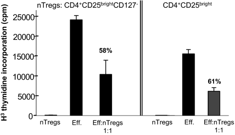
Thawed PBMCs were stained with anti-CD4 FITC (clone SK3; BD Pharmingen) and anti-CD25 PE (clone 2A3; BD Pharmingen). CD4+CD25bright (top 1–2%) and CD4+CD25− fractions were sorted with a fluorescence-activated cell sorter (FACS; BD FACSvantage) (for gating strategy, see Fig. 2). Sorted CD4+CD25− and CD4+CD25bright T-cells had a purity of 96–98% both from type 1 diabetic patients (before and during rapamycin treatment) and from normal donors. Sorting CD4+CD127lowCD25bright T-cells provided a population of nTregs with suppressive ability similar to those sorted as CD4+CD25bright cells, contrary to what has previously been published (Fig. 1) (6). We therefore concluded that there was no need to sort nTregs based on the expression of CD127.
FIG. 2.
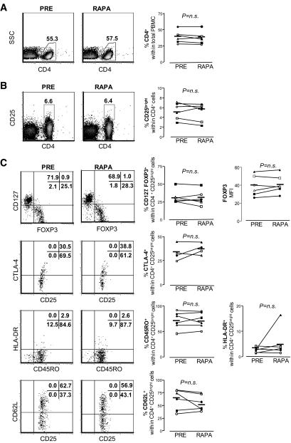
Percentages and phenotype of circulating CD4+ T-cells in type 1 diabetic patients before and during rapamycin treatment. PBMCs isolated from type 1 diabetic patients before (PRE) and during (RAPA) rapamycin treatment were stained with the indicated mAbs and analyzed by FACS. Representative plots of samples collected and analyzed from patient Hsr-064-ITA-rp04, before and during rapamycin therapy, are shown on the left. Numbers indicate how many cells express each marker. Graphs including analyses performed in all patients (each distinguishable by a specific symbol, see Table 1) are shown on the right, and the solid line represents the average level of type 1 diabetes PRE and type 1 diabetes RAPA samples. Statistical analysis is shown in each graph. A: Percentages of total CD4+ T-cells within PBMCs. B: Percentages of CD25bright T-cells within the CD4+ T-cell compartment. C: Percentages of CD127−FOXP3+, CTLA-4+, CD45RO+, HLA-DR+, and CD62L+ cells within the CD4+CD25bright T-cells. FOXP3 mean fluorescence intensity of CD4+CD25bright CD127− FOXP3+ cells is shown for both PRE and RAPA samples.
FIG. 1.
In vitro suppressive function of FACS-sorted CD4+CD25brightCD127− and CD4+CD25bright T-cells. FACS-sorted 104 effector CD4+CD25− T-cells (Eff.) were activated polyclonally with magnetic beads coated with anti-CD3+CD28 mAbs alone or in the presence of equal amounts of FACS-sorted autologous CD4+CD25brightCD127− nTregs (left) or CD4+CD25bright nTregs (right). Cell proliferation was assessed in all cultures at day 4 after addition of [3H]thymidine for the last 18 h of culture. Percentage of suppression is indicated. One representative experiment of four is shown.
Suppression assay.
FACS-sorted CD4+CD25− T-cells were plated at 1 × 10⁴ cells per well (in 200 μl X-vivo 15 [Cambrex-Biowhittaker] supplemented with 10% human serum AB [Sigma], 100 units/ml penicillin, and 100 units/ml streptomycin) in 96 round-bottom plates and stimulated with anti CD3/CD28-coupled beads (one bead per six cells) (Invitrogen-Dynal, Oslo, Norway). FACS-sorted CD4+CD25bright T-cells were added at a 1:1 ratio (CD4+CD25−:CD4+CD25bright). T-cell proliferation was assessed at day 4, after addition of [3H]thymidine for the last 18 h of culture (1 μCi per well) (Amersham, Buckingham, U.K.). Suppressive activity of CD4+CD25bright T-cells was measured as inhibition of cell proliferation of CD4+CD25− T-cells compared with proliferation of CD4+CD25− T-cells stimulated in the absence of nTregs. Historical data from our laboratory demonstrate that repeated measures of suppression using normal donor cells is relatively consistent (data not shown). In one patient, the suppression assay was performed using the CFSE dilution assay. Briefly, 1 × 10⁵ FACS-sorted CD4+CD25− T-cells were stained with CFSE (Molecular Probes, Eugene, OR) as described previously (18). Following the same protocol as for CFSE staining, 1 × 10⁵ FACS-sorted CD4+CD25high T-cells were first stained with SNARF (Molecular Probes) and were subsequently mixed with an equal number of CFSE+CD4+CD25− T-cells in round-bottom 96-well plates precoated with 10 μg/ml anti-CD3 and 1 μg/ml soluble anti-CD28 mAbs (BD Biosciences). Seven days later, the cells were collected and analyzed by FACS. The proportion of CFSE+ (FL-1) T-cells proliferating in vitro was calculated by gating on lymphocytes plus alive cells (TOPRO− FL-4) (Molecular Probes) and by excluding SNARF+ (FL-2) cells. The number of gated cells (events) in a given cycle (division: n) was divided by 2 raised to power n, to calculate the percentage of original precursor cells from which they arose. The sum of original precursors from division 1–6 represents the number of precursors cells that proliferated. The percentage of CFSE+ divided cells was calculated by ([no. of precursors that proliferated1–6/no. of total precursors0–6] × 100) (18). The percentage of CD4+ T-cells CFSE+ cells divided in the presence of CD4+CD25high T-cells was compared with the percentage of CD4+ CFSE+ divided T-cells in the absence of nTregs.
Cytokine detections.
FACS-sorted CD4+CD25− and CD4+CD25bright T-cells were plated at 1 × 10⁴ cells per well (in 200 μl X-vivo 15 supplemented with 10% human serum AB, 100 units/ml penicillin, and 100 units/ml streptomycin) in 96 round-bottom plates and stimulated with anti CD3/CD28-coupled beads (one bead per six cells) (Invitrogen-Dynal). Culture supernatant was collected 3 days after activation and frozen at −80°C. Detection of IL-2, interferon-γ (IFN-γ), tumor necrosis factor-α (TNF-α), IL-4, IL-5, and IL-10 was performed using a cytometric bead array kit (BD Pharmingen) according to the manufacturer's instructions.
Statistical analysis.
Comparisons between patients and normal donors were performed using Student's t test. Comparisons between samples collected before and samples collected during rapamycin treatment were performed using Student's paired t test. In all cases, two-tailed P < 0.05 was considered significant. Analyses were performed using the Prism V4.03 software (GraphPad, San Diego, CA).
RESULTS
To define whether rapamycin monotherapy modifies number and phenotype of circulating nTregs, PBMCs isolated from type 1 diabetic patients before and during rapamycin treatment were thawed and tested for the expression of regulatory-cell markers (6,25,26). The percentage of circulating CD4+ T-cells was not altered by rapamycin treatment (Fig. 2A). Similarly, the percentage of CD25bright T-cells, within the CD4+ T-cell subset, did not change during rapamycin monotherapy compared with that before treatment (Fig. 2B). Markers of regulatory T-cells (namely, FOXP3, CD127, CTLA-4, HLA-DR, CD45RO, and CD62L) were also similarly expressed by CD4+CD25bright T-cells isolated before and during rapamycin monotherapy (Fig. 2C).
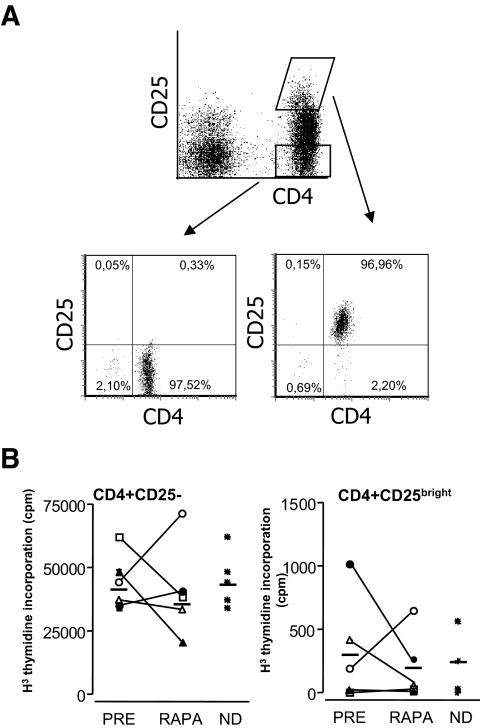
To determine whether rapamycin therapy modifies the functional features of peripheral CD4+CD25− effector T-cells and CD4+CD25bright T-cells (which should be enriched for nTregs), their proliferative capacity, cytokine production profile, and suppressive function were tested in vitro. The two T-cell subsets were first purified from type 1 diabetic patients before and during rapamycin monotherapy and from age-matched normal donors by flow cytometry (Fig. 3A). Purified CD4+CD25bright T-cells were all FOXP3+ (data not shown). FACS-sorted T-cells were activated in vitro by T-cell receptor–mediated polyclonal stimulation. Type 1 diabetic CD4+CD25− effector T-cells proliferated to a similar extent to those isolated from normal donors, irrespective of rapamycin therapy (Fig. 3B). CD4+CD25bright T-cells from type 1 diabetic subjects were anergic both before and during rapamycin treatment, as were those isolated from normal donors (Fig. 3B), and their proliferative capacity was restored on addition of exogenous IL-2 (data not shown). These data show that rapamycin does not alter the proliferative capacity of effector T-cells isolated from type 1 diabetic patients. In addition, the anergic phenotype of CD4+CD25bright T-cells (i.e., [H3]thymidine incorporation ≤1,000 cpm upon activation) purified from both type 1 diabetic patients (before and during rapamycin treatment) and normal donors suggests that the sorted T-cells comprise mainly nTregs that are not contaminated with activated CD4+CD25+ effector T-cells with high proliferative capacity.
FIG. 3.
Proliferative capacity of FACS-sorted CD4+CD25− and CD4+CD25bright T-cells. A: PBMCs from type 1 diabetes before and during rapamycin treatment and from normal donors were FACS sorted upon staining with anti-CD4 and -CD25 mAbs. One representative gating strategy and purity of CD4+CD25− and CD4+CD25bright T-cells isolated from patient Hsr-066-ITA-rp06 before rapamycin therapy are shown. B: The two sorted T-cell subsets (CD4+CD25− T-cells on the left and CD4+CD25bright T-cells on the right) were activated polyclonally with beads coated with anti-CD3+CD28 mAbs, and cell proliferation was assessed at day 4 after addition of [3H]thymidine for the last 18 h of culture. Each patient is identifiable by a specific symbol, and each star represents a different normal donor. The solid line represents the average levels of T-cell proliferation of samples from type 1 diabetic patients pretreatment (PRE), type 1 diabetic patients during treatment (RAPA), and normal donors (ND). The cells were considered anergic when T-cell proliferation was ≤1,000 counts per minute (cpm).
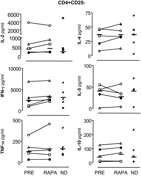
To ascertain whether rapamycin monotherapy alters the cytokine production profile of CD4+CD25− effector T-cells and CD4+CD25bright nTregs, the sorted CD4+ T-cell subsets were activated polyclonally, and supernatants were collected for cytokine measurements. The ability of type 1 diabetes CD4+CD25− effector T-cells to produce both Th1 (i.e., IL-2, IFN-γ, and TNF-α) and Th2 (i.e., IL-4, IL-5, and IL-10) cytokines was similar before and during rapamycin monotherapy, and the cytokine levels were comparable with those of CD4+CD25− T-cells isolated from normal donors (Fig. 4). Sorted CD4+CD25bright T-cells, due to their anergic state, did not produce significant levels of any of the tested cytokines, irrespective of rapamycin treatment (data not shown). Overall, effector CD4+CD25− T-cells isolated from type 1 diabetic patients are functionally similar before and during rapamycin monotherapy, in terms of proliferative ability and cytokine production profile, and are not different from those isolated from normal donors. The same holds true for CD4+CD25bright nTregs.
FIG. 4.
Cytokine production profile of FACS-sorted CD4+CD25− T-cells. FACS-sorted CD4+CD25− T-cells isolated from PBMCs of type 1 diabetic patients before (PRE) and during (RAPA) rapamycin treatment and from normal donors (ND) were activated polyclonally (5 × 10⁴ cells/ml) with beads coated with anti-CD3+CD28 mAbs, and supernatants were collected 72 h after activation. The indicated cytokines were evaluated by cytokine-bead array. Each patient is identifiable by a specific symbol, and each star represents a different normal donor. The solid line represents average levels of cytokines produced by type 1 diabetes PRE, type 1 diabetes RAPA, and ND CD4+CD25− T-cells.
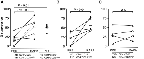
Finally, the ability of sorted CD4+CD25bright nTregs to suppress proliferation of CD4+CD25− effector T-cells was tested in vitro. CD4+CD25bright nTregs isolated from type 1 diabetic subjects during rapamycin monotherapy suppressed syngeneic effector T-cells significantly more than those isolated before treatment (Fig. 5A, compare pretreatment samples and samples during rapamycin treatment). The average level of nTreg suppression in type 1 diabetic patients before rapamycin therapy was significantly reduced in comparison with that of normal donors (Fig. 5A, compare pretreatment samples and normal donors). On the contrary, type 1 diabetes nTregs isolated during rapamycin therapy suppressed to the same levels of normal donor nTregs (Fig. 5A, compare patients during rapamycin treatment and normal donors). Type 1 diabetes nTregs isolated during rapamycin treatment had an increased suppressive capacity compared with those isolated before treatment also when normal donor CD4+CD25− effector T-cells were used as responder cells (Fig. 5B). These data indicate that the increased suppressive ability observed during rapamycin monotherapy is due to an improved capacity of type 1 diabetes nTregs to suppress proliferation of CD4+CD25− effector T-cells rather than to an increased susceptibility of type 1 diabetes CD4+CD25− effector T-cells to be suppressed in vitro on in vivo exposure to rapamycin. The observation that CD4+CD25bright nTregs isolated from normal donors suppressed type 1 diabetes CD4+CD25− effector T-cells isolated before and during rapamycin treatment to a similar extent (Fig. 5C) further supports the hypothesis that rapamycin monotherapy directly improves nTregs suppressive activity rather than modifying the susceptibility of effector T-cells to be suppressed.
FIG. 5.
In vitro suppressive function of FACS-sorted CD4+CD25bright T-cells. FACS-sorted effector CD4+CD25− T-cells (104 cells) were activated polyclonally with beads coated with anti-CD3+CD28 mAbs in the presence of equal amounts of FACS-sorted CD4+CD25bright T-cells. Cell proliferation was assessed in all cultures at day 4 after addition of [3H]thymidine for the last 18 h of culture except for cells isolated from one patient (dashed line) that were tested by CFSE dilution assay. Each patient is identifiable by a specific symbol, and each star represents a different normal donor. The solid line represents average levels of suppression of CD4+CD25bright T-cells pretreatment in type 1 diabetic patients (PRE), during treatment in type 1 diabetic patients (RAPA), and in normal donors (ND). A: Syngeneic suppression assays in which effector CD4+CD25− T-cells and CD4+CD25bright nTregs were isolated from the same type 1 diabetic patients and the same normal donor. B: Allogeneic suppression assays in which effector CD4+CD25− T-cells were isolated from normal donors (ND) while CD4+CD25bright nTregs were isolated from type 1 diabetic patients before (PRE) and during (RAPA) rapamycin treatment. C: Allogeneic suppression assays in which effector CD4+CD25− T-cells were isolated from type 1 diabetic patients before (PRE) and during (RAPA) rapamycin treatment while CD4+CD25bright nTregs were isolated from normal donors.
DISCUSSION
We have shown that rapamycin monotherapy in long-term type 1 diabetic patients does not alter the frequency and total number of circulating CD4+CD25brightFOXP3+CD127low/neg nTregs. Similarly, the ability of rapamycin-exposed nTregs to proliferate and produce cytokines in vitro is unaffected by the therapy. Interestingly, peripheral nTregs isolated from type 1 diabetic patients during rapamycin treatment have an intrinsic improved capacity to suppress proliferation of syngeneic and allogeneic CD4+CD25− effector T-cells, compared with that before treatment. Rapamycin therapy, therefore, refits nTreg suppressive activity, but it does not directly change effector T-cells.
Rapamycin administration to pre-diabetic NOD mice leads to expansion of nTregs that accumulate in the pancreatic lymph nodes and block diabetes development (19). Similarly, C57BL/6 mice receiving rapamycin for 14 days show an enhanced ratio between nTregs and CD4+ T-cells in the spleen and thymus (27). In humans, the frequency of circulating nTregs in renal transplant recipients is preserved under rapamycin treatment, whereas it is significantly decreased during therapy with calcineurin inhibitors (28). Similarly, we observed that rapamycin monotherapy preserves the number of circulating nTregs. In humans, a large expansion of peripheral nTregs has been observed only in renal transplant recipients receiving rapamycin as maintenance immunosuppressive therapy after profound T-cell depletion with Campath-1H (22). The absence of such expansion in patients receiving the calcineurin inhibitor cyclosporine A (22) suggests that acute lymphopenia and calcineurin-mediated signaling are important prerequisites for peripheral nTreg expansion in humans.
Rapamycin treatment causes G1 arrest in a variety of cell types, including T-cells (29). However, we demonstrated that, in contrast to CD4+CD25− effector T-cells, human nTregs are resistant to the antiproliferative effect of rapamycin in vitro (20). This antiproliferative effect is operational as long as rapamycin is present in the culture, because restimulation of rapamycin-exposed CD4+CD25− effector T-cells in the absence of rapamcyin leads to normal T-cell proliferation (Fig. 2B; 20).
The existence of CD4+CD25+ nTregs with defective suppressor function in type 1 diabetic patients has been demonstrated in some studies (9–11), whereas others showed that type 1 diabetes CD4+CD25+ Tregs are as suppressive as those of normal donors (12). Among the factors that could account for the discrepancies in these studies is the different purity of the tested Tregs. Studies have used FACS-sorted CD4+CD25bright T-cells (12) or bead-purified CD4+CD25+ T-cells (9–11), and it is possible that a defect in CD4+CD25+ nTregs was due to confounding effects from contaminating T effector cells when bead-purified T-cells were used. To reduce this risk, we FACS-sorted both CD4+CD25bright T-cells (falling in the top 1–2% of CD25+ cells) and CD4+CD25− effector T-cells from type 1 diabetic patients and normal donors. Functional data, in terms of cell proliferation and cytokine production profile of both CD4+CD25− and CD4+CD25bright T-cells, prove that in the current study, we are comparing similar T-cell subsets isolated from type 1 diabetic subjects before and during rapamycin treatment and from normal donors. Using these highly purified T-cells, we show that the suppressive function of nTregs freshly isolated from type 1 diabetic patients before rapamycin therapy is significantly reduced compared with that of normal donors. Although the number of patients was low, some patients had nTregs that were completely absent in suppressive function as determined by current protocols. Moreover, quantitative suppression by nTregs isolated from normal donors was relatively similar between subjects, whereas it varied markedly between type 1 diabetic patients. Thus, type 1 diabetic subjects appear to be variable in their freshly isolated peripheral nTreg function.
Increased and restored suppressive function of nTregs was observed in type 1 diabetic patients undergoing rapamycin monotherapy. These results are in accordance to our in vitro data, which demonstrated that peripheral CD4+ T-cells isolated from type 1 diabetic subjects and expanded in vitro with rapamycin have the same suppressive ability as those isolated from normal donors (20). The improved nTreg activity during rapamycin therapy is unlikely due to differences in numbers of bona fide nTregs (i.e., FOXP3+CD127low/neg) in the type 1 diabetic CD4+CD25bright T-cells isolated during rapamycin treatment compared with that before treatment because they are equally present, irrespective of rapamycin therapy. FOXP3 can be upregulated in T-cells on activation, and this might influence their suppressive function (30,31). We tested whether nTregs isolated during rapamycin treatment differ in their ability to upregulate FOXP3 on in vitro stimulation. Increased FOXP3 expression in RAPA-nTregs on in vitro activation was observed in only one of six patients (data not shown), indicating that this was unlikely to explain the rapamycin-associated increased nTreg suppression. Other explanations include heterogeneity of the Treg subsets in the sorted CD4+CD25bright T-cells before and during rapamycin treatment. It has been shown that rapamycin allows the in vitro generation of inducible human CD4+ Tregs that are CD25bright (21,32), and it is possible that rapamycin treatment leads to an increased representation of this subset in the subsequently purified T-cells. Alternatively, rapamycin monotherapy might modulate some molecule/s crucial for nTreg function. Gene expression profiling of T-cells exposed to rapamycin in vivo is currently ongoing to address this question.
Overall, these data show that rapamycin can refit nTreg function in vivo in long-term type 1 diabetic patients and can therefore influence the consequent islet transplant outcome. However, the current use of anti-CD25 mAb therapy (daclizumab) may abrogate this tolerogenic effect of rapamcyin. We observed that type 1 diabetic patients undergoing islet transplantation and receiving daclizumab have a massive depletion of circulating CD4+CD25+FOXP3+ T-cells even months after treatment (P.M. and M.B., unpublished observation). At present, one of the major challenges in transplantation is therefore to carefully design new combinational therapies so as not to abrogate the observed positive effects of rapamycin on nTreg function.
Acknowledgments
This work was supported by the Italian Telethon Foundation and Juvenile Diabetes Research Foundation Grants JT-01 and GJT04014.
We thank Alessandra Ferraro (San Raffaele Telethon Institute for Gene Therapy) for performing the suppressive experiments comparing CD4+CD25brightCD127− and CD4+CD25bright nTregs.
Published ahead of print at http://diabetes.diabetesjournals.org on 16 June 2008.
E.B. is currently affiliated with the Center for Regenerative Therapies-Dresden, Dresden University of Technology, Dresden, Germany.
The costs of publication of this article were defrayed in part by the payment of page charges. This article must therefore be hereby marked “advertisement” in accordance with 18 U.S.C. Section 1734 solely to indicate this fact.
REFERENCES
- 1.Atkinson MA, Eisenbarth GS: Type 1 diabetes: new perspectives on disease pathogenesis and treatment. Lancet 358 :221 –229,2001 [DOI] [PubMed] [Google Scholar]
- 2.Roep BO: Standardization of T-cell assays in type I diabetes: Immunology of Diabetes Society T-cell Committee. Diabetologia 42 :636 –637,1999 [DOI] [PubMed] [Google Scholar]
- 3.Monti P, Scirpoli M, Rigamonti A, Mayr A, Jaeger A, Bonfanti R, Chiumello G, Ziegler AG, Bonifacio E: Evidence for in vivo primed and expanded autoreactive T cells as a specific feature of patients with type 1 diabetes. J Immunol 179 :5785 –5792,2007 [DOI] [PubMed] [Google Scholar]
- 4.Sakaguchi S, Ono M, Setoguchi R, Yagi H, Hori S, Fehervari Z, Shimizu J, Takahashi T, Nomura T: Foxp3+ CD25+ CD4+ natural regulatory T cells in dominant self-tolerance and autoimmune disease. Immunol Rev 212 :8 –27,2006 [DOI] [PubMed] [Google Scholar]
- 5.Sakaguchi S: Naturally arising Foxp3-expressing CD25+CD4+ regulatory T cells in immunological tolerance to self and non-self. Nat Immunol 6 :345 –352,2005 [DOI] [PubMed] [Google Scholar]
- 6.Liu W, Putnam AL, Xu-Yu Z, Szot GL, Lee MR, Zhu S, Gottlieb PA, Kapranov P, Gingeras TR, Fazekas de St Groth B, Clayberger C, Soper DM, Ziegler SF, Bluestone JA: CD127 expression inversely correlates with FoxP3 and suppressive function of human CD4+ T reg cells. J Exp Med 203 :1701 –1711,2006 [DOI] [PMC free article] [PubMed] [Google Scholar]
- 7.Seddiki N, Santner-Nanan B, Martinson J, Zaunders J, Sasson S, Landay A, Solomon M, Selby W, Alexander SI, Nanan R, Kelleher A, Fazekas de St Groth B: Expression of interleukin (IL)-2 and IL-7 receptors discriminates between human regulatory and activated T cells. J Exp Med 203 :1693 –1700,2006 [DOI] [PMC free article] [PubMed] [Google Scholar]
- 8.Baecher-Allan C, Viglietta V, Hafler DA: Human CD4+CD25+ regulatory T cells. Semin Immunol 16 :89 –98,2004 [DOI] [PubMed] [Google Scholar]
- 9.Brusko TM, Wasserfall CH, Clare-Salzler MJ, Schatz DA, Atkinson MA: Functional defects and the influence of age on the frequency of CD4+ CD25+ T-cells in type 1 diabetes. Diabetes 54 :1407 –1414,2005 [DOI] [PubMed] [Google Scholar]
- 10.Lindley S, Dayan CM, Bishop A, Roep BO, Peakman M, Tree TI: Defective suppressor function in CD4+CD25+ T-cells from patients with type 1 diabetes. Diabetes 54 :92 –99,2005 [DOI] [PubMed] [Google Scholar]
- 11.Glisic-Milosavljevic S, Waukau J, Jailwala P, Jana S, Khoo HJ, Albertz H, Woodliff J, Koppen M, Alemzadeh R, Hagopian W, Ghosh S: At-risk and recent-onset type 1 diabetic subjects have increased apoptosis in the CD4+CD25+ T-cell fraction. PLoS ONE 2 :e146 ,2007 [DOI] [PMC free article] [PubMed] [Google Scholar]
- 12.Putnam AL, Vendrame F, Dotta F, Gottlieb PA: CD4+CD25high regulatory T cells in human autoimmune diabetes. J Autoimmun 24 :55 –62,2005 [DOI] [PubMed] [Google Scholar]
- 13.Masteller EL, Warner MR, Tang Q, Tarbell KV, McDevitt H, Bluestone JA: Expansion of functional endogenous antigen-specific CD4+CD25+ regulatory T cells from nonobese diabetic mice. J Immunol 175 :3053 –3059,2005 [DOI] [PubMed] [Google Scholar]
- 14.Tang Q, Henriksen KJ, Bi M, Finger EB, Szot G, Ye J, Masteller EL, McDevitt H, Bonyhadi M, Bluestone JA: In vitro-expanded antigen-specific regulatory T cells suppress autoimmune diabetes. J Exp Med 199 :1455 –1465,2004 [DOI] [PMC free article] [PubMed] [Google Scholar]
- 15.Tarbell KV, Petit L, Zuo X, Toy P, Luo X, Mqadmi A, Yang H, Suthanthiran M, Mojsov S, Steinman RM: Dendritic cell-expanded, islet-specific CD4+ CD25+ CD62L+ regulatory T cells restore normoglycemia in diabetic NOD mice. J Exp Med 204 :191 –201,2007 [DOI] [PMC free article] [PubMed] [Google Scholar]
- 16.Roncarolo MG, Battaglia M: Regulatory T-cell immunotherapy for tolerance to self antigens and alloantigens in humans. Nat Rev Immunol 7 :585 –598,2007 [DOI] [PubMed] [Google Scholar]
- 17.Abraham RT, Wiederrecht GJ: Immunopharmacology of rapamycin. Annu Rev Immunol 14 :483 –510,1996 [DOI] [PubMed] [Google Scholar]
- 18.Battaglia M, Stabilini A, Roncarolo MG: Rapamycin selectively expands CD4+CD25+FoxP3+ regulatory T cells. Blood 105 :4743 –4748,2005 [DOI] [PubMed] [Google Scholar]
- 19.Battaglia M, Stabilini A, Draghici E, Migliavacca B, Gregori S, Bonifacio E, Roncarolo MG: Induction of tolerance in type 1 diabetes via both CD4+CD25+ T regulatory cells and T regulatory type 1 cells. Diabetes 55 :1571 –1580,2006 [DOI] [PubMed] [Google Scholar]
- 20.Battaglia M, Stabilini A, Migliavacca B, Horejs-Hoeck J, Kaupper T, Roncarolo MG: Rapamycin promotes expansion of functional CD4+CD25+FOXP3+ regulatory T cells of both healthy subjects and type 1 diabetic patients. J Immunol 177 :8338 –8347,2006 [DOI] [PubMed] [Google Scholar]
- 21.Valmori D, Tosello V, Souleimanian NE, Godefroy E, Scotto L, Wang Y, Ayyoub M: Rapamycin-mediated enrichment of T cells with regulatory activity in stimulated CD4+ T cell cultures is not due to the selective expansion of naturally occurring regulatory T cells but to the induction of regulatory functions in conventional CD4+ T cells. J Immunol 177 :944 –949,2006 [DOI] [PubMed] [Google Scholar]
- 22.Noris M, Casiraghi F, Todeschini M, Cravedi P, Cugini D, Monteferrante G, Aiello S, Cassis L, Gotti E, Gaspari F, Cattaneo D, Perico N, Remuzzi G: Regulatory T cells and T cell depletion: role of immunosuppressive drugs. J Am Soc Nephrol 18 :1007 –1018,2007 [DOI] [PubMed] [Google Scholar]
- 23.Maffi P, De Taddeo F, Bertuzzi F, Piemonti L, Magistretti P, Fiorina P, Pozzi P, Nano R, Venturini M, Del Maschio A, Secchi A: Islet transplantation alone: effect of sirolimus pre-transplant treatment on clinical outcome (Abstract). Am J Transplant 6 (Suppl. 2):341 ,2006 [Google Scholar]
- 24.Shapiro AM, Lakey JR, Ryan EA, Korbutt GS, Toth E, Warnock GL, Kneteman NM, Rajotte RV: Islet transplantation in seven patients with type 1 diabetes mellitus using a glucocorticoid-free immunosuppressive regimen. N Engl J Med 343 :230 –238,2000 [DOI] [PubMed] [Google Scholar]
- 25.Baecher-Allan C, Wolf E, Hafler DA: MHC class II expression identifies functionally distinct human regulatory T cells. J Immunol 176 :4622 –4631,2006 [DOI] [PubMed] [Google Scholar]
- 26.Biagi E, Di Biaso I, Leoni V, Gaipa G, Rossi V, Bugarin C, Renoldi G, Parma M, Balduzzi A, Perseghin P, Biondi A: Extracorporeal photochemotherapy is accompanied by increasing levels of circulating CD4+CD25+GITR+Foxp3+CD62L+ functional regulatory T-cells in patients with graft-versus-host disease. Transplantation 84 :31 –39,2007 [DOI] [PubMed] [Google Scholar]
- 27.Qu Y, Zhang B, Zhao L, Liu G, Ma H, Rao E, Zeng C, Zhao Y: The effect of immunosuppressive drug rapamycin on regulatory CD4+CD25+Foxp3+T cells in mice. Transpl Immunol 17 :153 –161,2007 [DOI] [PubMed] [Google Scholar]
- 28.San Segundo D, Ruiz JC, Fernandez-Fresnedo G, Izquierdo M, Gomez-Alamillo C, Cacho E, Benito MJ, Rodrigo E, Palomar R, Lopez-Hoyos M, Arias M: Calcineurin inhibitors affect circulating regulatory T cells in stable renal transplant recipients. Transplant Proc 38 :2391 –2393,2006 [DOI] [PubMed] [Google Scholar]
- 29.Schmelzle T, Hall MN: TOR, a central controller of cell growth. Cell 103 :253 –262,2000 [DOI] [PubMed] [Google Scholar]
- 30.Walker MR, Kasprowicz DJ, Gersuk VH, Benard A, Van Landeghen M, Buckner JH, Ziegler SF: Induction of FoxP3 and acquisition of T regulatory activity by stimulated human CD4+CD25- T cells. J Clin Invest 112 :1437 –1443,2003 [DOI] [PMC free article] [PubMed] [Google Scholar]
- 31.Pillai V, Ortega SB, Wang CK, Karandikar NJ: Transient regulatory T-cells: a state attained by all activated human T-cells. Clin Immunol 123 :18 –29,2007 [DOI] [PMC free article] [PubMed] [Google Scholar]
- 32.Nikolaeva N, Bemelman FJ, Yong SL, van Lier RA, ten Berge IJ: Rapamycin does not induce anergy but inhibits expansion and differentiation of alloreactive human T cells. Transplantation 81 :445 –454,2006 [DOI] [PubMed] [Google Scholar]


