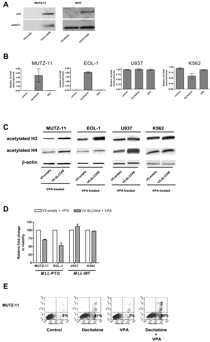
Figure 2.
Epigenetic silencing of SLC5A8 and functional consequences of forced expression of SLC5A8 in MLL-PTD+ cell lines. (A) Whole cell lysates were prepared and immunoblotting performed as described in “Methods.” Anti-SLC5A8 antibody (A-15) was purchased from Santa Cruz Biotechnology (Santa Cruz, CA). Nucleofections with V5-empty and V5-SLC5A8 expression vectors and immunoblotting to detect the V5-epitope were performed as described in “Methods.” (B) The demethylating agent, decitabine, but not the histone deacetylase inhibitor, VPA activates SLC5A8 transcription in MLL-PTD+ AML cell lines. Cell lines were incubated in the absence or presence of the hypomethylating reagent decitabine (2.5 μM) or VPA (1 mM) for 48 or 24 hours, respectively. SLC5A8 mRNA levels were measured by real time RT-PCR using SYBR Green dye for detection (Prism 7700 SDS, Applied Biosystems, Foster City, CA). Primers were SLC5A8RT-for, 5′-TCCGAGGTCTACCGTTTTG-3′ and SLC5A8RT-rev, 5′-GGGCAGGGCATAAA-TAAC-3′. The ΔΔCt method of relative quantification was carried out. SLC5A8 mRNA levels were normalized to 18S rRNA levels. Results are presented as relative SLC5A8 transcript levels (means ± SD). (C) Forced SMCT1 expression enhances VPA-induced acetylation of histone H3 and H4. Immunoblot analyses for total acetylated histones H3 and H4 were carried out on empty vector or V5-SLC5A8 vector transfected cells. Twenty-four hours after nucleofection, 1 mM VPA was added to the cultures for an additional 24 hours. Immunoblot detection of β-actin was used as a loading control. (D) Overexpression of SMCT1 sensitizes MLL-PTD+ cells to the growth inhibitory effects of VPA. Cell lines were incubated for an additional 24 hours with or without VPA (1 mM) beginning 24 hours posttransfection. The effect on viable cell numbers was measured using the trypan blue exclusion assay and is depicted as a fold-change in relation to the appropriate VPA-treated, empty-vector transfected cells. Error bars represent SD. (E) The sequential combination of decitabine followed by VPA results in enhanced apoptosis in the MLL-PTD+ cell lines. Cells were treated as described above, except with the inclusion of a sequential combination of decitabine (48 hours) followed by VPA (24 hours), and then harvested for staining with annexin V/propidium iodide and fluorescence-activated cell sorting analysis. The percentage of cells undergoing early apoptosis (lower right quadrant) is indicated.

