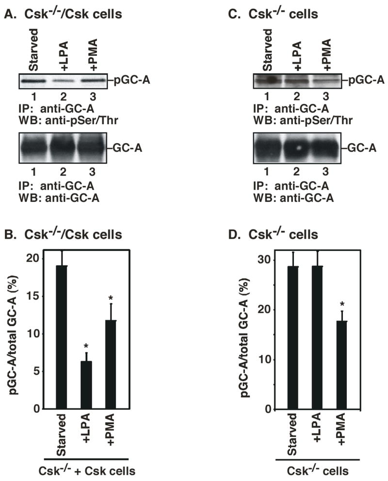
Figure 6.

LPA-induced decrease of Ser/Thr phosphorylation of GC-A depends on Csk. A. Csk−/−/Csk cells were serum-starved. Cells were then treated with LPA or PMA. Cell lysates were immunoprecipitated with anti-GC-A antibody. After SDS-PAGE, the filters were probed with anti-phospho-Ser/Thr antibody (top panel) or anti-GC-A antibody (bottom panel). B. The intensity of each band in A was quantified by an image analyzer. The percentage of phosphorylated GC-A is corrected by the amount of the total GC-A. C. Csk−/− cells were serum-starved. Cells were then treated with LPA or PMA. Cell lysates were immunoprecipitated with anti-GC-A antibody. After SDS-PAGE, the filters were probed with anti-phospho-Ser/Thr antibody (top panel) or anti-GC-A antibody (bottom panel). D. The intensity of each band in C was quantified by an image analyzer. The percentage of phosphorylated GC-A is corrected by the amount of the total GC-A. * indicates an significant difference from the untreated cells (t-test: p<0.05). Data represent the means ± S.D. of three experiments.
