Abstract
Diabetics have a higher incidence of urinary tract infection (UTI), are infected with a broader range of uropathogens, and more commonly develop serious UTI sequelae than nondiabetics. To better study UTI in the diabetic host, we created and characterized a murine model of diabetic UTI using the pancreatic islet β-cell toxin streptozocin in C3H/HeN, C3H/HeJ, and C57BL/6 mouse backgrounds. Intraperitoneal injections of streptozocin were used to initiate diabetes in healthy mouse backgrounds, as defined by consecutive blood glucose levels of >250 mg/dl. UTIs caused by uropathogenic Escherichia coli (UTI89), Klebsiella pneumoniae (TOP52 1721), and Enterococcus faecalis (0852) were studied, and diabetic mice were found to be considerably more susceptible to infection. All three uropathogens produced significantly higher bladder and kidney titers than buffer-treated controls. Uropathogens did not have as large an advantage in the Toll-like receptor 4-defective C3H/HeJ diabetic mouse, arguing that the dramatic increase in colonization seen in C3H/HeN diabetic mice may partially be due to diabetic-induced defects in innate immunity. Competition experiments demonstrated that E. coli had a significant advantage over K. pneumoniae in the bladders of healthy mice and less of an advantage in diabetic bladders. In the kidneys, K. pneumoniae outcompeted E. coli in healthy mice but in diabetic mice E. coli outcompeted K. pneumoniae and caused severe pyelonephritis. Diabetic kidneys contained renal tubules laden with communities of E. coli UTI89 bacteria within an extracellular-matrix material. Diabetic mice also had glucosuria, which may enhance bacterial replication in the urinary tract. These data support that this murine diabetic UTI model is consistent with known characteristics of human diabetic UTI and can provide a powerful tool for dissecting this infection in the multifactorial setting of diabetes.
Urinary tract infections (UTIs), which include infections of the bladder (cystitis) and kidney (pyelonephritis), affect primarily women and are responsible for nearly 13 million annual office visits in the United States (16). One-third of women will experience a recurrent infection within 3 to 6 months of the initial episode (19), and 44% will experience a recurrence within 1 year (21). These infections are most commonly caused by the gram-negative bacterium Escherichia coli, which is responsible for 80 to 85% of community-acquired UTIs; however, there are numerous other pathogens capable of infecting the urinary tract (20, 47). Uropathogenic E. coli (UPEC) employs a wide array of virulence factors to successfully colonize and survive within the urinary tract, including adhesive organelles, such as type 1, P, F1C, and S pili (2, 22, 40, 41); iron acquisition/transport systems (51); hemolysin (39); and flagella (27, 53). It has recently been found that UPEC has the ability to invade bladder urothelial cells and replicate to form intracellular bacterial communities largely protected from host innate immunity (1, 9, 23, 48). Bacteria disperse from these intracellular bacterial communities, some in filamentous morphology, which subverts elimination by polymorphonuclear leukocytes (PMN) and allows for further dissemination throughout the urinary tract (24, 48).
Diabetes mellitus is the most common endocrine disease, and worldwide incidence of this ailment is increasing (38). Type 1 diabetes is an autoimmune disorder by which insulin-producing islet β cells are destroyed by one's own immune system (15). Patients with type 1 diabetes, even with proper management and glycemic control, can develop a variety of diabetic sequelae, including retinopathy, neuropathy, nephropathy, and numerous cardiovascular complications. Additionally, diabetics are more prone to infection and these infections are more severe than in nondiabetics (6, 44).
The urinary tract is the most common site of infection in the diabetic host (30). Diabetics are more likely to have asymptomatic and symptomatic bacteriuria (14, 42). Acute pyelonephritis is approximately 10 times more common in the diabetic population (37). In addition to a higher risk of developing UTI, diabetic women have an increased risk of developing complications of UTI, such as emphysematous cystitis, abscess formation, and renal papillary necrosis (42). Although UPEC remains the predominant etiologic agent of UTI in diabetic individuals, infections by Klebsiella species (17, 29), enterococci (5, 29), Acinetobacter species (36), group B streptococci (35), fungi (26), and other less common uropathogens are more prevalent in diabetic women. Many hypotheses have been attributed to the increase of UTI in diabetic women, such as glucosuria, impaired immune cell function, or functional abnormalities of the urinary tract; however, these theories have not been fully tested or confirmed in an animal model of UTI (43). While there are multiple murine models of diabetes, including the model of nonobese diabetic mice (31), few have been used to effectively investigate diabetic UTI. This study presents a streptozocin (STZ)-induced murine model of diabetic UTI. This model is consistent with epidemiologic observations of diabetic UTI and thus will greatly assist in understanding the physiological and molecular mechanisms underlying uropathogenesis in the diabetic setting. Using this model, we discovered that diabetic mice are more prone to UTI and that differences in virulence observed in the kidney and bladder are dependent on the bacterial strain and host background.
MATERIALS AND METHODS
Bacterial strains and growth conditions.
Clinical strains used in this study were UTI89, a UPEC cystitis isolate (34); UTI89 hk::comGFP, a kanamycin-resistant and green fluorescent protein-expressing strain of UTI89 (53); TOP52 1721 (abbreviated TOP52), a Klebsiella pneumoniae cystitis isolate (49); and 0852, an Enterococcus faecalis UTI isolate (25). Bacteria were cultured at 37°C in Luria-Bertani (LB) broth (UTI89 and TOP52) or brain heart infusion broth (0852). UTI89 hk::comGFP growth media also contained 50 μg/ml kanamycin.
Induction of diabetes in mice.
To develop a diabetic mouse model of UTI, we gave 4- to 5-week-old female C3H/HeN (National Cancer Institute), C3H/HeJ (Jackson Laboratories), or C57BL/6 (Jackson Laboratories) mice two to three intraperitoneal (i.p.) injections of STZ (Sigma-Aldrich) to induce pancreatic islet β-cell death. Mice were weighed prior to injections, and STZ was freshly dissolved in dilution buffer (0.1 M sodium citrate, pH 4.5, with HCl, stored at 4°C) and filter sterilized. To induce diabetes, mice were given 0.1-ml i.p. injections of 200 mg STZ/kg of body weight by use of a Precision Sure-Dose 1/2-ml3 syringe with a 30-gauge, 3/8-in. needle. Dilution buffer-injected mice were used as healthy controls. All mice were fed a normal diet. Blood glucose levels were measured daily beginning 5 days after the second i.p. injection. STZ-injected mice with glucose levels of <250 were given a third STZ i.p. injection. This method consistently resulted in 80 to 85% penetrance of diabetes in STZ-injected mice. Blood glucose levels were measured for each mouse, and a mouse was considered diabetic after two consecutive readings of >250 mg/dl blood glucose.
Mouse infections, competitions, and organ titers.
Eight-week-old female diabetic or healthy control mice were infected by transurethral catheterization as previously described (33). Static bacterial cultures were started from freezer stocks, grown at 37°C for 18 h, and then subcultured at 1:250 (for UTI89 and TOP52) or 1:100 (for 0852) into fresh media. These subcultures were then grown statically at 37°C for 18 h (for UTI89 and TOP52) or 2 to 3 h (for 0852), pelleted, resuspended in phosphate-buffered saline, and diluted appropriately to yield 50 μl inocula (for UTI89 and TOP52) or 200 μl inocula (for 0852) of 1 × 10⁷ to 2 × 10⁷ CFU unless otherwise indicated. For competition experiments, 107 CFU each of K. pneumoniae TOP52 and E. coli UTI89 hk::comGFP were inoculated together in a total volume of 50 μl. To quantify bacteria present in mouse organs, bladders and kidneys were aseptically harvested at the indicated times postinfection, homogenized in phosphate-buffered saline, serially diluted, and plated onto LB plates, brain heart infusion broth plates, or LB-kanamycin (50 μg/ml) and BBL CHROMagar orientation media (BD Diagnostics) for competition experiments. All studies were approved by the Animal Studies Committee at Washington University School of Medicine.
Mouse urine collection and analysis.
Urine samples were collected from diabetic and healthy mice by bladder massage over a sterile 1.5-ml Eppendorf tube prior to bacterial inoculation. Urine samples were analyzed for glucose, protein, ketones, and specific gravity by use of Multistix Pro 10 LS urine reagent test strips (Bayer) according to the manufacturer's instructions.
Histology and electron microscopy.
Infected mouse bladders and kidneys were aseptically removed, fixed in neutral buffered formalin, and paraffin embedded. Sections were stained with hematoxylin and eosin and examined using an Olympus BX51 light microscope (Olympus America). For transmission electron microscopy, glutaraldehyde-fixed kidneys were harvested and processed as previously described (32). Sections were viewed on a JEOL 1200 EX transmission electron microscope (JEOL USA) at an 80-kV accelerating voltage.
Urine growth curves.
Overnight UTI89 and TOP52 shaking LB cultures were subcultured at 1:100 into filter-sterilized urine specimens from healthy volunteers with and without 2% glucose supplementation. Samples were grown with shaking at 37°C, and optical density at 600 nm (OD600) readings were taken at various time points. Doubling times (td) were calculated as follows: td = ln 2 (t2 − t1)/(ln OD2 − ln OD1).
Statistical analysis.
Competitive indices were calculated as follows: (UTI89 hk::comGFPout/UTI89 hk::comGFPin)/(TOP52out/TOP52in). The Wilcoxon signed-rank test was used to compare the log of competitive indices to a theoretical mean of zero. Continuous variables were compared using the Mann-Whitney U test since these variables were not normally distributed. Values below the limit of detection were assigned this minimum value for statistical analyses. All tests were two tailed, and a P value of less than 0.05 was considered significant. These analyses were performed using GraphPad Prism (GraphPad software, version 4.03). To calculate 50% infective dose (ID50) values, the Reed and Muench mathematical technique (46) was used and infection was defined as organs with bacterial titers above the limit of detection (20 CFU) at 72 h postinfection.
RESULTS
Diabetic mice have increased susceptibility to UPEC infection and higher bacterial burden than nondiabetics.
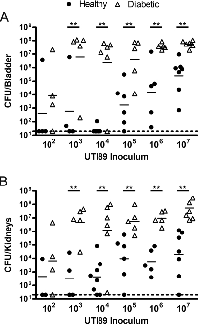
It is widely known that diabetics are more susceptible to UTI than nondiabetics. To determine if STZ-induced diabetic C3H/HeN mice are more susceptible to UTI than buffer-treated control mice, various doses of the UPEC cystitis isolate UTI89 were inoculated by transurethral catheterization. After 72 h of infection, mice were sacrificed and bladder and kidney bacterial titers were determined (Fig. 1A). Inocula of 107 (P = 0.0006), 106 (P = 0.0079), 105 (P = 0.0173), 104 (P = 0.0080), and 103 (P = 0.0317) CFU of E. coli UTI89 all resulted in significantly higher bladder titers in diabetic mice than in buffer-treated controls. The majority of healthy buffer-treated mice inoculated with 103 or 104 CFU of UTI89 were able to efficiently clear infection after 72 h; however, almost all diabetic mice had significant infection even with these relatively low inocula. Defining infection as a titer at 72 h greater than 20 CFU (minimum level of detection), the ID50 of E. coli UTI89 was 1.97 × 10⁴ CFU in healthy murine bladders and was less than 100 CFU (approximately 68.1) in diabetic bladders. Kidney titers were also significantly higher in diabetic mice than in buffer-treated control mice after inoculation with 107 (P = 0.0006), 106 (P = 0.0079), 105 (P = 0.0087), 104 (P = 0.0200), and 103 (P = 0.0159) CFU of E. coli UTI89 (Fig. 1B). By 72 h after inoculation with 103 CFU of UTI89, all diabetic mice had significant kidney titers while only half of the healthy mice remained infected. The ID50 of UTI89 was 2.08 × 10³ CFU in healthy murine kidneys and was less than 100 CFU (approximately 46.4) in diabetic kidneys.
FIG. 1.
Diabetic mice have increased susceptibility to E. coli UTI89 UTI compared to healthy mice. Female C3H/HeN diabetic mice and buffer-treated control mice were infected with various inocula of UTI89, a UPEC cystitis isolate. At 72 h postinfection, bladders (A) and kidneys (B) were harvested and homogenized and CFU were determined. Titer data are combined from three independent experiments. Short bars represent geometric means of each group, and dotted lines represent limits of detection. The symbol ** indicates significant P values of less than 0.05.
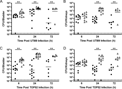
The kinetics of urinary tract colonization by E. coli UTI89 were compared between diabetic mice and healthy controls (Fig. 2A and B). UTI89 efficiently colonized mouse bladders as early as 6 h postinfection in both healthy and diabetic mice (Fig. 2A). The bacterial load in infected bladders of healthy mice decreased to a geometric mean of 9.2 × 10³ CFU per bladder by 72 h postinfection. Diabetic mouse bladders, on the other hand, retained high levels of bacterial colonization at 72 h postinfection. Similar patterns of persistent high-level colonization throughout the course of infection were seen in infected kidneys of diabetic mice (Fig. 2B).
FIG. 2.
Time course of E. coli UTI89 and K. pneumoniae TOP52 bladder and kidney infections in healthy and diabetic mice. Female C3H/HeN diabetic mice (Δ) and buffer-treated control mice (•) were inoculated with 107 CFU of the UPEC isolate UTI89 (A and B) or with 107 CFU of the K. pneumoniae isolate TOP52 (C and D) by transurethral catheterization. For organ titers, bladders (A and C) and kidneys (B and D) were harvested at various time points postinfection and CFU were calculated. Graphs of bacterial burden of E. coli UTI89 in the bladder (A) and kidneys (B) and K. pneumoniae TOP52 in the bladder (C) and kidneys (D) are shown. Titer data are combined from three independent experiments. Short bars represent geometric means of each group, and dotted lines represent limits of detection. The symbol ** indicates significant P values of less than 0.05.
These data show that diabetic mice are more susceptible to infection by the UPEC isolate UTI89. UTI89 had a considerably lower ID50 in the diabetic background than in healthy control mice. UTI89 also had significantly higher bladder titers at 6, 24, and 72 h postinfection in diabetic mice than in healthy mice. Additionally, diabetic mice had higher UTI89 titers in the kidneys than did healthy mice, especially at later time points in infection.
Diabetic mice infected with K. pneumoniae or E. faecalis have higher burdens of infection than nondiabetic mice.
Diabetics are more likely to get UTIs caused by non-UPEC uropathogens than are nondiabetic individuals. To determine how non-UPEC uropathogens perform in the diabetic mouse model over time, we inoculated diabetic and buffer-treated control mice with either the K. pneumoniae isolate TOP52 (Fig. 2C and D) or the E. faecalis isolate 0852 (Fig. 3) in their respective murine models of UTI. As early as 6 h postinfection, the bacterial load of K. pneumoniae TOP52 was 100-fold higher in the bladders of diabetic C3H/HeN mice than in those of healthy C3H/HeN mice (Fig. 2C) (P = 0.0005). This difference between healthy and diabetic bladder TOP52 bacterial burdens was further exaggerated to greater than 1,000-fold by 24 h postinfection (P = 0.0007). K. pneumoniae TOP52 showed similar levels of infection in the kidneys of diabetic and healthy mice at 6 h, but diabetic bacterial burdens were significantly higher by 24 h (P = 0.0007) and 72 h (P = 0.0043) postinfection.
FIG. 3.
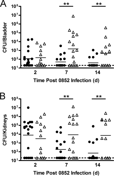
Time course of E. faecalis 0852 bladder and kidney infections in healthy and diabetic mice. Female C57BL/6 diabetic mice (Δ) and buffer-treated control mice (•) were inoculated with 107 CFU of the Enterococcus faecalis isolate 0852 by transurethral catheterization. Bladders and kidneys were harvested at various time points postinfection, and CFU were calculated. Graphs of bacterial burden of E. faecalis 0852 in the bladder (A) and in the kidneys (B) are shown. Titer data are combined from two independent experiments. Short bars represent geometric means of each group, and dotted lines represent limits of detection. The symbol ** indicates significant P values of less than 0.05. d, days.
In the C57BL/6 UTI model, the E. faecalis isolate 0852 showed no difference in bacterial titer at 2 days postinfection in the bladders (Fig. 3A) and kidneys (Fig. 3B) of diabetic and control mice. However, diabetic mouse kidneys had significantly higher bacterial burdens than buffer-treated control mouse kidneys at both 7 days (P = 0.0229) and 14 days (P = 0.0135) postinfection. E. faecalis 0852 titers were also significantly higher in diabetic mouse bladders than in control mouse bladders at 7 days (P = 0.0119) and 14 days (P = 0.0420) postinfection.
These data demonstrate that diabetic mice have greater burdens of UTI caused by the K. pneumoniae isolate TOP52 or the E. faecalis isolate 0852 than do healthy mice. The experiments involving E. faecalis in the C57BL/6 background also illustrate the versatility of STZ induction of diabetes and the ability to adapt this method to preexisting models of UTI characterized for specific uropathogens. Interestingly, the K. pneumoniae strain TOP52 has even larger differences between bladder bacterial loads of diabetic and control mice than does the E. coli strain UTI89 at 6 and 24 h postinfection. This greater advantage afforded to TOP52 in the diabetic background is consistent with the higher rates of K. pneumoniae cystitis observed in diabetic patients.
Advantages between UPEC and non-UPEC uropathogens shift in the diabetic host.
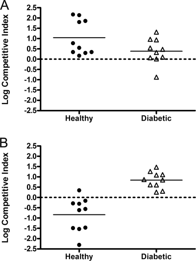
To directly compare UPEC to gram-negative non-UPEC uropathogens in the diabetic model, we conducted competition experiments in which 107 CFU E. coli UTI89 hk::comGFP and 107 CFU K. pneumoniae TOP52 were coinoculated into diabetic and buffer-treated C3H/HeN mice. UTI89 hk::comGFP has a kanamycin resistance cassette allowing for selection on antibiotic media. After 24 h, bladders and kidneys were harvested, titers of each pathogen were enumerated, and competitive indices were calculated. We then compared the log competitive indices of healthy and diabetic mice in the bladder (Fig. 4A) and kidney (Fig. 4B). A value of greater than zero indicates that UTI89 hk::comGFP outcompetes TOP52, while a value of less than zero indicates that TOP52 outcompetes UTI89 hk::comGFP. In the bladders of buffer-treated mice, the log of the competitive indices was significantly greater than zero (P = 0.0020), indicating that E. coli UTI89 hk::comGFP has a significant advantage over K. pneumoniae TOP52. In diabetic bladders, the log of competitive indices was also significantly greater than zero (P = 0.0371), albeit to a lesser degree than in healthy bladders. Thus, the competitive advantage of UTI89 hk::comGFP over TOP52 seems more pronounced in healthy bladders than in diabetic bladders. In the kidney, while K. pneumoniae TOP52 had a significant advantage over E. coli UTI89 hk::comGFP in the healthy background (P = 0.0137), UTI89 hk::comGFP substantially outcompeted TOP52 in the diabetic host (P = 0.0200).
FIG. 4.
Competition of E. coli UTI89 and K. pneumoniae TOP52 in the bladders and kidneys of healthy and diabetic mice. Female C3H/HeN diabetic mice (Δ) and buffer-treated control mice (•) were infected with 107 CFU each of E. coli UTI89 hk::comGFP and K. pneumoniae TOP52. After 24 h, bladders (A) and kidneys (B) were harvested, CFU of each pathogen were enumerated, and competitive indices [(UTI89 hk::comGFPout/UTI89 hk::comGFPin)/(TOP52out/TOP52in)] were calculated. A value of greater than zero indicates an E. coli UTI89 hk::comGFP advantage, while a value of less than zero indicates a K. pneumoniae TOP52 advantage. The logs of the competitive indices were significantly different than zero in all cases. Data are combined from two independent experiments. Bars represent means of each group, and dotted lines represent values at which each uropathogen competes equally.
An increased prevalence of non-UPEC strains causes cystitis in diabetic patients. In our model, the diabetic condition gave E. coli a lesser advantage over K. pneumoniae in the bladder, consistent with the increased prevalence of K. pneumoniae cystitis in diabetics. In the kidney, the situation was different. We found a dramatic shift in the kidney from an environment favoring K. pneumoniae in control mice to an environment favoring UPEC colonization in the diabetic setting.
UPEC causes marked interstitial pyelonephritis in diabetic mice.
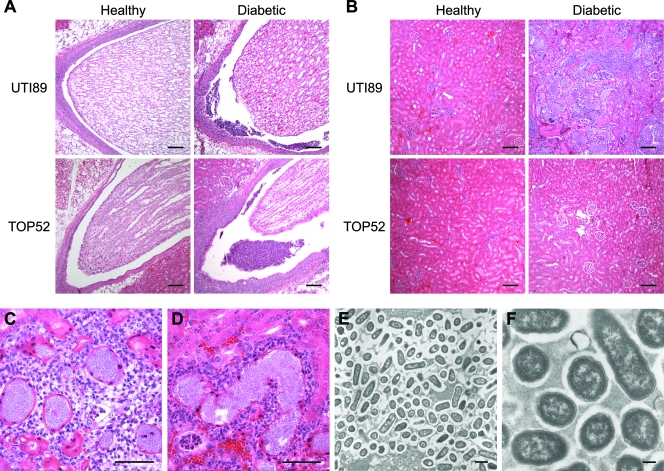
To further investigate the shift favoring an E. coli UTI89 competitive advantage in the diabetic kidney, we examined histologic hematoxylin and eosin-stained sections of single 72-h infections of E. coli UTI89 or K. pneumoniae TOP52. The renal pelvises (Fig. 5A) of buffer-treated control mice inoculated with UTI89 or TOP52 showed low levels of inflammation. The pelvises of infected diabetic mice infected with UTI89 or TOP52 were dilated and significantly inflamed, often with large sheets of PMN. The urothelium lining the pelvis was hyperplastic, with intraurothelial neutrophilia, and collections of bacteria were observed within the pelvic space. The kidney parenchyma (Fig. 5B) of healthy control mice infected with TOP52 or UTI89 and of diabetic TOP52-infected mice were largely unremarkable, with patent tubules and normal-appearing glomeruli. In contrast, the diabetic E. coli UTI89-infected kidney parenchyma displayed marked acute interstitial pyelonephritis with cortical and medullary involvement. There were multiple areas of abscessation, with widespread destruction of renal architecture. High-power views of these regions (Fig. 5C and D) showed marked inflammation that was largely neutrophilic in nature, with a small component of lymphocytes and plasma cells. Vast collections of extracellular bacteria filled the lumina of renal tubules. Intratubular and peritubular PMN were also observed.
FIG. 5.
E. coli UTI89 causes acute interstitial pyelonephritis in the kidneys of diabetic mice. Kidney sections from 72 h after infection with E. coli UTI89 or K. pneumoniae TOP52 were analyzed by light microscopy. (A) Renal pelvises from diabetic mice infected with either UTI89 or TOP52 revealed increased inflammatory cells, primarily PMN. (B) The kidney parenchyma appeared largely normal in healthy mice and in diabetic mice infected with TOP52; however, UTI89-infected kidneys showed marked histopathology with a loss of tissue architecture and a significant inflammatory infiltrate. (C and D) High-power magnification of UTI89-infected kidneys revealed large collections of bacteria filling the renal tubule lumina and collections of intratubular and peritubular PMN. (E and F) Electron microscopy of these renal tubules showed a tight collection of bacteria embedded in an extracellular-matrix material. Bars, 100 μm (A and B), 50 μm (C and D), 1 μm (E), and 0.2 μm (F).
To further characterize the collections of bacteria observed within the kidney tubules, transmission electron microscopy was performed (Fig. 5E and F). Bacteria were tightly packed between simple tubular epithelial cells of the kidney. An extracellular-matrix material was observed between bacteria. These UPEC collections had morphology and spacing similar to those of the intracellular bacterial communities formed in the bladder during cystitis.
Bladder histologies were similar between E. coli UTI89-infected and K. pneumoniae TOP52-infected bladders. In the healthy mouse bladders at 72 h, moderate acute inflammation and epithelial hyperplasia were observed. Diabetic mouse bladders had increased luminal bacteria and PMN compared to levels for buffer-treated controls (data not shown).
These findings demonstrate a significant pyelonephritic phenotype in the kidneys of UPEC-infected diabetic mice. This phenotype is specific to E. coli UTI89, as it was not observed for K. pneumoniae TOP52-infected diabetic kidneys, and is consistent with the significant advantage of UTI89 over TOP52 in the diabetic kidney.
Diabetes in TLR-4-deficient mice.
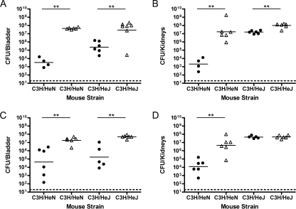
Many studies have argued that diabetics have a defect in host inflammatory cell function which may contribute to their increased infection rate. To further investigate whether innate host immunity or other factors irrespective of innate immunity play roles in the differences observed between diabetic and healthy infections, we inoculated diabetic and healthy C3H/HeJ and C3H/HeN female mice with E. coli UTI89 or K. pneumoniae TOP52 (Fig. 6). C3H/HeJ mice contain a mutation in the Toll-like receptor 4 (TLR-4) signaling domain and thus fail to transmit a signal. It has been shown that UPEC colonizes the bladders and kidneys of C3H/HeJ mice to significantly higher levels than those of C3H/HeN mice without inducing a significant neutrophil response early in infection (18, 50). Diabetic C3H/HeJ mice were more prone to infection by E. coli UTI89 at 72 h postinfection than healthy controls (Fig. 6A) (P = 0.0221). The presence of a diabetic advantage in C3H/HeJ mice suggests that additional TLR-4-independent factors may be important in diabetic UTI. However, the difference in UTI89 titers between healthy and diabetic bladders was greater in C3H/HeN mice (10,000-fold increase) than in C3H/HeJ mice (100-fold increase), suggesting that innate immune factors related to TLR-4-regulated processes may also be important. UTI89 had less than a 10-fold advantage in diabetic kidneys of C3H/HeJ mice (Fig. 6B) (P = 0.0023) at 72 h postinfection than in healthy controls, compared to the 10,000-fold advantage seen for C3H/HeN kidneys. Thus, TLR-4-regulated factors may account for much of the increase in bacterial burdens in diabetic mouse kidneys compared to those in healthy controls.
FIG. 6.
Differences in diabetic UTI in C3H/HeN and C3H/HeJ mice. Female C3H/HeN and C3H/HeJ diabetic mice (Δ) and buffer-treated control mice (•) were inoculated with 107 CFU of E. coli UTI89 (A and B) or 107 CFU of K. pneumoniae TOP52 (C and D). For organ titers, bladders (A and C) and kidneys (B and D) were harvested at 72 h postinfection and CFU were calculated. Graphs of bacterial burden of UTI89 in the bladders (A) and kidneys (B) and TOP52 in the bladders (C) and kidneys (D) of both mouse backgrounds are shown. Titer data are combined from two independent experiments. Short bars represent geometric means of each group, and dotted lines represent limits of detection. The symbol ** indicates significant P values of less than 0.05.
K. pneumoniae TOP52 had significantly higher titers in the bladders (Fig. 6C) of diabetic C3H/HeN (P = 0.0043) and C3H/HeJ (P = 0.0043) mice than in those of buffer-treated controls. Interestingly, the differences in geometric means of the 72-h-titer data were roughly equivalent in the C3H/HeN and C3H/HeJ backgrounds. Similarly to results for UTI89, while TOP52 displayed a significant advantage in the kidneys (Fig. 6D) of diabetic C3H/HeN mice compared to those of healthy C3H/HeN controls (P = 0.0043), TOP52 had no advantage in the kidneys of diabetic C3H/HeJ mice compared to those of healthy C3H/HeJ controls (P = 0.9307). Thus, TLR-4-regulated factors seemingly account for much of the increased advantage of TOP52 in the C3H/HeN kidney. Inducing diabetes in the C3H/HeJ mice produced no additional observable effects. Thus, much of the increase in C3H/HeN kidney colonization conferred upon inducing diabetes may be due to certain defects in TLR-4-regulated innate immune factors, although non-TLR-4-related factors may also be important.
Taken together, these experiments suggest that the diabetic phenotypes observed for both UPEC and non-UPEC organisms are likely multifactorial. A defect in TLR-4-regulated factors may play a role for diabetic mice. Nevertheless, other diabetic effects, irrespective of TLR-4, are also important in the ability of uropathogens to cause high burdens of infection in the urinary tract.
Urine growth curves.
Poorly regulated diabetic patients often spill glucose into their urine. To determine whether STZ-treated diabetic mice have glucosuria, we collected urine samples from diabetic and buffer-treated control mice. Urine test reagent strips were used to determine the glucose status and specific gravity of the mouse urine prior to inoculation with a uropathogen. Diabetic mouse urine consistently had ≥200 mg/dl (2%) glucose and a low specific gravity of 1.010. Healthy control mouse urine was negative for glucose and had a specific gravity of 1.030. Similar trace amounts of protein and ketones were found in diabetic and healthy mouse urine.
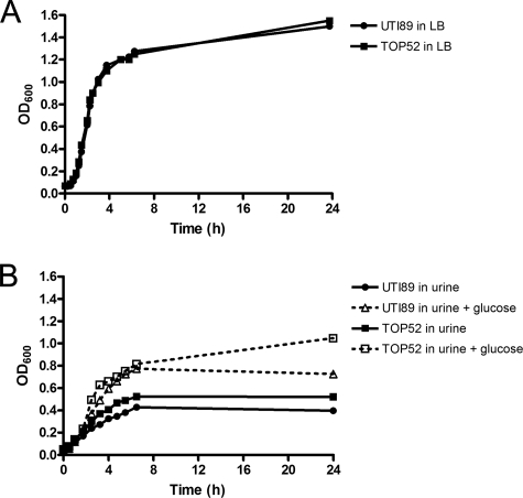
E. coli UTI89 and K. pneumoniae TOP52 grow equally well in LB, with doubling times of 34 min (Fig. 7A). To determine whether the glucosuria of the diabetic mice affects uropathogen growth, E. coli UTI89 and K. pneumoniae TOP52 were grown in filter-sterilized urine from healthy human subjects with and without 2% glucose supplementation (Fig. 7B). Optical density readings of both UTI89 and TOP52 correlated with CFU during logarithmic growth. The doubling times of both UTI89 (1.36 h with glucose and 2.24 without glucose, P = 0.0379) and TOP52 (1.05 h with glucose and 1.87 without glucose, P = 0.0262) were significantly shorter in glucose-supplemented urine. TOP52 displayed higher yields than UTI89 in urine with (P = 0.0286) and without (P = 0.0286) glucose supplementation.
FIG. 7.
Addition of glucose enhances E. coli UTI89 and K. pneumoniae TOP52 growth in urine. Growth of E. coli UTI89 and K. pneumoniae TOP52 over time was measured in LB (A) and filter-sterilized urine with and without the supplementation of 2% glucose (B). Growth curves shown are representative of three independent experiments.
These data show that STZ-induced diabetic mice have glucosuria similar to that of poorly controlled diabetics. This urine glucose may contribute to the increased bacterial burden within the urinary tracts of diabetic mice.
DISCUSSION
Models of STZ-induced diabetes have been used for decades (8, 28, 31); however, little has been done to study diabetic UTI. UTIs are more prevalent and more severe in the diabetic population than in the nondiabetic population, and murine models of infection that accurately mirror human infection are required to better understand disease (42). STZ-induced diabetic mice were more susceptible to UPEC UTI and had higher burdens of infection than buffer-treated controls. Remarkably low inocula of the UPEC strain UTI89 were able to effectively infect diabetic mice but were largely cleared from healthy mice. It is also known that diabetic patients are more likely to be infected with non-UPEC uropathogens (29). The K. pneumoniae strain TOP52 and the E. faecalis strain 0852 had significantly higher bacterial titers in the bladders and kidneys of STZ-induced diabetic mice than in those of healthy mice. E. coli UTI89 outcompeted K. pneumoniae TOP52 in both healthy and diabetic bladders. However, the advantage of UTI89 over TOP52 in the bladders of diabetic mice was reduced. This result reflects the clinical diabetic situation in which UPEC remains the predominant uropathogen, with an increased frequency of K. pneumoniae infection. In contrast, the advantage of UTI89 in the kidneys of diabetic mice over TOP52 was dramatic, since TOP52 outcompeted UTI89 in healthy kidneys. Finally, experiments with C3H/HeJ mice suggested that defects in the diabetic urinary tract to manage and clear bacterial infection are likely multifactorial, possibly involving TLR-4-regulated factors, glucosuria, and other unknown factors. All of these findings are consistent with human diabetic UTI epidemiologic data and suggest that this STZ-induced model of diabetic UTI can provide a valuable tool for studying this disease.
Histologic analysis of mouse kidneys revealed a severe pyelonephritic phenotype specific to E. coli UTI89 in the diabetic setting. Large extracellular bacterial biofilm-like communities were observed filling renal tubules. Bacteria within these communities were tightly packed within an extracellular matrix. Morphologically, these bacterial collections appeared similar to the UPEC biofilm-like intracellular bacterial communities observed within facet cells of the bladder (1, 48). Further studies are required to determine what enables UTI89 to form these communities within the renal tubules of the diabetic host.
There are numerous hypotheses for the enhanced ability of uropathogens to infect the urinary tracts of diabetic individuals (12). Poorly controlled diabetes often results in the spilling of glucose into the urine. Glucosuria increased the growth rates of UTI89 and TOP52 and has been shown to increase growth of numerous other uropathogenic isolates (10). Multiple studies have suggested that neutrophil dysfunction and differences in cytokine secretion may play important roles in diabetic UTI (11, 43, 52); however, data contradicting these claims have also been reported (3). We discovered that diabetes in C3H/HeJ mice resulted in significantly increased colonization of the bladders and kidneys by E. coli, albeit the effect was decreased compared to that in C3H/HeN mice. Additionally, the induction of diabetes resulted in no advantage for K. pneumoniae in the diabetic kidneys of C3H/HeJ mice. These results argue that both TLR-4-related and -unrelated factors may be defective in the setting of diabetes. TLR-4-regulated factors affected by diabetes may include the inability of neutrophils to effectively clear bacteria, as was seemingly the case for the diabetic kidneys infected by E. coli UTI89. UTI89 was able to establish massive extracellular biofilm-like collections even in the presence of a robust neutrophilic inflammatory infiltrate. Finally, studies have suggested that bacteria have increased adherence abilities in the urinary tracts of diabetics, possibly due to lower levels of urine Tamm-Horsfall protein in diabetics (4, 7, 45) or potential changes in the uroepithelial cells themselves (13). This murine model of diabetic UTI provides a powerful tool for dissecting these and other factors involved in diabetic uropathogenesis.
The STZ-induced diabetic UTI mouse model is extremely versatile. A type 1 diabetic syndrome can be induced in different murine backgrounds, allowing for the analysis of specific host factors in knockout or mutant mouse backgrounds. Additionally, uropathogens have been studied with various mouse strains and this method of STZ induction can be applied to the mouse strain best characterized for a given uropathogen. For example, we were able to induce diabetes and analyze infection by using the previously established mouse model of E. faecalis UTI in the C57BL/6 murine background (25). The STZ method of diabetic induction is extremely practical, with high levels of penetrance (80 to 85%) in a relatively short period of time (2 to 3 weeks), and can be well controlled with buffer i.p. injections in the identical mouse strain. While STZ produces a type-1-like, irreversible insult to pancreatic β-cells, leading to a severe diabetic phenotype in these mice (8), it may be possible to treat these mice with insulin or other agents to contrast UTI in the settings of controlled and uncontrolled diabetes. It is currently unclear whether the diabetic UTI phenotypes observed will be present in a euglycemic diabetic host. Additionally, this model could be used to test various UTI preventative measures, including vaccinations, which may be especially beneficial for this predisposed population.
This STZ-induced murine model of diabetic UTI is consistent with many known characteristics of human diabetic UTI, including increased susceptibility to infection and more-severe infection. Many of these traits of diabetic UTI are poorly understood and require an infection model to test hypotheses of diabetic uropathogenesis. Using this model, we discovered that diabetes affects E. coli, K. pneumoniae, and E. faecalis pathogenesis in diverse ways. Defects in TLR-4-dependent and -independent pathways appear to provide differential advantages to these pathogens. A dramatic decrease in the ID50 may explain the increased susceptibility of diabetics in general, and the ability of E. coli to establish biofilms in the diabetic kidney provides insights into the increased virulence seen in diabetics. This model of diabetic UTI is versatile and practical and can provide a powerful tool for dissecting UTI in the setting of diabetes.
Acknowledgments
We thank Michael Caparon, Peter Humphrey, and Helen Liapis for helpful discussions. We also thank Wandy Beatty for assistance with electron microscopy processing and imaging.
This work was supported by National Institutes of Health Office of Research on Women's Health: Specialized Center of Research on Sex and Gender Factors Affecting Women's Health grant R01 DK64540 and National Institute of Diabetes and Digestive and Kidney Diseases grant R01 DK051406.
Editor: V. J. DiRita
Footnotes
Published ahead of print on 21 July 2008.
REFERENCES
- 1.Anderson, G. G., J. J. Palermo, J. D. Schilling, R. Roth, J. Heuser, and S. J. Hultgren. 2003. Intracellular bacterial biofilm-like pods in urinary tract infections. Science 301105-107. [DOI] [PubMed] [Google Scholar]
- 2.Arthur, M., C. E. Johnson, R. H. Rubin, C. Arbeit, C. Campanelli, C. Kim, S. Steinbach, M. Agarwal, R. Wilkinson, and R. Goldstein. 1989. Molecular epidemiology of adhesin and hemolysin virulence factors among uropathogenic Escherichia coli. Infect. Immun. 57303-313. [DOI] [PMC free article] [PubMed] [Google Scholar]
- 3.Balasoiu, D., K. C. van Kessel, H. J. van Kats-Renaud, T. J. Collet, and A. I. Hoepelman. 1997. Granulocyte function in women with diabetes and asymptomatic bacteriuria. Diabetes Care 20392-395. [DOI] [PubMed] [Google Scholar]
- 4.Bernard, A. M., A. A. Ouled, R. R. Lauwerys, A. Lambert, and B. Vandeleene. 1987. Pronounced decrease of Tamm-Horsfall proteinuria in diabetics. Clin. Chem. 331264. [PubMed] [Google Scholar]
- 5.Boyko, E. J., S. D. Fihn, D. Scholes, L. Abraham, and B. Monsey. 2005. Risk of urinary tract infection and asymptomatic bacteriuria among diabetic and nondiabetic postmenopausal women. Am. J. Epidemiol. 161557-564. [DOI] [PubMed] [Google Scholar]
- 6.Carton, J. A., J. A. Maradona, F. J. Nuno, R. Fernandez-Alvarez, F. Perez-Gonzalez, and V. Asensi. 1992. Diabetes mellitus and bacteraemia: a comparative study between diabetic and non-diabetic patients. Eur. J. Med. 1281-287. [PubMed] [Google Scholar]
- 7.Dulawa, J., K. Jann, M. Thomsen, M. Rambausek, and E. Ritz. 1988. Tamm Horsfall glycoprotein interferes with bacterial adherence to human kidney cells. Eur. J. Clin. Investig. 1887-91. [DOI] [PubMed] [Google Scholar]
- 8.Ganda, O. P., A. A. Rossini, and A. A. Like. 1976. Studies on streptozotocin diabetes. Diabetes 25595-603. [DOI] [PubMed] [Google Scholar]
- 9.Garofalo, C. K., T. M. Hooton, S. M. Martin, W. E. Stamm, J. J. Palermo, J. I. Gordon, and S. J. Hultgren. 2007. Escherichia coli from urine of female patients with urinary tract infections is competent for intracellular bacterial community formation. Infect. Immun. 7552-60. [DOI] [PMC free article] [PubMed] [Google Scholar]
- 10.Geerlings, S. E., E. C. Brouwer, W. Gaastra, J. Verhoef, and A. I. Hoepelman. 1999. Effect of glucose and pH on uropathogenic and non-uropathogenic Escherichia coli: studies with urine from diabetic and non-diabetic individuals. J. Med. Microbiol. 48535-539. [DOI] [PubMed] [Google Scholar]
- 11.Geerlings, S. E., E. C. Brouwer, K. P. van Kessel, W. Gaastra, and A. M. Hoepelman. 2000. Cytokine secretion is impaired in women with diabetes mellitus. Adv. Exp. Med. Biol. 485255-262. [DOI] [PubMed] [Google Scholar]
- 12.Geerlings, S. E., R. Meiland, and A. I. Hoepelman. 2002. Pathogenesis of bacteriuria in women with diabetes mellitus. Int. J. Antimicrob. Agents 19539-545. [DOI] [PubMed] [Google Scholar]
- 13.Geerlings, S. E., R. Meiland, E. C. van Lith, E. C. Brouwer, W. Gaastra, and A. I. Hoepelman. 2002. Adherence of type 1-fimbriated Escherichia coli to uroepithelial cells: more in diabetic women than in control subjects. Diabetes Care 251405-1409. [DOI] [PubMed] [Google Scholar]
- 14.Geerlings, S. E., R. P. Stolk, M. J. Camps, P. M. Netten, J. B. Hoekstra, P. K. Bouter, B. Braveboer, T. J. Collet, A. R. Jansz, and A. M. Hoepelman. 2000. Asymptomatic bacteriuria can be considered a diabetic complication in women with diabetes mellitus. Adv. Exp. Med. Biol. 485309-314. [DOI] [PubMed] [Google Scholar]
- 15.Gillespie, K. M. 2006. Type 1 diabetes: pathogenesis and prevention. Can. Med. Assoc. J. 175165-170. [DOI] [PMC free article] [PubMed] [Google Scholar]
- 16.Griebling, T. L. 2007. Urinary tract infections in women, p. 587-620. In M. S. Litwin and C. S. Saigal (ed.), Urologic diseases in America. U.S. Department of Health and Human Services, Public Health Service, National Institutes of Health, National Institute of Diabetes and Digestive and Kidney Diseases. NIH publication no. 07-5512. U.S. Government Printing Office, Washington, DC.
- 17.Hansen, D. S., A. Gottschau, and H. J. Kolmos. 1998. Epidemiology of Klebsiella bacteraemia: a case control study using Escherichia coli bacteraemia as control. J. Hosp. Infect. 38119-132. [DOI] [PubMed] [Google Scholar]
- 18.Haraoka, M., L. Hang, B. Frendeus, G. Godaly, M. Burdick, R. Strieter, and C. Svanborg. 1999. Neutrophil recruitment and resistance to urinary tract infection. J. Infect. Dis. 1801220-1229. [DOI] [PubMed] [Google Scholar]
- 19.Hooton, T. M. 2001. Recurrent urinary tract infection in women. Int. J. Antimicrob. Agents 17259-268. [DOI] [PubMed] [Google Scholar]
- 20.Hooton, T. M., and W. E. Stamm. 1997. Diagnosis and treatment of uncomplicated urinary tract infection. Infect. Dis. Clin. N. Am. 11551-581. [DOI] [PubMed] [Google Scholar]
- 21.Ikaheimo, R., A. Siitonen, T. Heiskanen, U. Karkkainen, P. Kuosmanen, P. Lipponen, and P. H. Makela. 1996. Recurrence of urinary tract infection in a primary care setting: analysis of a 1-year follow-up of 179 women. Clin. Infect. Dis. 2291-99. [DOI] [PubMed] [Google Scholar]
- 22.Johnson, J. R. 1991. Virulence factors in Escherichia coli urinary tract infection. Clin. Microbiol. Rev. 480-128. [DOI] [PMC free article] [PubMed] [Google Scholar]
- 23.Justice, S. S., C. Hung, J. A. Theriot, D. A. Fletcher, G. G. Anderson, M. J. Footer, and S. J. Hultgren. 2004. Differentiation and developmental pathways of uropathogenic Escherichia coli in urinary tract pathogenesis. Proc. Natl. Acad. Sci. USA 1011333-1338. [DOI] [PMC free article] [PubMed] [Google Scholar]
- 24.Justice, S. S., D. A. Hunstad, P. C. Seed, and S. J. Hultgren. 2006. Filamentation by Escherichia coli subverts innate defenses during urinary tract infection. Proc. Natl. Acad. Sci. USA 10319884-19889. [DOI] [PMC free article] [PubMed] [Google Scholar]
- 25.Kau, A. L., S. M. Martin, W. Lyon, E. Hayes, M. G. Caparon, and S. J. Hultgren. 2005. Enterococcus faecalis tropism for the kidneys in the urinary tract of C57BL/6J mice. Infect. Immun. 732461-2468. [DOI] [PMC free article] [PubMed] [Google Scholar]
- 26.Kauffman, C. A., J. A. Vazquez, J. D. Sobel, H. A. Gallis, D. S. McKinsey, A. W. Karchmer, A. M. Sugar, P. K. Sharkey, G. J. Wise, R. Mangi, A. Mosher, J. Y. Lee, W. E. Dismukes, et al. 2000. Prospective multicenter surveillance study of funguria in hospitalized patients. Clin. Infect. Dis. 3014-18. [DOI] [PubMed] [Google Scholar]
- 27.Lane, M. C., C. J. Alteri, S. N. Smith, and H. L. Mobley. 2007. Expression of flagella is coincident with uropathogenic Escherichia coli ascension to the upper urinary tract. Proc. Natl. Acad. Sci. USA 10416669-16674. [DOI] [PMC free article] [PubMed] [Google Scholar]
- 28.Like, A. A., and A. A. Rossini. 1976. Streptozotocin-induced pancreatic insulitis: new model of diabetes mellitus. Science 193415-417. [DOI] [PubMed] [Google Scholar]
- 29.Lye, W. C., R. K. Chan, E. J. Lee, and G. Kumarasinghe. 1992. Urinary tract infections in patients with diabetes mellitus. J. Infect. 24169-174. [DOI] [PubMed] [Google Scholar]
- 30.MacFarlane, I. A., R. M. Brown, R. W. Smyth, D. W. Burdon, and M. G. FitzGerald. 1986. Bacteraemia in diabetics. J. Infect. 12213-219. [DOI] [PubMed] [Google Scholar]
- 31.Makino, S., K. Kunimoto, Y. Muraoka, Y. Mizushima, K. Katagiri, and Y. Tochino. 1980. Breeding of a non-obese, diabetic strain of mice. Jikken Dobutsu 291-13. [DOI] [PubMed] [Google Scholar]
- 32.Martinez, J. J., M. A. Mulvey, J. D. Schilling, J. S. Pinkner, and S. J. Hultgren. 2000. Type 1 pilus-mediated bacterial invasion of bladder epithelial cells. EMBO J. 192803-2812. [DOI] [PMC free article] [PubMed] [Google Scholar]
- 33.Mulvey, M. A., Y. S. Lopez-Boado, C. L. Wilson, R. Roth, W. C. Parks, J. Heuser, and S. J. Hultgren. 1998. Induction and evasion of host defenses by type 1-piliated uropathogenic Escherichia coli. Science 2821494-1497. [DOI] [PubMed] [Google Scholar]
- 34.Mulvey, M. A., J. D. Schilling, and S. J. Hultgren. 2001. Establishment of a persistent Escherichia coli reservoir during the acute phase of a bladder infection. Infect. Immun. 694572-4579. [DOI] [PMC free article] [PubMed] [Google Scholar]
- 35.Munoz, P., A. Llancaqueo, M. Rodriguez-Creixems, T. Pelaez, L. Martin, and E. Bouza. 1997. Group B streptococcus bacteremia in nonpregnant adults. Arch. Intern. Med. 157213-216. [DOI] [PubMed] [Google Scholar]
- 36.Ng, T. K., J. M. Ling, A. F. Cheng, and S. R. Norrby. 1996. A retrospective study of clinical characteristics of Acinetobacter bacteremia. Scand. J. Infect. Dis. Suppl. 10126-32. [PubMed] [Google Scholar]
- 37.Nicolle, L. E., D. Friesen, G. K. Harding, and L. L. Roos. 1996. Hospitalization for acute pyelonephritis in Manitoba, Canada, during the period from 1989 to 1992; impact of diabetes, pregnancy, and aboriginal origin. Clin. Infect. Dis. 221051-1056. [DOI] [PubMed] [Google Scholar]
- 38.Onkamo, P., S. Vaananen, M. Karvonen, and J. Tuomilehto. 1999. Worldwide increase in incidence of type I diabetes—the analysis of the data on published incidence trends. Diabetologia 421395-1403. [DOI] [PubMed] [Google Scholar]
- 39.Opal, S. M., A. S. Cross, P. Gemski, and L. W. Lyhte. 1990. Aerobactin and alpha-hemolysin as virulence determinants in Escherichia coli isolated from human blood, urine, and stool. J. Infect. Dis. 161794-796. [DOI] [PubMed] [Google Scholar]
- 40.Ott, M., J. Hacker, T. Schmoll, T. Jarchau, T. K. Korhonen, and W. Goebel. 1986. Analysis of the genetic determinants coding for the S-fimbrial adhesin (sfa) in different Escherichia coli strains causing meningitis or urinary tract infections. Infect. Immun. 54646-653. [DOI] [PMC free article] [PubMed] [Google Scholar]
- 41.Ott, M., H. Hoschutzky, K. Jann, I. Van Die, and J. Hacker. 1988. Gene clusters for S fimbrial adhesin (sfa) and F1C fimbriae (foc) of Escherichia coli: comparative aspects of structure and function. J. Bacteriol. 1703983-3990. [DOI] [PMC free article] [PubMed] [Google Scholar]
- 42.Patterson, J. E., and V. T. Andriole. 1997. Bacterial urinary tract infections in diabetes. Infect. Dis. Clin. N. Am. 11735-750. [DOI] [PubMed] [Google Scholar]
- 43.Patterson, J. E., and V. T. Andriole. 1995. Bacterial urinary tract infections in diabetes. Infect. Dis. Clin. N. Am. 925-51. [PubMed] [Google Scholar]
- 44.Pozzilli, P., and R. D. Leslie. 1994. Infections and diabetes: mechanisms and prospects for prevention. Diabet. Med. 11935-941. [DOI] [PubMed] [Google Scholar]
- 45.Rambausek, M., J. Dulawa, K. Jann, and E. Ritz. 1988. Tamm Horsfall glycoprotein in diabetes mellitus: abnormal chemical composition and colloid stability. Eur. J. Clin. Investig. 18237-242. [DOI] [PubMed] [Google Scholar]
- 46.Reed, L. J., and H. Muench. 1938. A simple method of estimating fifty percent endpoints. Am. J. Hyg. 27493-497. [Google Scholar]
- 47.Ronald, A. 2003. The etiology of urinary tract infection: traditional and emerging pathogens. Dis. Mon. 4971-82. [DOI] [PubMed] [Google Scholar]
- 48.Rosen, D. A., T. M. Hooton, W. E. Stamm, P. A. Humphrey, and S. J. Hultgren. 2007. Detection of intracellular bacterial communities in human urinary tract infection. PLoS Med. 4e329. [DOI] [PMC free article] [PubMed] [Google Scholar]
- 49.Rosen, D. A., J. S. Pinkner, J. M. Jones, J. N. Walker, S. Clegg, and S. J. Hultgren. 2008. Utilization of an intracellular bacterial community pathway in Klebsiella pneumoniae urinary tract infection and the effects of FimK on type 1 pilus expression. Infect. Immun. 763337-3345. [DOI] [PMC free article] [PubMed] [Google Scholar]
- 50.Schilling, J. D., S. M. Martin, C. S. Hung, R. G. Lorenz, and S. J. Hultgren. 2003. Toll-like receptor 4 on stromal and hematopoietic cells mediates innate resistance to uropathogenic Escherichia coli. Proc. Natl. Acad. Sci. USA 1004203-4208. [DOI] [PMC free article] [PubMed] [Google Scholar]
- 51.Snyder, J. A., B. J. Haugen, E. L. Buckles, C. V. Lockatell, D. E. Johnson, M. S. Donnenberg, R. A. Welch, and H. L. Mobley. 2004. Transcriptome of uropathogenic Escherichia coli during urinary tract infection. Infect. Immun. 726373-6381. [DOI] [PMC free article] [PubMed] [Google Scholar]
- 52.Stapleton, A. 2002. Urinary tract infections in patients with diabetes. Am. J. Med. 113(Suppl. 1A)80S-84S. [DOI] [PubMed] [Google Scholar]
- 53.Wright, K. J., P. C. Seed, and S. J. Hultgren. 2005. Uropathogenic Escherichia coli flagella aid in efficient urinary tract colonization. Infect. Immun. 737657-7668. [DOI] [PMC free article] [PubMed] [Google Scholar]


