

Abstract
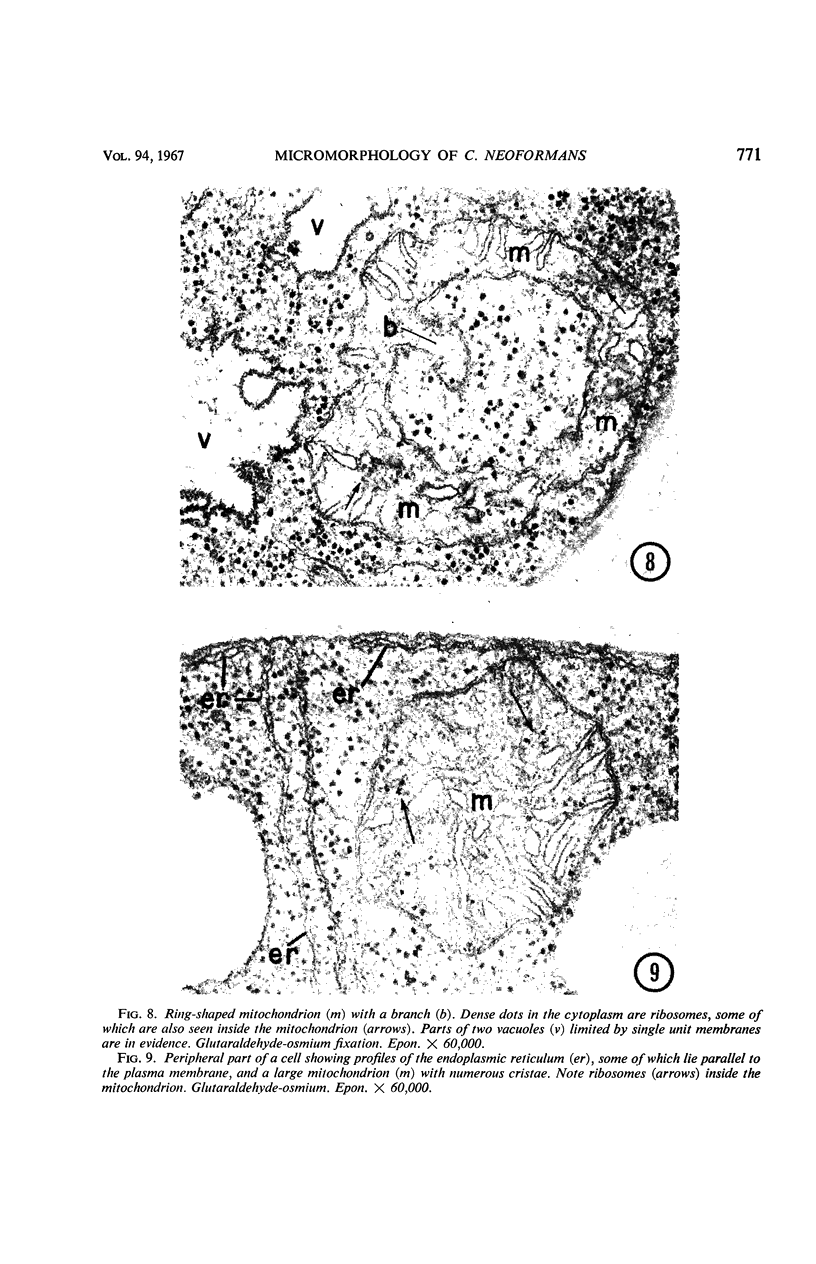
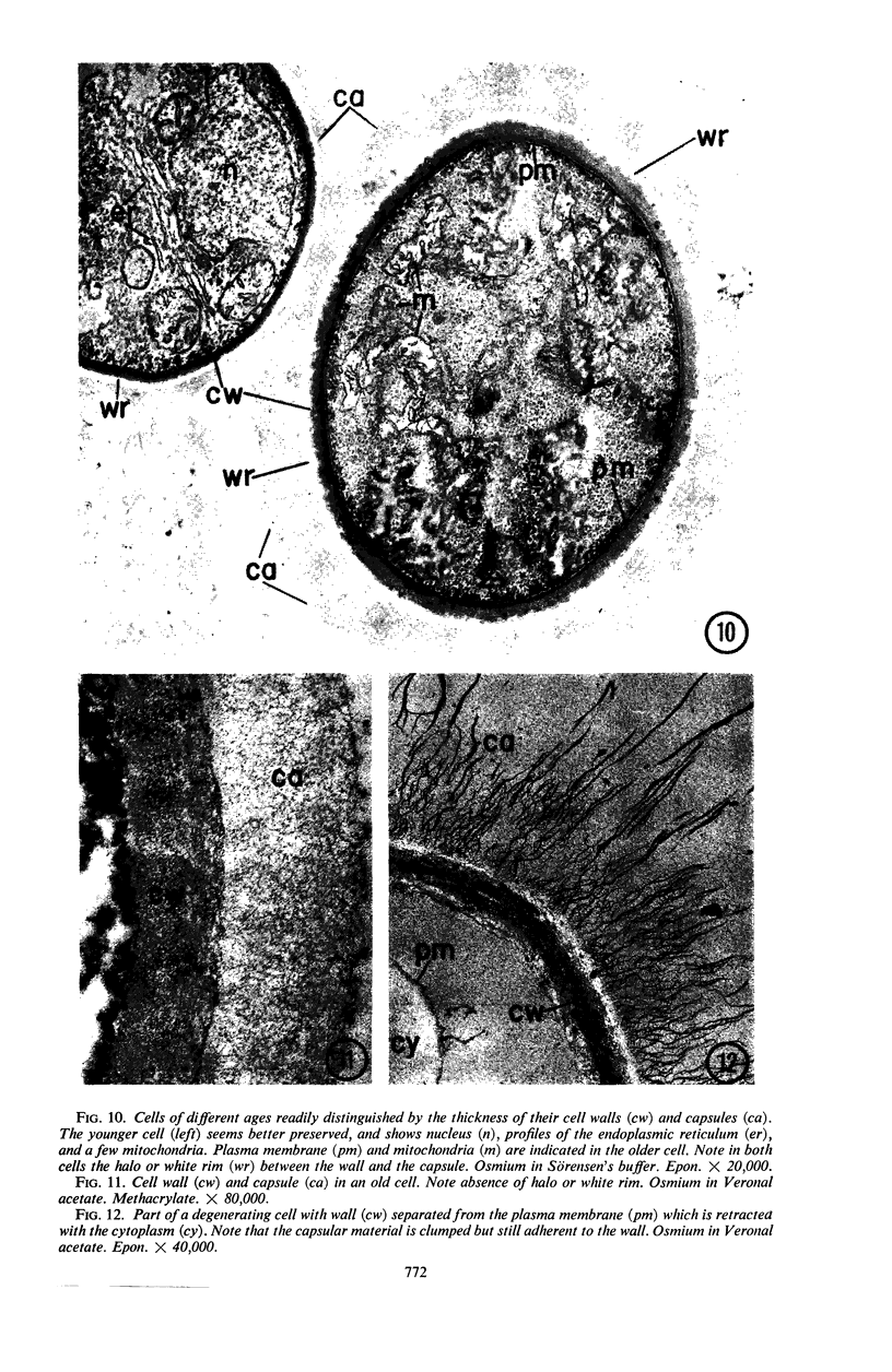
Fine details of the internal and external morphology of Cryptococcus neoformans as seen in ultrathin sections are described and illustrated with electron micrographs. The capsule characteristic of this species contained microfibrils (30 to 40 A in diameter) that appeared to radiate from the cell wall and to coil and intertwine in various directions. These thin, uniformly structured, electron-dense filaments are believed to represent complex polysaccharide molecules. The internal morphology of C. neoformans was in many ways similar to that of yeasts studied by other authors. The cell was uninucleate with a single nucleolus. The nuclear envelope, a pair of unit membranes interrupted by pores, was typical of that found in eucaryotic organisms. Smooth endoplasmic reticulum, mitochondria, vacuoles, storage granules, and ribosomes were consistent features of the cytoplasm. In addition, C. neoformans presented membranous organelles derived from the plasma membrane and comparable to bacterial mesosomes and mitochondria of an annulate type.
Full text
PDF


Images in this article


Selected References
These references are in PubMed. This may not be the complete list of references from this article.
- AGAR H. D., DOUGLAS H. C. Studies on the cytological structure of yeast: electron microscopy of thin sections. J Bacteriol. 1957 Mar;73(3):365–375. doi: 10.1128/jb.73.3.365-375.1957. [DOI] [PMC free article] [PubMed] [Google Scholar]
- BAKERSPIGEL A. SOME OBSERVATIONS ON THE CYTOLOGY OF CANDIDA ALBICANS. J Bacteriol. 1964 Jan;87:228–230. doi: 10.1128/jb.87.1.228-230.1964. [DOI] [PMC free article] [PubMed] [Google Scholar]
- CARBONELL L. M., POLLAK L. ULTRAESTRUCTURA DEL PARACOCCIDIOIDES BRASILIENSIS EN CULTIVOS DE LA FASE LEVADURIFORME. Mycopathol Mycol Appl. 1963 Jun 15;19:184–204. doi: 10.1007/BF02051247. [DOI] [PubMed] [Google Scholar]
- CONTI S. F., NAYLOR H. B. Electron microscopy of ultrathin sections of Schizosaccharomyces octosporus. I. Cell division. J Bacteriol. 1959 Dec;78:868–877. doi: 10.1128/jb.78.6.868-877.1959. [DOI] [PMC free article] [PubMed] [Google Scholar]
- CONTI S. F., NAYLOR H. B. Electron microscopy of ultrathin sections of Schizosaccharomyces octosporus. II. Morphological and cytological changes preceding ascospore formation. J Bacteriol. 1960 Mar;79:331–340. doi: 10.1128/jb.79.3.331-340.1960. [DOI] [PMC free article] [PubMed] [Google Scholar]
- EDWARDS M. R., HAZEN E. L., EDWARDS G. A. The fine structure of the yeast-like cells of Histoplasma in culture. J Gen Microbiol. 1959 Jun;20(3):496–503. doi: 10.1099/00221287-20-3-496. [DOI] [PubMed] [Google Scholar]
- EDWARDS M. R., STEVENS R. W. FINE STRUCTURE OF LISTERIA MONOCYTOGENES. J Bacteriol. 1963 Sep;86:414–428. doi: 10.1128/jb.86.3.414-428.1963. [DOI] [PMC free article] [PubMed] [Google Scholar]
- FITZ-JAMES P. C. Participation of the cytoplasmic membrane in the growth and spore fromation of bacilli. J Biophys Biochem Cytol. 1960 Oct;8:507–528. doi: 10.1083/jcb.8.2.507. [DOI] [PMC free article] [PubMed] [Google Scholar]
- Fuhs G. W. Symposium on the fine structure and replication of bacteria and their parts. I. Fine structure and replication of bacterial nucleoids. Bacteriol Rev. 1965 Sep;29(3):277–293. doi: 10.1128/br.29.3.277-293.1965. [DOI] [PMC free article] [PubMed] [Google Scholar]
- HASHIMOTO T., CONTI S. F., NAYLOR H. B. Studies of the fine structure of microorganisms. IV. Observations on budding Saccharomyces cerevisiae by light and electron microscopy. J Bacteriol. 1959 Mar;77(3):344–354. doi: 10.1128/jb.77.3.344-354.1959. [DOI] [PMC free article] [PubMed] [Google Scholar]
- HAWKER L. E. FINE STRUCTURE OF FUNGI AS REVEALED BY ELECTRON MICROSCOPY. Biol Rev Camb Philos Soc. 1965 Feb;40:52–92. doi: 10.1111/j.1469-185x.1965.tb00795.x. [DOI] [PubMed] [Google Scholar]
- Hyde J. M., Walkinshaw C. H. Ultrastructure of basidiospores and mycelium of Lenzites saepiaria. J Bacteriol. 1966 Oct;92(4):1218–1227. doi: 10.1128/jb.92.4.1218-1227.1966. [DOI] [PMC free article] [PubMed] [Google Scholar]
- LUCK D. J. THE INFLUENCE OF PRECURSOR POOL SIZE ON MITOCHONDRIAL COMPOSITION IN NEUROSPORA CRASSA. J Cell Biol. 1965 Mar;24:445–460. doi: 10.1083/jcb.24.3.445. [DOI] [PMC free article] [PubMed] [Google Scholar]
- LUFT J. H. Improvements in epoxy resin embedding methods. J Biophys Biochem Cytol. 1961 Feb;9:409–414. doi: 10.1083/jcb.9.2.409. [DOI] [PMC free article] [PubMed] [Google Scholar]
- LUFT J. H. Permanganate; a new fixative for electron microscopy. J Biophys Biochem Cytol. 1956 Nov 25;2(6):799–802. doi: 10.1083/jcb.2.6.799. [DOI] [PMC free article] [PubMed] [Google Scholar]
- McALEAR J. H., EDWARDS G. A. [Continuity of plasma membrane and nuclear membrane]. Exp Cell Res. 1959 Mar;16(3):689–692. doi: 10.1016/0014-4827(59)90139-9. [DOI] [PubMed] [Google Scholar]
- McClary D. O., Bowers W. D., Jr Structural differentiation of obligately aerobic and facultatively anaerobic yeasts. J Cell Biol. 1967 Feb;32(2):519–524. doi: 10.1083/jcb.32.2.519. [DOI] [PMC free article] [PubMed] [Google Scholar]
- PALADE G. E. A study of fixation for electron microscopy. J Exp Med. 1952 Mar;95(3):285–298. doi: 10.1084/jem.95.3.285. [DOI] [PMC free article] [PubMed] [Google Scholar]
- REYNOLDS E. S. The use of lead citrate at high pH as an electron-opaque stain in electron microscopy. J Cell Biol. 1963 Apr;17:208–212. doi: 10.1083/jcb.17.1.208. [DOI] [PMC free article] [PubMed] [Google Scholar]
- RIBI E., SALVIN S. B. Antigens from the yeast phase of Histoplasma capsulatum. I. Morphology of the cell as revealed by the electron microscope. Exp Cell Res. 1956 Apr;10(2):394–404. doi: 10.1016/0014-4827(56)90013-1. [DOI] [PubMed] [Google Scholar]
- ROBINOW C. F. On the plasma membrane of some bacteria and fungi. Circulation. 1962 Nov;26:1092–1104. doi: 10.1161/01.cir.26.5.1092. [DOI] [PubMed] [Google Scholar]
- Robinow C. F., Marak J. A fiber apparatus in the nucleus of the yeast cell. J Cell Biol. 1966 Apr;29(1):129–151. doi: 10.1083/jcb.29.1.129. [DOI] [PMC free article] [PubMed] [Google Scholar]
- SABATINI D. D., BENSCH K., BARRNETT R. J. Cytochemistry and electron microscopy. The preservation of cellular ultrastructure and enzymatic activity by aldehyde fixation. J Cell Biol. 1963 Apr;17:19–58. doi: 10.1083/jcb.17.1.19. [DOI] [PMC free article] [PubMed] [Google Scholar]
- Shadomy H. J., Utz J. P. Preliminary studies on a hyphaforming mutant of Cryptococcus neoformans. Mycologia. 1966 May-Jun;58(3):383–390. [PubMed] [Google Scholar]
- TSUKAHARA T. CYTOLOGICAL STRUCTURE OF CRYPTOCOCCUS NEOFORMANS. Jpn J Microbiol. 1963 Aug;7:53–60. doi: 10.1111/j.1348-0421.1963.tb00242.x. [DOI] [PubMed] [Google Scholar]
- VITOLS E., NORTH R. J., LINNANE A. W. Studies on the oxidative metabolism of Saccharomyces cerevisiae. I. Observations on the fine structure of the yeast cell. J Biophys Biochem Cytol. 1961 Mar;9:689–699. doi: 10.1083/jcb.9.3.689. [DOI] [PMC free article] [PubMed] [Google Scholar]