Abstract
Naturally occurring Newcastle disease virus (NDV) strains vary greatly in virulence, ranging from no apparent infection to severe disease causing 100% mortality in chickens. The viral determinants of NDV virulence are not completely understood. Cleavage of the fusion protein is required for the initiation of infection, and it acts as a determinant of virulence. The attachment protein HN was found to play a minor role in virulence. In this study, we have evaluated the role of the internal proteins (N, P, and L) in NDV virulence by using a chimeric reverse-genetics approach. The N, P, and L genes were exchanged individually between an avirulent NDV strain, LaSota, and an intermediate virulent NDV strain, Beaudette C (BC), and the N and P genes were also exchanged together. The recovered chimeric viruses were evaluated for their pathogenicity in the natural host, chickens. Our results showed that the pathogenicities of N and P chimeric viruses were similar to those of their respective parental viruses, indicating that the N and P genes probably play minor roles in virulence. However, replacement of the L gene of BC with that of LaSota significantly increased the pathogenicity of the L-chimeric virus, suggesting that the L gene probably contributes to the virulence of NDV. The L-chimeric BC virus was found to replicate at a 100-fold-higher level than its parental virus in chicken brain, suggesting that the increase in pathogenicity may be due to the increased replication level of the chimeric virus. Our findings offer new insights into the pathogenesis of NDV infection.
Newcastle disease virus (NDV) infects all species of birds but causes a highly contagious respiratory, enteric, or neurological disease in chickens (22). Newcastle disease is prevalent worldwide and causes severe economic losses in the poultry industry. NDV is a member of the genus Avulavirus in the family Paramyxoviridae (16). The genome of NDV is a nonsegmented, single-stranded, negative-sense RNA of 15,186 nucleotides (6, 14). It encodes at least six proteins: the nucleocapsid protein (N), the phosphoprotein (P), the matrix protein (M), the fusion protein (F), the hemagglutinin-neuraminidase protein (HN), and the large polymerase protein (L) (4, 29). Two additional proteins, V and W, are produced by RNA editing during P gene transcription (21, 27). The M, F, and HN proteins are associated with the virus envelope, and the latter two are the protective antigens of NDV. The virus genomic RNA is tightly encapsidated by the N protein and is associated with the P and L proteins. This encapsidated RNA is the template for transcription and replication (15). The N protein interacts with the viral polymerase (P-L) during genome expression and with the P protein during the assembly of the nucleocapsid. The P protein forms complexes with both the N and L proteins and also has a supplemental role in RNA synthesis. The L protein is the largest viral protein and contains all the catalytic activities associated with the viral polymerase (15). The V protein functions as an interferon antagonist, while the function of the W protein is not known (11).
NDV strains cause a wide variation of disease in chickens. Based on the severity of the disease in chickens, NDV strains are categorized into three pathotypes: lentogenic, mesogenic, and velogenic (2). Lentogenic strains causing subclinical infection or mild respiratory diseases are considered avirulent or of lesser virulence. Mesogenic strains are of intermediate virulence and cause respiratory infection with moderate mortality, while velogenic strains are highly virulent, causing 100% mortality in chickens. The virulence of NDV strains is determined by using three internationally accepted in vivo tests: (i) mean death time (MDT) in 9-day-old embryonated chicken eggs, (ii) intracerebral pathogenicity index (ICPI) in 1-day-old chicks, and (iii) intravenous pathogenicity index (IVPI) in 6-week-old chickens (1).
The viral determinants responsible for the variation in pathogenicity observed among NDV strains are not well understood. The amino acid sequence at the F protein cleavage site has been shown to be a major determinant of NDV virulence (8, 19). The cleavage of precursor protein F0 to F1 and F2 by host cell proteases is required for a progeny virus to become infective. F0 proteins of intermediate and highly virulent NDV strains have multiple basic residues at the cleavage site which are substrates for the furin family of proteases and are cleaved intracellularly by most tissue types. These strains replicate systemically, causing severe disease. F0 proteins of avirulent or less-virulent NDV strains mostly have a single basic residue at the cleavage site and are cleaved extracellularly by proteases found in the respiratory tract. This requirement limits the virus to replicate only in the respiratory tract, causing mild respiratory disease. In addition, the F1 subunits of virulent NDV strains begin with a phenylalanine residue, whereas most avirulent NDV strains have a leucine residue at this position. This observation has also been confirmed by using a reverse genetics method in which the monobasic residue of the F protein cleavage site of an avirulent NDV strain was replaced by multibasic residues of a virulent NDV strain, which resulted in increased virulence of the mutated virus (19, 23, 25). Hence, it is well established that the sequence at the F protein cleavage site determines the site of NDV replication and plays an important role in NDV pathogenesis, but the sequence at the F protein cleavage site does not determine the degree of virulence among lentogenic strains or among mesogenic or velogenic strains. The HN protein, which possesses both the receptor recognition and neuraminidase activities of the virus, has also been evaluated for its role in virulence by using a reverse-genetics approach (7, 12). It was shown that the HN protein determines tropism and plays a minimal role in virulence (12). However, in another study, replacement of the HN gene of a mesogenic NDV strain by HN genes of neurotropic or viscerotropic NDV strains failed to enhance the pathogenicity of the chimeric viruses (7). Furthermore, replacement of both the F and HN genes of a mesogenic NDV strain by F and HN genes of velogenic NDV strains also did not increase the virulence of the chimeric viruses, indicating that the F and HN genes and their homotypic interactions are not the major determinants of NDV virulence (7). These results suggest that a viral protein(s) other than F and HN probably contributes to the virulence of NDV.
For this report, we examined the individual contributions of internal proteins N, P, and L to the virulence of NDV. The reverse genetics method was used to generate chimeric NDVs in which the N, P, or L genes were exchanged individually between a lentogenic strain, LaSota, and a mesogenic strain, Beaudette C (BC). In addition, chimeric viruses were generated in which the N and P genes were exchanged together to allow homotypic protein interactions. The virulence of these chimeric viruses was determined by the MDT, ICPI, and IVPI tests. Our in vivo results indicated that the N and P proteins play a minimal role in NDV virulence but the L protein probably is associated with NDV virulence.
MATERIALS AND METHODS
Cells and viruses.
DF-1 cells (chicken embryo fibroblast cell line) were grown in Dulbecco's minimal essential medium (DMEM) with 10% fetal bovine serum (FBS) and maintained in DMEM with 5% FBS. HEp-2 cells (human epidermoid carcinoma cell line) were grown in Eagle's minimal essential medium containing 10% FBS and maintained in Eagle's minimal essential medium with 5% FBS. The modified vaccinia virus strain Ankara, expressing T7 RNA polymerase (a generous gift of Bernard Moss, National Institutes of Health), was grown in primary chicken embryo fibroblast cells in DMEM with 10% FBS. A moderately pathogenic (mesogenic) NDV strain, BC, and a lentogenic vaccine strain, LaSota, were received from National Veterinary Services Laboratory (Ames, IA). The NDV strains were grown in the allantoic cavities of 9-day-old embryonated chicken eggs.
Construction of full-length chimeric BC/LaSota antigenomic cDNAs.
The construction of full-length antigenomic cDNAs of NDV strains BC (pBC) and LaSota (pLaSota) has been described previously (10, 13). These full-length cDNA clones were used to recover the recombinant viruses rBC and rLaSota, respectively (10, 13).
For this study, chimeric BC viruses containing the N, P, or L gene of strain LaSota in place of their own gene and the reciprocal chimeric LaSota viruses containing the N, P, or L gene of strain BC were generated (Fig. 1). The published nucleotide sequences of NDV strains BC (GenBank accession no. AF064091) and LaSota (GenBank accession no. NC_002617) were used for the construction of chimeric viruses. To exchange the L gene between pBC and pLaSota, a unique restriction site, PacI, was created in the 3′ untranslated region (UTR) of the L gene on full-length cDNAs of pBC and pLaSota (Fig. 1A). Briefly, the AflII-RsrII fragment of the L gene was amplified from both pBC and pLaSota in two steps and subcloned into pGEM-7Z (+) (Promega, Madison, WI). In the first step, fragment AflII-BamHI of the L gene was PCR amplified by using the primers Xba-AflF (5′-GCT CTA GAC TTA AGA AAC ATA CGC AAA GAG-3′) and BamPac/L− (5′-CTG GAT CCA TAA TTT AAT TAA ATC AAC AAG AAT ACA ATT GGC C-3′) and then subcloned between the XbaI and BamHI sites of pGEM-7Z (+), resulting in pGEM-7Z (AflII-PacI). The second fragment, AflII-RsrII, was PCR amplified by using primer Pac/B (5′-CTG GAT CCA TAG TTT AAT TAA ATC ACC AAG GAT ACA ATT GGC C-3′) for rBC or Pac/L (5′-CTG GAT CCA TAA TTT AAT TAA ATC AAC AAG AAT ACA ATT GGC C-3′) for rLaSota and BamRsr− (5′-CTG GAT CCG GAC CGC GAG GAG GTG GAG ATG-3′) and subcloned into the plasmid pGEM-7Z (AflII-PacI) between the PacI and BamHI sites, creating AflII-RsrII fragments of both pBC and pLaSota, each containing a PacI site in the 3′ UTR of the L gene. The introduced PacI sites are shown by underlining. The mutated AflII-RsrII fragments were excised from pGEM-7Z and replaced with their corresponding counterparts in the full-length antigenome cDNAs of pBC and pLaSota. These full-length antigenomes of pBC and pLaSota were digested with AgeI and PacI to exchange the L gene. The full-length cDNA plasmid of rBC with the L gene of rLaSota in place of its own L gene was designated pBCLaSoL, whereas the full-length cDNA plasmid of rLaSota with the L gene of rBC in place of its own L gene was designated pLaSoBCL.
FIG. 1.
Schematic representation (not drawn to scale) of strategies for exchange of N, P, or L gene between the pBC and pLaSota backbones. (A) Unique restriction site PacI was introduced after the L gene ORF and before the trailer. The L genes were exchanged between pBC and pLaSota by using AgeI and PacI without the trailer region. (B) Unique restriction sites AsiSI, between the N and P genes, and PmeI, between the P and M genes, were created on both pBC and pLaSota cDNAs. These sites were used to exchange the P gene. (C) The NP genes were exchanged between pBC and pLaSota by using AscI and AsiSI sites. (D) The N and P genes were exchanged in combination by using the restriction sites AscI and PmeI. The positions of restriction sites are indicated by the numbers. Le, leader; Tr, trailer; N, nucleocapsid protein; P, phosphoprotein; M, matrix protein; F, fusion protein; HN, hemagglutinin-neuraminidase protein; L, large polymerase protein.
To exchange the N and/or P gene, we introduced a unique restriction site, AsiSI, in the 3′ UTR of the N gene and a PmeI site in the 3′ UTR of the P gene in the full-length cDNAs of both pBC and pLaSota (Fig. 1B). To introduce the AsiSI site between the N and P genes, AscI-SacII fragments of pBC and pLaSota were PCR amplified and cloned into pGEM-7Z (+). Briefly, PCR was performed by using primers AscI-T7-F (5′-ATT CGG CGC GCC TAA TAC GAC TCA CTA TAG GG-3′) and 1645ASiSI-R (5′-CGC AAA TGC AGC GAT CGC CTA CGG GTG AGG ATA TTG GAT GA-3′) to get PCR product A and primers 1645ASiSIF (5′-GGC TAG GCG ATC GCT CGA TTT GCG GCC CTA TAT GAC CAC-3′) and BC/LaSo2400R (5′-GGG CGG CCT TGA CTT GGT TCT GCG GTC-3′) to get PCR product B. The introduced AsiSI sites are shown by underlining. Then both PCR products, A and B, were used to generate the final AscI-SacII fragment containing the AsiSI site by overlap PCR using the primers AScI-T7F and BC/LaSo2400R and subsequently cloned into full-length cDNAs of both pBC and pLaSota. Similarly, to introduce the PmeI site between the P and M genes, SacII-NotI fragments of both pBC and pLaSota were PCR amplified and cloned into pGEM-7Z (+). Briefly, PCR was performed by using the forward primer NP1225 (5′-GCA GCA AGG AGA GGC CTG GCA-3′) and reverse primer 3210BC-PmeIR (5′-TAG CTA GTT TAA ACA CGG TTG CGC GAT CAT TCA GTG GGG-3′) to generate PCR product C and forward primer 3210BC-PmeF (5′-GCG CAA CCG TGT TTA AAC TAG CTA CAT TAA GGA TTA AGA-3′) and reverse primer BC/LaSo 4970R (5′-TGT ATC AGA GCT GCG GCC GCT GTT ATT TG-3′ to generate PCR product D. The introduced PmeI sites are shown by underlining. Then, both PCR products, C and D, were used to generate the final SacII-NotI fragment containing the PmeI site by overlap PCR using primers NP1225 and BC/LaSo4970R. The resulting PCR product was cloned into full-length cDNAs of both pBC and pLaSota, creating the unique restriction sites AsiSI and PmeI. All the full-length cDNA clones were sequenced in their entirety to confirm the presence of unique sites and absence of any undesired mutations. The N gene open reading frame (ORF) was exchanged by using the AscI and AsiSI sites, and the P gene ORF was exchanged by using the AsiSI and PmeI sites (Fig. 1C). pBC bearing the N gene of LaSota instead of its own N gene was designated pBCLaSoN, whereas pLaSota bearing the N gene of BC instead of its own N gene was designated pLaSoBCN. Likewise, the pBC backbone containing the P gene of LaSota was designated pBCLaSoP, and the pLaSota backbone containing the P gene of BC was designated pLaSoBCP. In order to construct chimeric cDNAs where both the N and P gene ORFs were exchanged between BC and LaSota, the AscI and PmeI sites were used, and the cDNAs were designated pBCLaSoP+N and pLaSoBCP+N, respectively (Fig. 1D). All the exchanged regions in the full-length cDNAs were sequenced by dideoxynucleotide sequencing to confirm the presence of desired gene swaps.
Recovery of chimeric rNDVs.
Chimeric recombinant NDVs (rNDVs) were recovered by cotransfection of each NDV chimeric full-length cDNA plasmid, along with support plasmids encoding the N, P, and L proteins, into HEp-2 cells (Table 1). In each transfection, the respective support plasmids were used, as shown in Table 1. Simultaneously, HEp-2 cells were infected with recombinant vaccinia virus Ankara, which produces T7 RNA polymerase. Four days after transfection, the cell culture supernatant was used to recover the chimeric rNDVs by either passaging in DF-1 cells until a virus-specific cytopathic effect (CPE) appeared or injecting it into the allantoic cavities of 9-day-old embryonated chicken eggs until the allantoic fluid showed NDV-specific HA (12). The medium of cells transfected with rLaSota chimeric cDNAs was supplemented with 10% allantoic fluid as a source of exogenous proteases.
TABLE 1.
Combinations of full-length and support plasmids used to recover chimeric virusesa
| Recovered virus | Full-length plasmid | Support plasmids
|
||
|---|---|---|---|---|
| pN | pP | pL | ||
| rBC | pBC | BC | BC | BC |
| rBCLaSoL | pBCLaSoL | BC | BC | LaSota |
| rBCLaSoP | pBCLaSoP | BC | LaSota | BC |
| rBCLaSoN | pBCLaSoN | LaSota | BC | BC |
| rBCLaSoP+N | pBCLaSoP+N | LaSota | LaSota | BC |
| rLaSota | pLaSota | LaSota | LaSota | LaSota |
| rLaSoBCL | pLaSoBCL | LaSota | LaSota | BC |
| rLaSoBCP | pLaSoBCP | LaSota | BC | LaSota |
| rLaSoBCN | pLaSoBCN | BC | LaSota | LaSota |
| rLaSoBCP+N | pLaSoBCP+N | BC | BC | LaSota |
Full-length cDNAs and various combinations of support plasmids were used to transfect HEp-2 cells. The support plasmid heterologous to full-length cDNA is shown in bold.
Sequence analysis of chimeric viruses.
Total RNAs were isolated from virus-infected DF-1 cells by using TRIzol (Invitrogen, Carlsbad, CA) according to the manufacturer's instructions. The extracted total RNA samples were reverse-transcribed by using a Superscript II reverse transcriptase kit (Invitrogen) to obtain first-strand cDNAs. For L-gene-chimeric viruses, the first-strand cDNA synthesis was carried out by using primer HN7957 (5′-CGA GTG AGT TCA AGC AGT ACC AAA GC-3′). The cDNA products generated were used for PCR using primers HN7957 and HN/L− (5′-TGT CTG CTG AGA ATG AGG TG-3′) for strain BC and primers L6400 (5′-GGG TCT TAG AGT CGA AGA TCT C-3′) and 15186R (5′-ACC AAA CAA AGA TTT GG-3′) for strain LaSota. Similarly, for N- and/or P-chimeric viruses, the first-strand cDNA synthesis was carried out by using primer AscI-T7F (5′-ATT CGG CGC GCC TAA TAC GAC TCA CTA TAG GG-3′) for the N gene and primer NP1225 (5′-GCA GCA AGG AGA GGC CTG GCA-3′) for the P gene. The cDNAs generated were then PCR amplified by using primers NP196 (5′-GAC CAG AAG ATA GGT GGA AC-3′) and BC/LaSo2400R (5′-GGG CGG CCT TGA CTT GGT TCT GCG GTC-3′) for the N gene and primers NP1225 (5′-GCA GCA AGG AGA GGC CTG GCA-3′) and BC/LaSo7970R (5′-TGT ATC AGA GCT GCG GCC GCT GTT ATT TG-3′) for the P gene. All the PCR products were purified by using a PCR purification kit (Qiagen, Valencia, CA) and then sequenced with an ABI 3100 DNA sequencer (Applied Biosystems Inc, CA).
Growth characteristics of chimeric viruses in DF-1 cells and in chicken embryos.
The growth kinetics of N-, P-, and L-chimeric viruses, along with their respective parental viruses, were determined by using multicycle growth curves in DF-1 cells and in chicken embryos. DF-1 cells in duplicate wells of six-well plates were infected with viruses at a multiplicity of infection (MOI) of 0.01 PFU. After 1 h of adsorption, the cells were washed with DMEM and then covered with DMEM containing 5% FBS at 37°C in 5% CO2. The medium of cells infected with rLaSota or its chimeric viruses was supplemented with 10% allantoic fluid. Supernatant samples were collected and replaced with an equal volume of fresh medium at 8-h intervals until 64 h postinfection. The titers of virus in the samples were quantified by plaque assay in DF-1 cells using an 0.8% methylcellulose overlay. The cells infected with rLaSota or chimeric LaSota viruses were supplemented with 1 μg/ml N-acetyl trypsin (Sigma-Aldrich). The infected cells were incubated at 37°C for 3 to 4 days until the development of plaques. The cells were then fixed with methanol and stained with crystal violet for the enumeration of plaques. The average plaque diameter was calculated by taking measurements of 10 plaques for each virus.
To analyze the replication kinetics of chimeric viruses in chicken embryos, 9-day-old specific-pathogen-free (SPF) chicken embryos were infected with 103 PFU of virus per embryo through the chorioallantoic route. Three embryos were chilled at every 12-h interval, allantoic fluid samples were harvested, and the titers of virus in the samples were determined by plaque assay.
Pathogenicity studies.
The pathogenicity of the recovered chimeric viruses was determined by MDT test in 9-day-old embryonated chicken eggs, the ICPI test in 1-day-old chicks, and the IVPI test in 6-week-old chickens (1). SPF eggs or chickens were used in all experiments.
Furthermore, the pathogenicity of the L-chimeric viruses was evaluated in 6-week-old chickens by the natural route of infection. Briefly, groups of 10 6-week-old SPF chickens were inoculated with 106 PFU of virus per chicken via the occulonasal route. The birds were observed daily for clinical symptoms of disease until 10 days postinfection.
Growth kinetics of chimeric viruses in chicken neuronal tissue.
To compare the replication of the L-chimeric viruses in chicken brains, 10 1-day-old SPF chicks were inoculated with 50 μl of samples (103 PFU of virus/chick) via the intracerebral route. Two birds were sacrificed daily until 5 days postinoculation, and brain tissue samples were collected and snap-frozen on dry ice. The brain tissue samples were homogenized, and the virus titers in the tissue samples were determined by plaque assay in DF-1 cells.
RESULTS
Construction of cDNAs encoding chimeric NDV antigenomes.
To examine the role of internal proteins in NDV virulence, chimeric cDNAs were constructed in which the N, P, and L genes were individually exchanged between strains pBC and pLaSota. Two additional chimeric cDNAs were constructed in which both the N and P genes were exchanged together. The cloning strategy that was used to construct the chimeric antigenomic cDNAs is described in Materials and Methods and in Fig. 1. We introduced the unique restriction sites AsiSI, in the 3′ UTR of the N gene; PmeI, in the 3′ UTR of the P gene; and PacI, in the 3′ UTR of the L gene in pBC and pLaSota. The N gene was swapped between pBC and pLaSota by using the AscI and AsiSI sites; the P gene was swapped by using the AsiSI and PmeI sites; and the L gene was swapped by using the AgeI and PacI sites (Fig. 1). The N and P genes between pBC and pLaSota were swapped together by using the AsiSI and PmeI sites (Fig. 1D).
Sequence analysis of the chimeric cDNAs confirmed the presence of the specific gene exchange and absence of any undesired mutations. The introduction of the unique AsiSI, PmeI, and PacI sites resulted in 3, 3, and 1 nucleotide changes, respectively. These changes did not alter the total length in nucleotides of the chimeric antigenomic cDNAs.
Recovery of parental and chimeric NDVs.
The procedures previously described to recover rBC and rLaSota were used to recover parental and chimeric viruses (10, 13). During transfection and recovery, the respective support plasmids of either pBC or pLaSota were used along with their respective full-length cDNAs to prevent potential heterologous recombination in the transfected cells (Table 1). There were no noticeable differences in recovery efficiency between the chimeric viruses and their respective parental viruses. To obtain pure clones of viruses, the chimeric and parental viruses were triple-plaque purified before amplification in 9-day-old embryonated chicken eggs. To confirm that the recovered viruses were indeed chimeric viruses containing the exchanged genes, total cellular RNAs were extracted from infected DF-1 cells, and RT-PCRs were performed to amplify the region covering the N, P, or L gene, as well as the F gene, of NDV. The sequencing of amplified PCR products confirmed the presence of the introduced unique restriction site(s) and the exchange of N, P, or L gene(s) between rBC and rLaSota viruses. The recovered recombinant viruses were named as shown in Table 1. After five sequential passages in DF-1 cells and three passages in 9-day-old embryonated chicken eggs, the sequencing of the chimeric viruses did not show any changes or mutations in the exchanged genes. The nucleotide sequence spanning the F protein cleavage site was also determined to ensure that the amino acid sequence at the cleavage site was unaltered. Our results did not show any changes in the sequences of F protein cleavage sites of these chimeric viruses.
Growth of chimeric viruses in vitro.
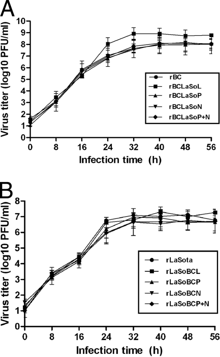
The multicycle growth of the chimeric and their parental viruses was compared in DF-1 cells. Briefly, DF-1 cells were infected with N-, P-, or L-chimeric virus at an MOI of 0.01, supernatant samples were harvested at 8-h intervals, and titration of the viruses was performed by plaque assay (Fig. 2). When the growth of strains rBCLaSoP, rBCLaSoN, and rBCLaSoP+N was compared to that of the parental rBC, no significant growth differences were observed. The N- and P-chimeric viruses in DF-1 cells replicated to titers and with kinetics that were similar to those of the parental rBC virus. These results suggested that the replacement of the N, P, or N and P genes of moderately virulent BC virus with the corresponding genes from avirulent LaSota virus did not affect the growth of chimeric viruses in DF-1 cells (Fig. 2A). However, the chimeric virus rBCLaSoL, in which the L gene of the LaSota virus was used to replace the L gene of the BC virus, showed 10-fold more growth than the parental rBC virus. There was no difference in growth kinetics between rBC and rBCLasoL in the early stage of virus replication (up to 16 h). However, a difference in growth kinetics was observed 16 h postinfection.
FIG. 2.
Multicycle growth kinetics of the parental and the chimeric NDVs in DF-1 cells. (A) BC viruses. (B) LaSota viruses. Six-well plates of DF-1 cell monolayers were infected with the virus at an MOI of 0.01 PFU per cell for 1 h. The cells were washed with phosphate-buffered saline and then overlaid with DMEM containing 5% FBS at 37°C in 5% CO2. Supernatant samples were collected at 8-h intervals until 56 h postinfection. The medium of all LaSota backbone viruses contained 10% fresh allantoic fluid. The six-well plates were replaced with equal volumes of fresh medium. Virus yields at different time points were determined by plaque assay. Error bars show standard deviations.
In the case of N-, P-, and L-chimeric viruses in the LaSota backbone, all the chimeric viruses replicated to titers similar to those of their parental rLaSota virus, indicating that the replacement of the N, P, or L gene of the LaSota virus with the corresponding gene of the BC virus did not affect the growth of the chimeric viruses in DF-1 cells (Fig. 2B).
The CPE and plaque sizes of the N-, P-, and L-chimeric viruses were compared to those of their respective parental viruses in DF-1 cells. It was observed that all the N- and P-chimeric viruses except rBCLaSoL produced CPE similarly to their parental virus; rBCLaSoL produced faster and more-extensive CPE than its parental rBC. There were no observable differences in the plaque sizes of the N-, P-, and L-chimeric viruses of both the rBC and rLaSota strains in comparison to the plaque sizes of their respective parental viruses (data not shown).
Growth of chimeric viruses in chicken embryos.
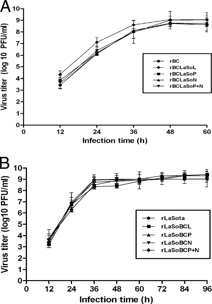
The in vivo growth kinetics assay of the chimeric viruses in 9-day-old chicken embryos showed that the chimeric virus rBCLaSoL grew 1 log more than the parental rBC up until 36 h postinoculation and gradually reached the same level of growth as rBC after 48 h of infection. However, we did not observe marked growth differences in the chimeric viruses rBCLaSoP, rBCLaSoN, and rBCLaSoP+N in comparison to the growth of the parental rBC (Fig. 3A). These results indicated that the reciprocal exchange of N, P, or N and P genes between BC and LaSota viruses did not alter the replication of the chimeric viruses compared to that of their parental viruses, except for rBCLaSoL, in which the presence of the L gene of LaSota in BC in place of its own L increased the replication of the chimeric virus at the early stage of infection. All the LaSota backbone chimeric viruses, rLaSoBCP, rLaSoBCN, rLaSoBCL, and rLaSoBCP+N, grew at the same rate as their parental rLaSota virus, indicating that the presence of the N, P, L, or P and N genes of BC virus in the LaSota backbone did not alter the replication of chimeric viruses in 9-day-old chicken embryos (Fig. 3B).
FIG. 3.
Multicycle growth kinetics of parental and chimeric NDVs in 9-day-old chicken embryos. (A) BC backbone. (B) LaSota backbone. Embryos were inoculated with 103 PFU of virus via the allantoic route. Three embryos were chilled at every 12-h interval up to 60 h (rBC backbone) and 96 h (rLaSota backbone). Allantoic fluids were collected, and virus yields were determined by plaque assay in DF-1 cells. Error bars show standard deviations.
Pathogenicity of parental and chimeric viruses in chicken embryos.
To determine the effects of seven nucleotide changes made to introduce unique restriction sites into the viral genomes of newly generated rBC and rLaSota viruses, MDT tests were performed in 9-day-old embryonated chicken eggs. Our results showed that the MDT values of newly generated BC and Laota viruses were similar to those of their respective parental viruses, indicating that the nucleotide changes in the 3′ UTR of the N, P, and L genes did not alter the pathogenicity of the parental viruses (data not shown).
The pathogenicity of the N-, P-, and L-chimeric viruses, along with their respective parental viruses, was evaluated in 9-day-old chicken embryos by MDT test. The MDT tests were performed twice as test 1 and test 2 (Table 2). The MDT results showed that the rBCLaSoL strain took 53 h and 56 h in test 1 and 2, respectively, to kill all the embryos, compared to 62 h and 60 h for the rBC strain. The MDT values for rBCLaSoP were 64 h and 60 h in test 1 and test 2, respectively, similar to those of the parental rBC. In test 1 and test 2, the MDT values for the BCLaSoN strain were 60 h and 62 h, whereas the MDTs for rBCLaSoP+N were 58 h and 61 h, respectively. The MDT values for rLaSoBCL were 115 h and 114 h, compared to 106 h and 110 h for rLaSota in the respective tests. The MDTs for rLaSoBCP were 114 h and 116 h in test 1 and test 2, respectively. The MDT values for rLaSoBCN were 109 h and 114 h and for rLaSoBCP+N were 108 h and 112 h in test 1 and 2, respectively. These MDT values indicated that when the N or P gene was exchanged individually or in combination between the avirulent NDV strain LaSota and the moderately virulent strain BC, it did not significantly affect the pathogenicity of the chimeric viruses in comparison to that of their respective parental viruses. However, replacement of the L gene of the BC virus with the L gene of LaSota changed the MDT value of the mesogenic BC strain to the level of a velogenic strain.
TABLE 2.
Pathogenicity studies of recombinant chimeric viruses in 9-day-old chicken embryos by MDT testa
| Virus | MDT (h)
|
|
|---|---|---|
| Test 1 | Test 2 | |
| rBC | 62 | 60 |
| rBCLaSoL | 53 | 56 |
| rBCLaSoP | 64 | 60 |
| rBCLaSoN | 60 | 62 |
| rBCLaSoP+N | 58 | 61 |
| rLaSota | 106 | 110 |
| rLaSoBCL | 115 | 114 |
| rLaSoBCP | 114 | 116 |
| rLaSoBCN | 109 | 114 |
| rLaSoBCP+N | 108 | 112 |
The MDT duration is more than 90 h for lentogenic strains, 60 to 90 h for mesogenic strains, and under 60 h for velogenic strains (1).
Pathogenicity of chimeric viruses in 1-day-old chicks.
The pathogenicity of chimeric viruses in 1-day-old chicks in the ICPI test showed that the rBCLaSoL strain had ICPI values of 1.70 and 1.80 in test 1 and test 2, respectively, whereas its mesogenic parental strain rBC had ICPI values of 1.45 and 1.49 in test 1 and 2, respectively (Table 3). The rBCLaSoP and rBCLaSoN strains showed ICPI values of 1.24 and 1.31, respectively, slightly lower than that of their parental rBC strain, which had an ICPI value of 1.45. Interestingly, the ICPI value of rBCLaSoP+N was 1.44, similar to the ICPI value of the parental rBC strain. These results indicated that the N and P proteins probably do not play any role in NDV pathogenicity, and a slight decrease observed in the pathogenicity of pBCLaSoN and pBCLaSoP could be due to the heterotypic interaction of N and P proteins. The ICPI values of rLaSota, rLaSoBCL, rLaSoBCP, rLaSoBCN, and rLaSoBCP+N were 0.00, indicating that the N and P genes of pBC did not increase the pathogenicity of chimeric rLaSota viruses above the detection level.
TABLE 3.
Pathogenicity studies of recombinant chimeric viruses in 1-day-old chickens by ICPI testa
| Virus | ICPI
|
|
|---|---|---|
| Test 1 | Test 2 | |
| rBC | 1.45 | 1.49 |
| rBCLaSoL | 1.70 | 1.80 |
| rBCLaSoP | 1.24 | ND |
| rBCLaSoN | 1.31 | ND |
| rBCLaSoP+N | 1.44 | ND |
| rLaSota | 0.00 | 0.00 |
| rLaSoBCL | 0.00 | 0.00 |
| rLaSoBCP | 0.00 | 0.00 |
| rLaSoBCN | 0.00 | 0.00 |
| rLaSoBCP+N | 0.00 | 0.00 |
The ICPI values for velogenic strains approach the maximum score of 2.00, whereas lentogenic strains give values close to 0.
Pathogenicity of L-chimeric viruses in 6-week-old chickens.
The pathogenicity of L-chimeric viruses was evaluated further in 6-week-old chickens by IVPI test at two different times, and the results showed that the IVPI values of the rBCLaSoL strain were 2.26 and 2.33 in test 1 and test 2, respectively, in comparison to values of 2.06 and 2.00 for its parental rBC strain (Table 4). The IVPI values for rLaSota and rLaSoBCL were 0.00. These results further indicated that replacement of the L gene of the BC strain with the L gene of the LaSota strain increased the pathogenicity of the chimeric virus rBCLaSoL.
TABLE 4.
Pathogenicity studies of L-chimeric viruses in 6-week-old chickens by IVPI testa
| Virus | IVPI
|
|
|---|---|---|
| Test 1 | Test 2 | |
| rBC | 2.06 | 2.00 |
| rBCLaSoL | 2.26 | 2.33 |
| rLaSota | 0.00 | 0.00 |
| rLaSoBCL | 0.00 | 0.00 |
The IVPI values for velogenic strains approach the maximum score of 3.00, whereas lentogenic strains give values close to 0.0.
We further evaluated the pathogenicity of L-chimeric viruses in 6-week-old chickens by inoculating 106 PFU of virus/bird through the natural route of infection. Birds inoculated with rBCLaSoL first exhibited symptoms of sickness and paralysis at 3 days postinfection. Similarly, birds inoculated with the parental rBC virus also exhibited sickness and paralysis at 3 days postinfection. However, as the disease progressed, the onsets of paralysis and death were more rapid in the case of birds inoculated with rBCLaSoL than in those birds inoculated with rBC (Table 5). By day 7 postinfection, all 10 birds inoculated with rBCLaSoL were dead, whereas none of the birds inoculated with rBC died. In the case of rLaSota and rLaSoBCL, birds were either mildly sick or healthy throughout the course of the study. These results reinforced our observation that there is a gain in virulence when the L gene of BC is replaced by the L gene of LaSota.
TABLE 5.
Pathogenicity of L-chimeric viruses in 6-week-old chickens inoculated via the occulonasal routea
| Virus | Status | Day
|
|||||||||
|---|---|---|---|---|---|---|---|---|---|---|---|
| 1 | 2 | 3 | 4 | 5 | 6 | 7 | 8 | 9 | 10 | ||
| rBC | Normal | 10 | 10 | 6 | 6 | 4 | 4 | 4 | 4 | 4 | 4 |
| Sick | 3 | 3 | 2 | 2 | 2 | 2 | 2 | 2 | |||
| Paralyzed | 1 | 1 | 4 | 4 | 4 | 4 | 4 | 4 | |||
| Dead | |||||||||||
| rBCLaSoL | Normal | 10 | 10 | ||||||||
| Sick | 9 | ||||||||||
| Paralyzed | 1 | 6 | 1 | 1 | |||||||
| Dead | 4 | 9 | 9 | 10 | 10 | 10 | 10 | ||||
| rLaSota | Normal | 10 | 10 | 10 | 10 | 10 | 10 | 10 | 10 | 10 | 10 |
| Sick | |||||||||||
| Paralyzed | |||||||||||
| Dead | |||||||||||
| rLaSoBCL | Normal | 10 | 10 | 10 | 10 | 10 | 10 | 10 | 10 | 10 | 10 |
| Sick | |||||||||||
| Paralyzed | |||||||||||
| Dead | |||||||||||
Groups of 10 6-week-old chickens were inoculated with approximately 106 PFU of virus per bird and observed for signs of disease and mortality for 10 days.
Level of replication of L-chimeric viruses in chicken neuronal tissue.
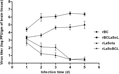
The levels of replication of L-chimeric viruses was compared to those of their parental viruses in the brains of 1-day-old chicks (Fig. 4). Ten chicks were injected intracerebrally with 103 PFU of chimeric virus per chick. Two chicks were sacrificed daily, brain samples were collected, and the virus titers were determined by plaque assay. The chimeric virus rBCLaSoL replicated at a level 2 to 3 log titers higher than that of the parental rBC in the first 3 days postinoculation and continued to replicate at a higher rate than rBC (Fig. 4). However, the titers of rLaSota and rLaSoBCL in chicken brain gradually decreased with time, probably due to inability of the viruses to grow in neuronal tissues. This was expected as the lentogenic NDV strains with a monobasic residue at the F protein cleavage sites require trypsin-like proteases for their activation which are not found in the neuronal tissues. These results suggested that the increased pathogenicity of the chimeric virus rBCLaSoL could be due to its level of growth in the neuronal tissues being higher than that of its parental rBC virus.
FIG. 4.
The growth kinetics of parental and L-chimeric NDV strains in the brains of 1-day-old chicks. SPF 1-day-old chicks were inoculated intracerebrally with 103 PFU of virus/chick. Two birds were sacrificed each day after infection; brains were collected and homogenized, and virus titrated by plaque assay in DF-1 cells. Error bars show standard deviations.
DISCUSSION
NDV strains show a continuous spectrum of virulence for chickens. Currently, the mechanism(s) responsible for the variations in virulence of NDV strains is not fully understood. Much attention has been focused on the amino acid sequence at the F protein cleavage site. Lentogenic strains have F proteins with two basic residues at the cleavage site. They are activated by proteases secreted from cells present only in the respiratory and intestinal tissues. Mesogenic and velogenic strains have F proteins with multibasic residues at the cleavage site. They are activated by the furin family of enzymes that are present in cells of most tissue types. Thus, based on the sequence at the F protein cleavage site, NDV strains could be divided into two F protein cleavage groups, one group containing a monobasic residue at the cleavage site, represented by lentogenic strains, and the other group containing multibasic residues at the cleavage site, represented by mesogenic and velogenic strains. However, the pathogenicity varies widely among strains in each group, suggesting that a viral factor(s) other than the F protein cleavage site is responsible for the variations in the pathogenicity of NDV. The attachment protein, HN, was found to play a role in tissue tropism but played a minor role in the virulence of NDV (7, 12).
In this study, we examined the role of internal proteins (N, P, and L) in the pathogenicity of NDV. Reverse genetics was used to exchange the N, P, or L gene between a lentogenic strain, LaSota, and a mesogenic strain, BC. The caveat has to be added that the strain LaSota has a monobasic F protein cleavage site and the BC has a multibasic F protein cleavage site. Therefore, it would be necessary to analyze the gain or loss in virulence of each chimeric virus in its respective F protein cleavage group. Ideally, to eliminate any effects of the sequence at the F protein cleavage site on viral pathogenicity, one would require both viruses to be from the same F protein cleavage group. However, this was not possible due to not having reverse genetic systems for two NDV strains varying greatly in pathogenicity but belonging to the same F protein cleavage group.
We transferred the N, P, and L genes individually between rLaSota and rBC viruses. This was done to assess the effect of each gene alone on the replication and pathogenicity of the chimeric virus. Since the N and P proteins of paramyxoviruses are known to interact, these two genes were also replaced in combination (5, 9). We were able to readily recover N, P, and L replacement chimeric viruses, indicating that all three internal-protein genes are compatible between NDV strains LaSota and BC. This result was not unexpected since there are high levels of amino acid sequence identity of the N gene (97.9%), P gene (97.5%), and L gene (99.1%) between strains LaSota and BC (17, 26, 30). All the chimeric viruses replicated efficiently in cell cultures and in chicken embryos, suggesting that the replacement of the internal proteins N and P had little or no effect on virus growth in vitro and in vivo. However, it was surprising that the chimeric rBC in which the L gene was replaced with that of the rLaSota virus resulted in improved growth both in cell culture and the chicken embryos. There are precedents where the replacement of an internal protein has resulted in similar or improved growth of the chimeric virus in cell cultures (3, 24). It is possible that the L protein of LaSota virus functions more efficiently in the context of other BC virus proteins than does the L protein of BC virus. Alternatively, the L protein of LaSota virus probably functions better by interacting more efficiently with some unknown cellular factor(s) that is required for RNA synthesis. The L gene of BC virus did not have any effect on LaSota virus. It is possible that the L gene of BC virus has a minor effect on the replication of LaSota virus which could not be detected in our growth kinetics studies.
The results of the analysis of the pathogenicity of the chimeric viruses by using three pathogenicity tests showed that replacing the L protein of BC virus with that of the LaSota virus significantly increased the virulence of the chimeric virus. These results suggested that the L polymerase protein of NDV is a determinant of virulence. The viral polymerase protein, PB2, of influenza virus has also been shown to contribute to host range and pathogenicity (31). Furthermore, attenuation of measles virus has also been associated with the P and L genes (28).
Our results indicate that the L gene is not the only determinant that is responsible for the virulence of NDV. Some velogenic NDV strains have an ICPI value of 2.0 (1), whereas BC virus with the LaSota L gene has an ICPI value of 1.70. Furthermore, recombinant LaSota virus with an altered polybasic sequence at the F protein cleavage site has an ICPI value of 1.3 (20, 23, 25). These results indicate that, in addition to the cleavage site sequence of the F protein and the L gene, other viral proteins and/or nucleotide sequences in the genomic RNA of NDV contribute to virulence.
The molecular mechanism for increased virulence caused by the L protein of LaSota virus is not known. However, our growth kinetics studies of the L-chimeric viruses in chicken brain showed that the rBCLaSoL virus in which the L gene has been replaced with that of LaSota virus replicated 100-fold more efficiently than the parental rBC virus. It is possible that the L protein of the LaSota virus causes increased RNA synthesis, which leads to higher levels of virus replication in vitro and in vivo. A higher level of virus replication in vivo can lead to a lethal outcome as the replicating virus overwhelms the host immune response. The results of a previous study showed that reduced RNA synthesis was associated with reduced virulence of NDV (18). Therefore, it will be interesting to compare the RNA synthesis activities of the L proteins of BC and LaSota viruses by using a minireplicon system.
The rBC chimeric viruses in which the N or P gene was replaced with the corresponding gene of the rLaSota virus showed a slight decrease in virulence. However, when both the N and P genes of the rBC virus were replaced with those of the rLaSota virus, the virulence of the chimeric virus was similar to that of the parental rBC virus. This result indicated that the slight decrease in virulence of N- or P-chimeric virus was probably due to heterologous N and P protein interaction. Although in this study we did not examine the virulence of chimeric virus in which the N, P, and L proteins were replaced in combination, it will be interesting to analyze such chimeric viruses in the future. In this study, we were not able to determine the virulence of rLaSota backbone chimeric viruses because the virulence was below the detection level of our pathogenicity tests.
In summary, we have shown that the L protein of NDV is a determinant of virulence and is probably responsible for the degree of virulence seen among NDV strains. Our results suggest that the virulence of NDV is a multigenic trait. The L protein, in addition to the amino acid sequences at the F protein cleavage site, determines the overall virulence of an NDV strain. It is also possible that additional viral genes might be contributing to the virulence of NDV. Further studies are needed to identify the specific amino acid residues in the L protein that are responsible for the virulence. The identification of virulence determinants is of considerable importance because of the potential to generate better live NDV vaccines. It may also be possible to apply these findings to other paramyxoviruses.
Acknowledgments
We thank Daniel Rockemann, Yongqi Yan, and Govindarajan Dhanasekaran for their excellent technical assistance and Ireen Dryburgh-Barry for her critical reading of the manuscript.
Footnotes
Published ahead of print on 11 June 2008.
REFERENCES
- 1.Alexander, D. J. 1989. Newcastle disease, p. 114-120. In H. G. Purchase, L. H. Arp, C. H. Domermuth, and J. E. Pearson (ed.), A laboratory manual for the isolation and identification of avian pathogens, 3rd ed. American Association for Avian Pathologists, University of Pennsylvania, Kennett Square, PA.
- 2.Alexander, D. J. 1997. Newcastle disease and other avian Paramyxoviridae infection, p. 541-569. In B. W. Calnek (ed.), Diseases of poultry, 10th ed. Iowa State University Press, Ames, IA.
- 3.Bailly, J. E., J. M. McAuliffe, A. P. Durbin, W. R. Elkins, P. L. Collins, and B. R. Murphy. 2000. A recombinant human parainfluenza virus type 3 (PIV3) in which the nucleocapsid N protein has been replaced by that of bovine PIV3 is attenuated in primates. J. Virol. 743188-3195. [DOI] [PMC free article] [PubMed] [Google Scholar]
- 4.Chambers, P., N. S. Millar, S. G. Platt, and P. T. Emmerson. 1986. Nucleotide sequence of the gene encoding the matrix protein of Newcastle disease virus. Nucleic Acids Res. 149051-9061. [DOI] [PMC free article] [PubMed] [Google Scholar]
- 5.Curran, J., J. B. Marq, and D. Kolakofsky. 1995. An N-terminal domain of the Sendai paramyxovirus P protein acts as a chaperone for the NP protein during the nascent chain assembly step of genome replication. J. Virol. 69849-855. [DOI] [PMC free article] [PubMed] [Google Scholar]
- 6.de Leeuw, O., and B. Peeters. 1999. Complete nucleotide sequence of Newcastle disease virus: evidence for the existence of a new genus within the subfamily Paramyxovirinae. J. Gen. Virol. 80131-136. [DOI] [PubMed] [Google Scholar]
- 7.Estevez, C., D. King, B. Seal, and Q. Yu. 2007. Evaluation of Newcastle disease virus chimeras expressing the hemagglutinin-neuraminidase protein of velogenic strains in the context of a mesogenic recombinant virus backbone. Virus Res. 129182-190. [DOI] [PubMed] [Google Scholar]
- 8.Gotoh, B., Y. Ohnishi, N. M. Inocencio, E. Esaki, K. Nakayama, P. J. Barr, G. Thomas, and Y. Nagai. 1992. Mammalian subtilisin-related proteinases in cleavage activation of the paramyxovirus fusion glycoprotein: superiority of furin/PACE to PC2 or PC1/PC3. J. Virol. 666391-6397. [DOI] [PMC free article] [PubMed] [Google Scholar]
- 9.Homann, H. E., W. Willenbrink, C. J. Buchholz, and W. J. Neubert. 1991. Sendai virus protein-protein interactions studied by a protein-blotting protein-overlay technique: mapping of domains on NP protein required for binding to P protein. J. Virol. 651304-1309. [DOI] [PMC free article] [PubMed] [Google Scholar]
- 10.Huang, Z., S. Krishnamurthy, A. Panda, and S. K. Samal. 2001. High-level expression of a foreign gene from the most 3′-proximal locus of a recombinant Newcastle disease virus. J. Gen. Virol. 821729-1736. [DOI] [PubMed] [Google Scholar]
- 11.Huang, Z., S. Krishnamurthy, A. Panda, and S. K. Samal. 2003. Newcastle disease virus V protein is associated with viral pathogenesis and functions as an alpha interferon antagonist. J. Virol. 778676-8685. [DOI] [PMC free article] [PubMed] [Google Scholar]
- 12.Huang, Z., A. Panda, S. Elankumaran, D. Govindarajan, D. D. Rockemann, and S. K. Samal. 2004. The hemagglutinin-neuraminidase protein of Newcastle disease virus determines tropism and virulence. J. Virol. 784176-4184. [DOI] [PMC free article] [PubMed] [Google Scholar]
- 13.Krishnamurthy, S., Z. Huang, and S. K. Samal. 2000. Recovery of a virulent strain of Newcastle disease virus from cloned cDNA: expression of a foreign gene results in growth retardation and attenuation. Virology 278168-182. [DOI] [PubMed] [Google Scholar]
- 14.Krishnamurthy, S., and S. K. Samal. 1998. Nucleotide sequences of the trailer, nucleocapsid protein gene and intergenic regions of Newcastle disease virus strain Beaudette C and completion of the entire genome sequence. J. Gen. Virol. 792419-2424. [DOI] [PubMed] [Google Scholar]
- 15.Lamb, R. A., and G. D. Parks. 2007. Paramyxoviridae: the viruses and their replication, p. 1449-1496. In D. M. Knipe and P. M. Howley (ed), Fields virology, 5th ed. Wolters Kluwer-Lippincott Williams & Wilkins, Philadelphia, PA.
- 16.Lamb, R. A., P. L. Collins, D. Kolakofsky, J. A. Melero, Y. Nagai, M. B. A. Oldstone, C. R. Pringle, and B. K. Rima. 2005. Family Paramyxoviridae, p. 655-668. In C. M. Fauquet (ed.), Virus taxonomy: the classification and nomenclature of viruses. The eighth report of the International Committee on Taxonomy of Viruses. Elsevier Academic Press, San Diego, CA.
- 17.Locke, D. P., H. S. Sellers, J. M. Crawford, S. Schultz-Cherry, D. J. King, R. J. Meinersmann, and B. S. Seal. 2000. Newcastle disease virus phosphoprotein gene analysis and transcriptional editing in avian cells. Virus Res. 6955-68. [DOI] [PubMed] [Google Scholar]
- 18.Madansky, C. H., and M. A. Bratt. 1981. Noncytopathic mutants of Newcastle disease virus are defective in virus-specific RNA synthesis. J. Virol. 37317-327. [DOI] [PMC free article] [PubMed] [Google Scholar]
- 19.Nagai, Y. 1995. Virus activation by host proteinases. A pivotal role in the spread of infection, tissue tropism and pathogenicity. Microbiol. Immunol. 391-9. [DOI] [PubMed] [Google Scholar]
- 20.Panda, A., Z. Huang, S. Elankumaran, D. D. Rockemann, and S. K. Samal. 2004. Role of fusion protein cleavage site in the virulence of Newcastle disease virus. Microb. Pathog. 361-10. [DOI] [PMC free article] [PubMed] [Google Scholar]
- 21.Peeters, B., P. Verbruggen, F. Nelissen, and O. de Leeuw. 2004. The P gene of Newcastle disease virus does not encode an accessory X protein. J. Gen. Virol. 852375-2378. [DOI] [PubMed] [Google Scholar]
- 22.Peeters, B. P., Y. K. Gruijthuijsen, O. S. de Leeuw, and A. L. Gielkens. 2000. Genome replication of Newcastle disease virus: involvement of the rule-of-six. Arch. Virol. 1451829-1845. [DOI] [PubMed] [Google Scholar]
- 23.Peeters, B. P., O. S. de Leeuw, G. Koch, and A. L. Gielkens. 1999. Rescue of Newcastle disease virus from cloned cDNA: evidence that cleavability of the fusion protein is a major determinant for virulence. J. Virol. 735001-5009. [DOI] [PMC free article] [PubMed] [Google Scholar]
- 24.Pham, Q. N., S. Biacchesi, M. H. Skiadopoulos, B. R. Murphy, P. L. Collins, and U. J. Buchholz. 2005. Chimeric recombinant human metapneumoviruses with the nucleoprotein or phosphoprotein open reading frame replaced by that of avian metapneumovirus exhibit improved growth in vitro and attenuation in vivo. J. Virol. 7915114-15122. [DOI] [PMC free article] [PubMed] [Google Scholar]
- 25.Romer-Oberdorfer, A., O. Werner, J. Veits, T. Mebatsion, and T. C. Mettenleiter. 2003. Contribution of the length of the HN protein and the sequence of the F protein cleavage site to Newcastle disease virus pathogenicity. J. Gen. Virol. 843121-3129. [DOI] [PubMed] [Google Scholar]
- 26.Seal, B. S., J. M. Crawford, H. S. Sellers, D. P. Locke, and D. J. King. 2002. Nucleotide sequence analysis of the Newcastle disease virus nucleocapsid protein gene and phylogenetic relationships among the Paramyxoviridae. Virus Res. 83119-129. [DOI] [PubMed] [Google Scholar]
- 27.Steward, M., I. B. Vipond, N. S. Millar, and P. T. Emmerson. 1993. RNA editing in Newcastle disease virus. J. Gen. Virol. 742539-2547. [DOI] [PubMed] [Google Scholar]
- 28.Takeda, M., A. Kato, F. Kobune, H. Sakata, Y. Li, T. Shioda, Y. Sakai, M. Asakawa, and Y. Nagai. 1998. Measles virus attenuation associated with transcriptional impediment and a few amino acid changes in the polymerase and accessory proteins. J. Virol. 728690-8696. [DOI] [PMC free article] [PubMed] [Google Scholar]
- 29.Wilde, A., C. McQuain, and T. Morrison. 1986. Identification of the sequence content of four polycistronic transcripts synthesized in Newcastle disease virus infected cells. Virus Res. 577-95. [DOI] [PubMed] [Google Scholar]
- 30.Wise, M. G., H. S. Sellers, R. Alvarez, and B. S. Seal. 2004. RNA-dependent RNA polymerase gene analysis of worldwide Newcastle disease virus isolates representing different virulence types and their phylogenetic relationship with other members of the paramyxoviridae. Virus Res. 10471-80. [DOI] [PubMed] [Google Scholar]
- 31.Wright, P. F., G. Neumann, and Y. Kawaoka. 2007. Orthomyxoviruses, p. 1691-1740. In D. M. Knipe and P. M. Howley (ed.), Fields virology, 5th ed. Wolters Kluwer-Lippincott Williams & Wilkins, Philadelphia, PA.


