Abstract
The utility of high-resolution magic-angle spinning (HR-MAS) NMR for studying drug delivery in whole tissues was explored by dosing female Sprague–Dawley rats with topical or injectable benzoic acid (BA). In principle, HR-MAS NMR permits the detection of both intra- and extracellular compounds. This is an advantage over the previous detection of topically applied BA using microdialysis coupled to HPLC/UV as microdialysis samples only the extracellular space. Skin and muscle samples were analyzed by 1H HR-MAS NMR, and BA levels were determined using an external standard solution added to the sample rotor. One to two percent of the BA topical dose was detected in the muscle, showing that BA penetrated through the dermal and subcutaneous layers. Since BA was not detected in the muscle in the microdialysis studies, the NMR spectra revealed the intracellular localization of BA. The amount of BA detected in muscle after subcutaneous injection correlated with the distance from the dosing site. Overall, the results suggest that HR-MAS NMR can distinguish differences in the local concentration of BA varying with tissue type, dosage method, and tissue proximity to the dosing site. The results illustrate the potential of this technique for quantitative analysis of drug delivery and distribution and the challenges to be addressed as the method is refined.
Nuclear magnetic resonance (NMR) spectroscopy has played a significant role in the growth of metabonomics, an area of clinical research that aims to measure the total metabolic response of an organism to a stimulus such as a drug or toxicant or a physiological change such as a disease state.1–3 The effects of the stimulus can be evaluated by measuring levels of specific analytes or through pattern recognition to permit the identification of a fingerprint of efficacy or toxicity.1–3 Spectral analysis of the individual components that comprise the fingerprint reveals the identity of drug metabolites4 and disease biomarkers.5,6 The major advantages of NMR spectroscopy for metabonomics studies over other techniques, such as mass spectrometry,7,8 are the universal nature of NMR detection, ease of quantitation, and the ability to analyze both biofluids and tissues to understand the local versus systemic effects of a stimulus.9,10 Such an integrated metabonomics approach is an ongoing focus of this collaborative effort. Although NMR is generally less sensitive than other spectroscopic techniques, recent technological developments have reduced sample mass requirements and experiment times significantly.11,12
Tissue analysis by NMR spectroscopy is possible with a technique known as high-resolution magic-angle spinning (HR-MAS). HR-MAS NMR spectroscopy evolved from solid-state NMR techniques13,14 as a method to analyze 1H spectra of molecules attached to solid-phase synthesis resins.15 The nature of the tissue or resin matrix gives rise to broad signals in the NMR spectrum because of sample heterogeneity and the inability of molecules in the matrix to tumble isotropically. With HR-MAS, the sample is inserted in a specially designed NMR probe that tilts the sample at an angle relative to the static magnetic field. By spinning the sample at this angle (54.73°), resonance line widths in the spectra of solid and semisolid materials are dramatically reduced.
Many types of human and animal tissues have been analyzed with HR-MAS, including brain,16,17 heart,18 liver,19,20 and kidney.19,21–23 Different tissue types vary in their chemical composition and thus give rise to unique spectra, as illustrated in Figure 1 for rat muscle and skin. The improved resolution afforded by HR-MAS has made NMR useful for the temporal and spatial localization of endogenous metabolites and disease biomarkers.2 This information complements metabolic profiles of biofluids that reflect the overall systemic state of the organism. One of the major applications of tissue analysis by HR-MAS NMR spectroscopy is the diagnosis and monitoring of cancer.16,21,23,24 Tumor samples often contain elevated lipid concentrations compared to healthy controls,23,24 which are not always detected in cancer cell extracts.24 Although tissue excision is required, the intra- and extracellular contents of cells are analyzed directly by HR-MAS avoiding the risk of losing important bioanalytical information because of extraction or dilution of this information because of systemic transport.
Figure 1.
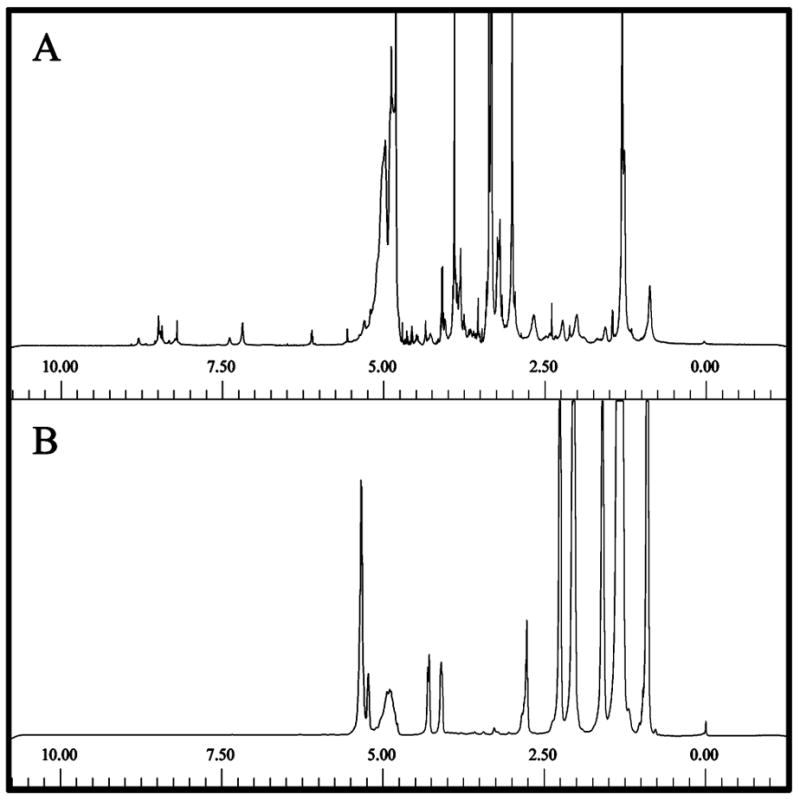
Representative 1H HR-MAS NMR spectra of rat tissue acquired with presaturation of the solvent resonance and a 50 ms CPMG (Carr–Purcell–Meiboom–Gill) train (τ = 500 μs, n = 50) to improve resolution of the endogenous small molecules. Many endogenous components are detected in muscle (A) while the peaks observed in skin (B) arise from fatty acids and lipids.46,47 In both tissues, the spectral region from 5.00 to 10.00 ppm is relatively free of resonances from the sample matrix, facilitating detection of exogenous aromatic compounds.
Another potential, but relatively unexplored, application of HR-MAS NMR tissue analysis is for drug delivery and distribution studies. Formulation is a critical part of the drug development process affecting the ADME (absorption, distribution, metabolism, and excretion) properties of a drug. While oral formulation is the preferred route for the administration of many drugs for humans, topical, inhalation, intramuscular, and intravenous routes are also widely used. Spectral profiles of animal tissues obtained by HR-MAS NMR spectroscopy after treatment with a drug in different formulations may reflect quantitative differences in the ADME properties of the drug.
Microdialysis is a minimally invasive method for localized, in vivo sampling of hydrophilic small molecules in the extracellular space surrounding a probe implanted in a live animal.25,26 Benzoic acid is an excellent model compound to use in investigating the potential of HR-MAS NMR spectroscopy for drug delivery and distribution studies because of its high skin penetration (1.8 ± 0.6 mM)27 and chemical stability. The utility of NMR for drug quantitation in whole tissues was explored by dosing female Sprague–Dawley rats with topical or subcutaneous benzoic acid (BA) and then analyzing excised skin and muscle by 1H HR-MAS.28 NMR has previously been used to analyze rat cerebral spinal fluid sampled by microdialysis to determine the effects of a neurotoxin on endogenous components,29,30 and magnetic resonance microscopy has been used to image the probe/tissue interface.31 Although we are currently engaged in a metabonomics evaluation of microdialysis samples using microcoil NMR analysis to evaluate the effects of oxidative stress,32 we believe that this is the first report of a comparison of the levels of a drug or drug model compound in tissues detected by microdialysis sampling with HPLC/UV analysis and HR-MAS NMR spectroscopy of intact tissue samples.
EXPERIMENTAL SECTION
Rat Tissue Samples
Tissue samples were obtained from female Fuzzy (preliminary studies) or Sprague–Dawley (dosing studies) rats. The control 1H NMR spectra of undosed animals were indistinguishable for both types of rats. All rats were 7–8 months old and weighed 250–300 g. Procedures for animal care are described elsewhere27 and were conducted in accordance with the Principles of Laboratory Animal Care (NIH publication no. 85-23, revised 1985).
The topical formulation consisted of 10.0 mg benzoic acid (Fisher Scientific, Fair Lawn, NJ) in 50.0 μL propylene glycol (99.5% pure, Aldrich, Milwaukee, WI). The dosed animals were anesthetized according to procedures described previously,27 and then the entire dose was administered directly to each rat’s skin (left hind leg region, Figure 2A). In previous microdialysis studies, a maximum steady-state concentration of benzoic acid was reached in the skin after 5 h.27 Therefore, 5 h after application of the dose, the animal was sacrificed and the tissue was excised for NMR analysis.
Figure 2.

Sampling procedure.28 (A) The BA was sampled in the anesthetized rat after dosing using a microdialysis probe (indicated by the arrow). (B) Hind leg tissue was excised after the animal was sacrificed. A section approximately 8 mm × 5 mm × 10 mm was initially cut from the center of the tissue surrounding the microdialysis probe. Smaller pieces (≤ 3 mm wide) of skin (white) or muscle (dark) were cut from this section of tissue and placed into the sample rotor (C). (D) Histology slide of rat tissue (20× magnification). Dermis (1), subcutaneous layer (2), and muscle (3) are clearly visible by color contrast in the slide as well as to the naked eye. The hole in the dermis is from a microdialysis probe, which was actually implanted in the muscle for these studies.
The injectable formulation consisted of 2.04 mg benzoic acid dissolved in 1.00 mL phosphate buffered saline, pH 7.5. The entire dose was injected subcutaneously after the rat was anesthetized. After dosing, the concentration of BA in the muscle was monitored directly by microdialysis sampling and HPLC/UV detection. The details of the surgical procedure for microdialysis sampling and the chromatographic system for detection of BA have been described by McDonald and Lunte.27,33 The concentration peaked 45 min after dosing, at which time the animal was sacrificed and the tissue was excised for analysis.
The tissue samples were excised from the left hind leg of all rats at and surrounding the dosing site (Figure 2). After excision, small pieces of tissue were cut and placed into the cylindrical NMR sample rotor (ZrO2, 18 mm × 3 mm i.d.). Twenty microliters of D2O (99.9% atom D, Cambridge Isotope Laboratories, Cambridge, MA) were added to the rotor to provide a deuterium lock signal in the NMR spectrometer. (For samples analyzed quantitatively, this D2O contained a known concentration of the internal standard TSP, 3-(trimethylsilyl)propionic-2,2,3,3-d4 acid, sodium salt, Aldrich.) The rotor was capped tightly, and the sample was analyzed immediately. For tissue samples, extra care was taken to analyze fresh samples to avoid degradation that could cause misinterpretation of the results.34
NMR Spectroscopy
1H spectra for the tissue samples were acquired at ambient temperature on a 500 MHz DRX spectrometer equipped with a g-HR MAS 500 SB BL4 probe (Bruker BioSpin Corporation, Billerica, MA). At the time of these studies, the HR-MAS probe was not equipped with a temperature control unit. Prior to tissue analysis, the magic angle was calibrated by analyzing a potassium bromide (Fisher Scientific) sample spun at 6 kHz. The tissue samples were spun at 5 kHz and 1024 scans were acquired into 32 768 points across a spectral width of 6009.615 Hz unless otherwise noted. The number of scans was selected to obtain a maximum signal-to-noise ratio (S/N) on an experimental time scale (1 h 20 min) during which the tissue spectrum was observed to not change significantly. Presaturation of the HOD resonance (4.78 ppm) was achieved using a low-power pulse (55 dB) prior to the acquisition of the NMR signal.35–37 An exponential function equivalent to 0.3 Hz line broadening was applied prior to Fourier transformation, and all postacquisition processing was performed using XWIN NMR 3.1 (Bruker BioSpin Corporation). The CPMG (Carr–Purcell–Meiboom–Gill) experiment was used to suppress the broad spectral components arising from proteins and lipids in the tissue background on the basis of their more rapid rates of T2 relaxation.38–42 The CPMG pulse sequence effectively improves resolution of resonances arising from small molecules by incorporating a 180° pulse train after the initial 90° radio frequency pulse: 90°-(τ-180°-τ)n-Acquire. In preliminary experiments, the total pulse train was 50 ms (τ = 500 μs, n = 50), which was observed to significantly improve the resolution of the small molecule resonances that are overlapped with broad resonances (e.g., resonance c in Figure 3). For the dosing studies, the CPMG conditions were changed slightly to synchronize the 180° pulses with the rotor period (τ = 400 μs, n = 60, total pulse train time = 48 ms).43
Figure 3.
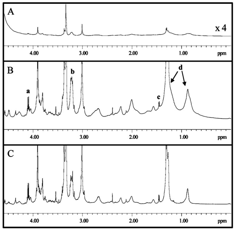
1H HR-MAS NMR spectra of rat muscle tissue (256 scans each). (A) In the 1H spectrum acquired without water suppression, because of the limited dynamic range of the detector, the intense HOD resonance makes it difficult to detect the less intense resonances above the spectral noise. (B) When a presaturation pulse is used to selectively suppress the HOD resonance, endogenous compounds are identified on the basis of their chemical shifts: a, lactate (δ 4.10 ppm); b, choline/phosphocholine (δ 3.21 ppm); c, alanine (δ 1.45 ppm); and d, lipids (δ 1.30 ppm, 0.87 ppm).20,46,47 (C) When a 50 ms CPMG train is incorporated into the experiment (τ = 500 μs, n = 50) in addition to presaturation, the resonances of the protein and lipid components are suppressed and the small molecule resonances are detected with improved resolution.
Quantitation
Since the NMR signal arises from the nuclei (e.g., protons) themselves, the area underneath each signal is proportional to the number of nuclei. Therefore, the signal area is directly proportional to the concentration (number of moles) of the analyte.44 To quantitate benzoic acid in rat tissue, the integral of the downfield BA doublet (7.90 ppm, 2 protons) was compared to the TSP singlet (0.00 ppm, 9 protons). Twenty microliters of 31.0 mM (rat 1) or 62.3 mM (rats 2–4) TSP in D2O was added to the rotor containing the tissue samples as an internal standard for quantitation. Spectra of tissue from untreated rats and D2O only (no TSP added) showed no background interference for the expected TSP resonance at 0.00 ppm. On the basis of the known molar amount of TSP added to the rotor and the measured benzoic acid and TSP integrals, the molar amount and hence the mass of BA detected in the tissue samples were calculated. While we recognize that careful attention to T1 and T2 relaxation times and S/N are required for absolute quantitation,44 they do not affect a relative comparison of the integrals measured in these samples. Relaxation measurements were not practical for these tissue samples, especially given our lack of a temperature control unit for the HR-MAS probe. Since the peak shapes of the BA and TSP resonances used for quantitation were similar, differential T2 effects were neglected in this proof-of-concept study. Careful relaxation measurements would be required to provide absolute quantitative analyses.
RESULTS AND DISCUSSION
Figure 4 shows the 1H NMR spectra of control and topically dosed rat tissue. By comparing Figure 4A and B, it is apparent that the upfield doublet of BA occurs in a spectral region free of background resonances from rat muscle. A similar result was observed for rat skin. The lack of spectral overlap between BA and background muscle resonances facilitates both the detection and quantitation of this model drug compound. Figure 1 shows that for both rat muscle and skin, the spectral region downfield of 5.00 ppm is relatively free of resonances arising from the tissue matrix.
Figure 4.
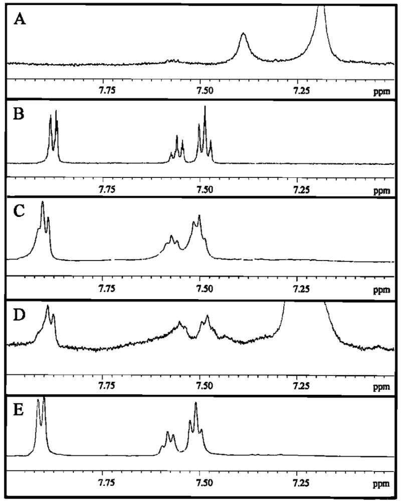
Detection of BA in rat tissue after a topical dose (10.0 mg benzoic acid in 50 μL propylene glycol) by 1H HR-MAS with CPMG in 1 h 20 min (τ = 400 μs, n = 60). (A) Muscle tissue of an undosed animal. (B) 1 mM BA solution only, showing that the BA resonances occur in spectral regions with minimal interference from the tissue spectral background. (C) Skin (106.3 mg) of dosed rat 2. (D) Muscle (90.7 mg) of rat 2. (E) Skin (111.1 mg) of rat 3. The vertical scale of each spectrum is maximized accordingly to visualize the BA peaks. The masses of tissue indicated are tissue wet weights.
The spectra in Figure 4C and D obtained on tissue samples collected 5 h after applying a topical dose of BA directly to the rat’s skin (10.0 mg in 50 μL propylene glycol) show that BA is readily detectable in both rat skin and muscle. The muscle sampled was directly beneath the skin where the drug was applied. To reach the muscle, BA must penetrate through the dermis and subcutaneous layers (Figure 2D). As the benzoic acid penetrates, it diffuses, leading to a reduction in the intensity of the BA resonance detected in the muscle (Figure 4D) versus the skin (Figure 4C).
The BA resonances in the skin spectrum of rat 3 (Figure 4E) are much sharper than those observed in the skin and muscle spectra of rat 2 (Figure 4C and D). Line shape differences may arise from a variety of sources. Some of these differences may be related to the homogeneity of the magnetic field that is adjusted by shimming before each experiment. Tissue samples can be challenging to shim reproducibly in a reasonable amount of time (5–10 min), and the shimming quality depends on how well the sample rotor is packed. The line shape differences in Figure 4 may also reveal the localization of BA to different environments, such as the skin surface (for BA that did not penetrate) and the intra- and extracellular environments (for BA that did penetrate into the skin). Such differences cannot be distinguished by microdialysis, which only samples the extracellular fluid.
The S/N and line shape of the skin spectrum obtained for rat 3 (Figure 4E) were sufficient to allow acquisition of a TOCSY (totally correlated spectroscopy) spectrum (Figure 5). The coupling between the benzoic acid aromatic protons (highlighted by the box in Figure 5) is easily detected. As illustrated by Figure 5, the identification of cross-peaks in the two-dimensional (2D) spectrum demonstrates the potential for obtaining useful information about the drug or its metabolites in tissues by 2D NMR experiments, especially if some of the analyte resonances are overlapped with tissue background in the 1D 1H spectrum. For example, the structural changes occurring during the metabolism of a prodrug could be identified by 2D NMR methods.
Figure 5.
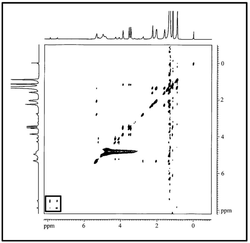
TOCSY spectrum of rat skin after dosing with 10.0 mg BA in 50 μL propylene glycol. The coupling between the benzoic acid ring protons is indicated with the box. Sixteen scans were acquired for each of 256 increments in F1 and a 60-ms TOCSY mixing time was used.
A limitation when performing 2D experiments, however, is that they may take several hours. Some of the analytical information in the metabolic profile may be compromised because of enzymatic degradation of the tissue or diffusion of the analyte on this time scale. This problem was encountered in these studies as no temperature control unit for the HR-MAS probe was available. This limited the experiment times for rat muscle and skin tissue to ~2 h or less.
Figure 6 shows the muscle tissue spectra after subcutaneous injection of 2.04 mg benzoic acid in 1.00 mL of phosphate buffered saline. Because of poor solubility in aqueous buffer, the mass of drug delivered was reduced by a factor of 5 relative to the topical dose (and the concentration was reduced 100-fold). A microdialysis probe implanted in the muscle beneath the injection site and an HPLC–UV system was used to determine when the BA concentration reached a maximum in the muscle. Forty-five minutes postinjection, the BA concentration peaked, and the animal was sacrificed. Figure 6 shows that BA is detectable in the muscle of this animal, but the S/N of the resulting spectra is low. The S/N appears to be slightly better in Figure 6B compared to Figure 6C. The muscle yielding the spectrum in Figure 6B was sampled from muscle tissue just beneath the injection site in the subcutaneous layer. The muscle yielding the spectrum in Figure 6C was farther beneath the subcutaneous layer into which the BA was injected. The collective results for the topical and injectable doses of BA suggest that the HR-MAS NMR can detect local concentration differences of a model drug in tissues sampled at various sites with respect to the point of administration and is thus potentially useful for spatially profiling drug distribution.
Figure 6.
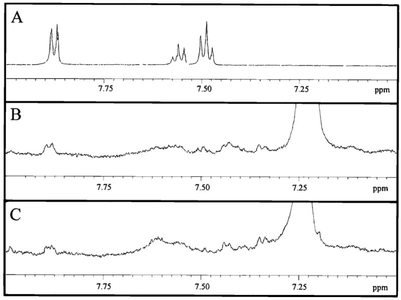
Detection of benzoic acid in rat tissue after an injectable dose (2.04 mg benzoic acid in 1.00 mL phosphate buffer) by 1H HR-MAS NMR spectroscopy. (A) Buffer solution containing 1 mM benzoic acid. (B) Muscle (100.9 mg) of dosed rat 4. (C) Muscle (84.1 mg) of the same animal located farther below the subcutaneous layer. Experimental conditions are the same as those listed in Figure 4. Spectra B and C are displayed on the same vertical scale, which is 32 times greater than spectrum A. The masses of tissue indicated are tissue wet weights.
Quantitation of tissue-specific drugs is important in assessing their bioavailability and efficacy. Quantitation by NMR spectroscopy is relatively simple because the area underneath the NMR signal is related to the number of protons giving rise to that signal and hence the molar amount of analyte.44 Therefore, calibration curves are not required and quantitation can be performed in a single experiment by adding an internal standard of known concentration. The main requirement for quantitation is that the standard and analyte of interest have well-resolved resonances and that the spectra have high S/N so that integration is precise and accurate.
The limits of quantitation (LOQ) and detection (LOD) were initially estimated by measuring the S/N of the 1H doublet (7.90 ppm) in spectra of BA solutions of the following concentrations: 1 μM, 10 μM, 100 μM, and 1000 μM. The measured S/N was then normalized for the number of scans acquired in the experimental time used in the rat tissue studies. On the basis of these calculations for the standard BA solutions, we estimated an LOQ of 40 μM and an LOD of 12 μM (0.5 μg and 0.2 μg, respectively, given that the volume of solution analyzed was 110 μL). Practically speaking, we expected these limits to be higher in muscle and skin samples because of the nature of the tissue matrix.
To quantitate BA in rat tissue, the integral of the aromatic doublet (7.90 ppm) was compared to the singlet of the internal standard TSP (0.00 ppm) added to the sample rotor. Both resonances were in spectral regions free from interference by other signals. The number of moles of benzoic acid (BA) detected was determined by eq 1, given that the number of moles of TSP added to the rotor was known.
| (1) |
The mass of BA was then calculated, using a molecular weight of 122.12 g/mol. The results are given in Table 1.
Table 1.
Quantitation of Benzoic Acid (BA) in Rat Tissue Analyzed by HR-MAS NMR Spectroscopy
| rat ID | tissue type | dosing method | μg BA detected | μg BA per mg tissuea | mg BA dosed | % of BA dose detected |
|---|---|---|---|---|---|---|
| 1 | muscle | topical | 230 | 2.6 | 10.4 | 2.2 |
| 2 | skin | topical | 306 | 2.9 | 10.0 | 3.1 |
| 2 | muscle | topical | 118 | 1.3 | 10.0 | 1.2 |
| 3 | skin | topical | 227 | 2.1 | 10.0 | 2.3 |
| 4 | muscle | injection | 12 | 0.1 | 2.04 | 0.6 |
| 4b | muscle | injection | 24 | 0.2 | 2.04 | 1.2 |
Calculated using the tissue wet weight in mg.
Tissue sampled closer to microdialysis probe.
Because BA is a low molecular weight, neutral compound that penetrates the skin, we expected it to disperse away from the dosage area into the surrounding tissue. In general, Table 1 shows that the mass of BA detected was routinely less than 3% of the amount dosed, showing that the local concentration of the compound in the tissue sampled is relatively low. The CPMG experiment used to acquire these data improved the general resolution of the tissue spectra but it also probably suppressed the intensity of resonances arising from BA bound to macromolecules, such as proteins or membranes. Thus, like the results obtained by microdialysis sampling, the BA levels determined by HR-MAS largely represent unbound drug. The mass of BA detected in skin sampled after topical application was ~230–300 μg, or 2.3–3.1% of the amount dosed. Because the volume of tissue could not be measured accurately, the concentration of BA detected by HR-MAS NMR was not determined. The concentration of BA detected in the skin using microdialysis sampling was 1.8 ± 0.6 mM,27 or 0.1% of the dose administered, a factor of 20–30 less than that detected by NMR. This result is not too surprising, considering that NMR samples the intra- and extracellular BA as well as any BA that had not penetrated into the dermis but remained on the skin surface. Since topically applied BA was detected in the muscle by NMR (Table 1), it is clear that some penetration occurred. The BA remaining on the skin surface could be removed in future experiments by tape stripping the top layer of the skin or washing prior to analysis. Additional sampling of muscle distal to the dosing site would facilitate the generation of spatial maps of drug distribution and provide more conclusive evidence regarding the extent and rate of drug distribution.
Furthermore, although BA has good skin penetrability,27,45 it was not detected above the LOD of the HPLC/UV method (0.3 μM) by microdialysis sampling in the extracellular fluid of the muscle tissue in the topical application studies.27 The results in Figure 4D and Table 1 show that BA was detected in the muscle by NMR. Because HR-MAS NMR detects compounds in both intracellular and extracelluar domains, these results suggest that a significant fraction of the BA is localized in the intracellular space. Investigating other active pharmaceutical ingredients and their formulations by the combination of microdialysis HPLC/UV and HR-MAS NMR could reveal the extent of intra- versus extracellular localization.
The mass of BA detected in the muscle after topical application varied by a factor of 2 for the animals studied. This may be related to variability between animals or to the poorer S/N of the muscle spectra compared to the skin spectra which would be improved by analysis at lower temperature because more scans could be acquired. Several factors are likely to affect spectral precision, including the time it takes to prepare the sample and set up the NMR experiment, the reproducibility with which the sample rotor is packed, the quality of the shimming, and the internal temperature of the sample during analysis. Although further analysis is needed to determine the true precision of the method, the results in Table 1 illustrate that despite some animal-to-animal variability, the results are internally consistent, that is, more BA is detected in the skin than the muscle for the topical dose.
After the topical dose, the average amount of BA detected in the skin and muscle was 2.2 ± 0.8 μg/mg of tissue sampled. More studies are needed to determine if this result implies a relatively even distribution of benzoic acid between skin and muscle. The muscle could contain more macromolecule-bound benzoic acid than the skin and the S/N was much less in the muscle spectra. For the injectable formulation, substantially lower amounts of BA were determined. Furthermore, it appears that the proximity of the tissue to the dosing site corresponds to the amount of BA measured. Twice as much BA was detected in muscle sampled closer to the injection site versus muscle deeper beneath the subcutaneous layer. The low S/N for benzoic acid observed in Figure 6B and C combined with the results presented in Table 1 indicate that the detection limit for benzoic acid in rat muscle is ~10–20 μg, using the CPMG method. As predicted, the practical LOD achieved was significantly higher than that calculated from HR-MAS analysis of BA solutions. A lower detection limit in tissue might be achievable when the analysis is performed at reduced temperature and the experimental time can be increased to improve S/N.
CONCLUSIONS
These experiments show the potential of HR-MAS NMR spectroscopy as an analytical tool for studying drug delivery and distribution in tissues. The results of these studies suggest that the technique can distinguish differences in local concentration of a model drug compound varying with tissue type, dosage method, and tissue proximity to the dosing site. Several challenges were encountered in this study that must be addressed in the subsequent refinement of this method. More tissue samples must be analyzed to distinguish the method’s precision versus animal-to-animal variability. Absolute quantitation of the unbound drug analyzed by the CPMG method will require calculating the losses in resonance intensity because of T2 relaxation for the analyte and standard in each of the tissues studied. Interference from the spectral background arising from macromolecules would be eliminated if the delivery of isotopically labeled drug compounds was studied, and thus the CPMG method would not be necessary for analysis of tissue spectra. However, generating labeled drugs can be costly and time-consuming, and therefore methods other than CPMG for obtaining quantitative, high-resolution tissue spectra should be explored. Distinguishing bound versus unbound drug and penetrated versus unpenetrated drug will also require the use of additional sample preparation methods and NMR experiments.
Despite these challenges, the results presented clearly suggest that HR-MAS NMR is a useful complement to existing methods for in vivo drug quantitation. When microdialysis sampling is coupled to HPLC-UV, relatively low limits of detection are achievable (~0.1 μM),27 but only chromophoric metabolites in the extracellular fluid surrounding the microdialysis probe can be detected. HR-MAS NMR analysis of rat tissue is a more universal approach to detecting the total concentration of the drug in both the tissue cells themselves and the extracellular space surrounding the tissue, providing concentrations are sufficiently high. Furthermore, the entire metabolic profile of the tissue can be obtained with a simple 1H spectrum, potentially allowing relative changes in other endogenous components to be determined and providing a means for assessing the therapeutic or toxic effects of the drug.
Although the detection limit of benzoic acid was ~10–20 μg in these studies, it could be improved by analyzing the sample at lower temperature for a longer time period. Depending on the detection limits achievable with improved instrumentation, a therapeutic dose may or may not be detectable in rat tissue. HR-MAS NMR spectroscopy should, however, be a useful method for toxicity studies, when the dosages are substantially higher. Metabolic profiling of tissue after inducing a toxic insult using a combination of microdialysis-HPLC/UV and HR-MAS analysis would provide insight into the mechanism of toxicity and therefore aid in characterizing potential drug side effects.
Acknowledgments
L.H.L. acknowledges the ACS Division of Analytical Chemistry for a fellowship sponsored by Eli Lilly and Company. The authors would like to thank the Lawrence Memorial Hospital, especially Dr. Mike Thompson, for their help with processing the histology slide. Steve Huhn of Bruker-Biospin Corporation also provided valuable technical assistance. A portion of this work was funded by the NIH (grant number 8 R01 EB00247).
References
- 1.Henry CM. Chem Eng News. 2002;80:66–70. [Google Scholar]
- 2.Lindon JC, Holmes E, Nicholson JK. Anal Chem. 2003;75:385A–391A. doi: 10.1021/ac031386+. [DOI] [PubMed] [Google Scholar]
- 3.Lindon JC, Nicholson JK, Holmes E, Everett JR. Concepts Magn Reson. 2000;12:289–320. [Google Scholar]
- 4.Everett JR. In: Bioanalytical approaches for drugs: including anti-asthmatics and metabolites. Reid E, Wilson ID, editors. Vol. 22. Royal Society of Chemistry; Cambridge, U.K: 1992. pp. 3–10. [Google Scholar]
- 5.Vion-Dury J, Nicoli F, Torri G, Torri J, Kriat M, Sciaky M, Davin A, Viout P, Confort-Gouny S, Cozzone PJ. Biochimie. 1992;74:801–807. doi: 10.1016/0300-9084(92)90062-j. [DOI] [PubMed] [Google Scholar]
- 6.Brindle JT, Nicholson JK, Schofield PM, Grainger DJ, Holmes E. Analyst. 2003;128:32–36. doi: 10.1039/b209155k. [DOI] [PubMed] [Google Scholar]
- 7.Shockcor JP, Unger SE, Wilson ID, Foxall PJD, Nicholson JK, Lindon JC. Anal Chem. 1996;68:4431–4435. doi: 10.1021/ac9606463. [DOI] [PubMed] [Google Scholar]
- 8.Wang W, Zhou H, Lin H, Roy S, Shaler TA, Hill LR, Norton S, Kumar P, Anderle M, Becker CH. Anal Chem. 2003;75:4818–4826. doi: 10.1021/ac026468x. [DOI] [PubMed] [Google Scholar]
- 9.Waters NJ, Holmes E, Williams A, Waterfield CJ, Farrant RD, Nicholson JK. Chem Res Toxicol. 2001;14:1401–1412. doi: 10.1021/tx010067f. [DOI] [PubMed] [Google Scholar]
- 10.Nicholson JK, Connelly J, Lindon JC, Holmes E. Nat Rev Drug Discovery. 2002;1:153–161. doi: 10.1038/nrd728. [DOI] [PubMed] [Google Scholar]
- 11.Keifer PA. Curr Opin Biotechnol. 1999;10:34–41. doi: 10.1016/s0958-1669(99)80007-x. [DOI] [PubMed] [Google Scholar]
- 12.Lindon JC, Nicholson JK. Trends Anal Chem. 1997;16:190–200. [Google Scholar]
- 13.Andrew ER, Bradbury A, Eades RG. Nature. 1958;4650:1659. [Google Scholar]
- 14.Lowe IJ. Phys Rev Lett. 1959;2:285–287. [Google Scholar]
- 15.Fitch WL, Detre G, Holmes CP. J Org Chem. 1994;59:7955–7956. [Google Scholar]
- 16.Barton SJ, Howe FA, Tomlins AM, Cudlip SA, Nicholson JK, Bell A, Griffiths JR. Magn Reson Mater Phys Biol Med. 1999;8:121–128. doi: 10.1007/BF02590529. [DOI] [PubMed] [Google Scholar]
- 17.Garrod S, Humpfer E, Spraul M, Connor SC, Polley S, Connelly J, Lindon JC, Nicholson JK, Holmes E. Magn Reson Med. 1999;41:1108–1118. doi: 10.1002/(sici)1522-2594(199906)41:6<1108::aid-mrm6>3.0.co;2-m. [DOI] [PubMed] [Google Scholar]
- 18.Griffin JL, Williams HJ, Sang E, Nicholson JK. Magn Reson Med. 2001;46:249–255. doi: 10.1002/mrm.1185. [DOI] [PubMed] [Google Scholar]
- 19.Garrod S, Humpfer E, Connor SC, Connelly JC, Spraul M, Nicholson JK, Holmes E. Magn Reson Med. 2001;45:781–790. doi: 10.1002/mrm.1106. [DOI] [PubMed] [Google Scholar]
- 20.Waters NJ, Holmes E, Waterfield CJ, Farrant RD, Nicholson JK. Biochem Pharmacol. 2002;64:67–77. doi: 10.1016/s0006-2952(02)01016-x. [DOI] [PubMed] [Google Scholar]
- 21.Tate AR, Foxall PJD, Holmes E, Moka D, Spraul M, Nicholson JK, Lindon JC. NMR Biomed. 2000;13:64–71. doi: 10.1002/(sici)1099-1492(200004)13:2<64::aid-nbm612>3.0.co;2-x. [DOI] [PubMed] [Google Scholar]
- 22.Moka D, Vorreuther R, Schicha H, Spraul M, Humpfer E, Lipinski M, Foxall PJD, Nicholson JK, Lindon JC. Anal Comm. 1997;34:107–109. doi: 10.1016/s0731-7085(97)00176-3. [DOI] [PubMed] [Google Scholar]
- 23.Moka D, Vorreuther R, Schicha H, Spraul M, Humpfer E, Lipinski M, Foxall PJD, Nicholson JK, Lindon JC. J Pharm Biomed Anal. 1998;17:125–132. doi: 10.1016/s0731-7085(97)00176-3. [DOI] [PubMed] [Google Scholar]
- 24.Tomlins AM, Foxall PJD, Lindon JC, Lynch MJ, Spraul M, Everett JR, Nicholson JK. Anal Commun. 1998;35:113–115. [Google Scholar]
- 25.Weiss DJ, Lunte CE, Lunte SM. Trends Anal Chem. 2000;19:606–616. [Google Scholar]
- 26.Hansen DK, Davies MI, Lunte SM, Lunte CE. J Pharm Sci. 1999;88:14–27. doi: 10.1021/js9801485. [DOI] [PMC free article] [PubMed] [Google Scholar]
- 27.McDonald S, Lunte C. Pharm Res. 2003;20:1827–1834. doi: 10.1023/b:pham.0000003381.10208.8c. [DOI] [PMC free article] [PubMed] [Google Scholar]
- 28.Lucas LH. PhD Thesis. University of Kansas; Lawrence, KS: 2004. p. 226. [Google Scholar]
- 29.Khandelwal P, Beyer CE, Lin Q, McGonigle P, Schechter LE, Bach AC., II J Neurosci Methods. 2004;133:181–189. doi: 10.1016/j.jneumeth.2003.10.012. [DOI] [PubMed] [Google Scholar]
- 30.Khandelwal P, Beyer CE, Lin Q, Schechter LE, Bach AC., II Anal Chem. 2004;76:4123–4127. doi: 10.1021/ac049812u. [DOI] [PubMed] [Google Scholar]
- 31.Stenken JA, Reichert WM, Klitzman B. Anal Chem. 2002;74:4849–4854. doi: 10.1021/ac020234w. [DOI] [PubMed] [Google Scholar]
- 32.Price KE, Vandaveer SS, Lunte CE, Larive CK. J Pharm Biomed Anal. 2005 doi: 10.1016/j.jpba.2005.02.034. in press. [DOI] [PMC free article] [PubMed] [Google Scholar]
- 33.Wilson SF. PhD Thesis. University of Kansas; Lawrence, KS: 2004. [Google Scholar]
- 34.Waters NJ, Garrod S, Farrant RD, Haselden JN, Connor SC, Connelly J, Lindon JC, Holmes E, Nicholson JK. Anal Biochem. 2000;282:16–23. doi: 10.1006/abio.2000.4574. [DOI] [PubMed] [Google Scholar]
- 35.Zuiderweg ERP, Hallenga K, Olejniczak ET. J Magn Reson. 1986;70:336–343. [Google Scholar]
- 36.Hore PJ. Methods Enzymol. 1989;176:64–77. [PubMed] [Google Scholar]
- 37.Hoult DI. J Magn Reson. 1976;21:337–347. [Google Scholar]
- 38.Meiboom S, Gill D. Rev Sci Instrum. 1958;29:688–691. [Google Scholar]
- 39.Carr HY, Purcell EM. Phys Rev. 1954;94:630–638. [Google Scholar]
- 40.Rabenstein DL. J Biochem Biophys Methods. 1984;9:277–306. doi: 10.1016/0165-022x(84)90013-7. [DOI] [PubMed] [Google Scholar]
- 41.Rabenstein DL, Millis KK, Strauss EJ. Anal Chem. 1988;60:1380A–1391A. doi: 10.1021/ac00175a001. [DOI] [PubMed] [Google Scholar]
- 42.Chin J, Chen A, Shapiro MJ. J Comb Chem. 2000;2:293–296. doi: 10.1021/cc0000038. [DOI] [PubMed] [Google Scholar]
- 43.Wieruszeski JM, Montagne G, Chessari G, Rousselot-Pailley P, Lippens G. J Magn Reson. 2001;152:95–102. doi: 10.1006/jmre.2001.2394. [DOI] [PubMed] [Google Scholar]
- 44.Lucas LH, Larive CK. In: Analysis and Purification Methods in Combinatorial Chemistry. Yan B, editor. John Wiley and Sons; New York: 2004. pp. 3–36. [Google Scholar]
- 45.Kitagawa S, Hosokai A, Kaseda Y, Yamamoto N, Kaneko Y, Matsuoka E. Int J Pharm. 1998;161:115–122. [Google Scholar]
- 46.Millis KK, Weybright P, Campbell N, Fletcher JA, Fletcher CD, Cory DG, Singer S. Magn Reson Med. 1999;41:257–267. doi: 10.1002/(sici)1522-2594(199902)41:2<257::aid-mrm8>3.0.co;2-n. [DOI] [PubMed] [Google Scholar]
- 47.Sitter B, Sonnewald U, Spraul M, Fjösne HE, Gribbestad IS. NMR Biomed. 2002;15:327–337. doi: 10.1002/nbm.775. [DOI] [PubMed] [Google Scholar]


