

Abstract
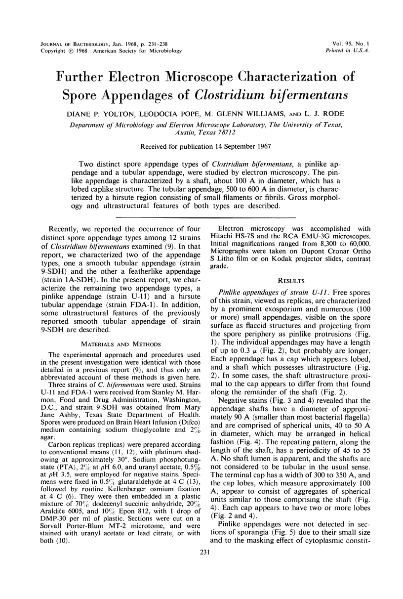
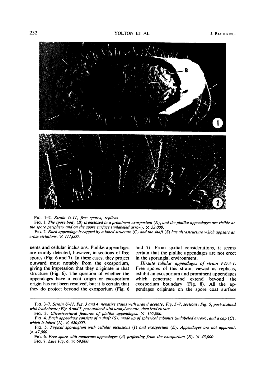
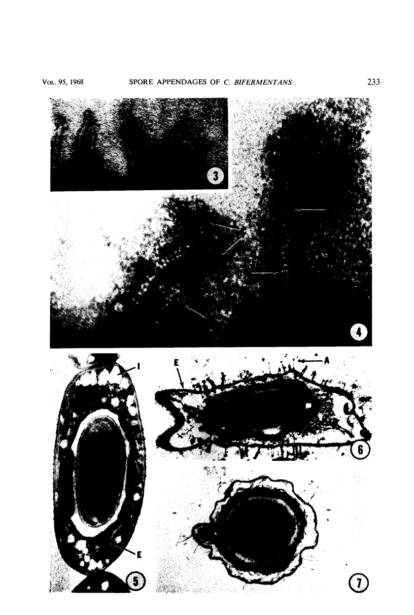
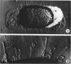
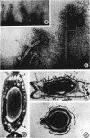
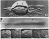
Two distinct spore appendage types of Clostridium bifermentans, a pinlike appendage and a tubular appendage, were studied by electron microscopy. The pinlike appendage is characterized by a shaft, about 100 A in diameter, which has a lobed caplike structure. The tubular appendage, 500 to 600 A in diameter, is characterized by a hirsute region consisting of small filaments or fibrils. Gross morphology and ultrastructural features of both types are described.
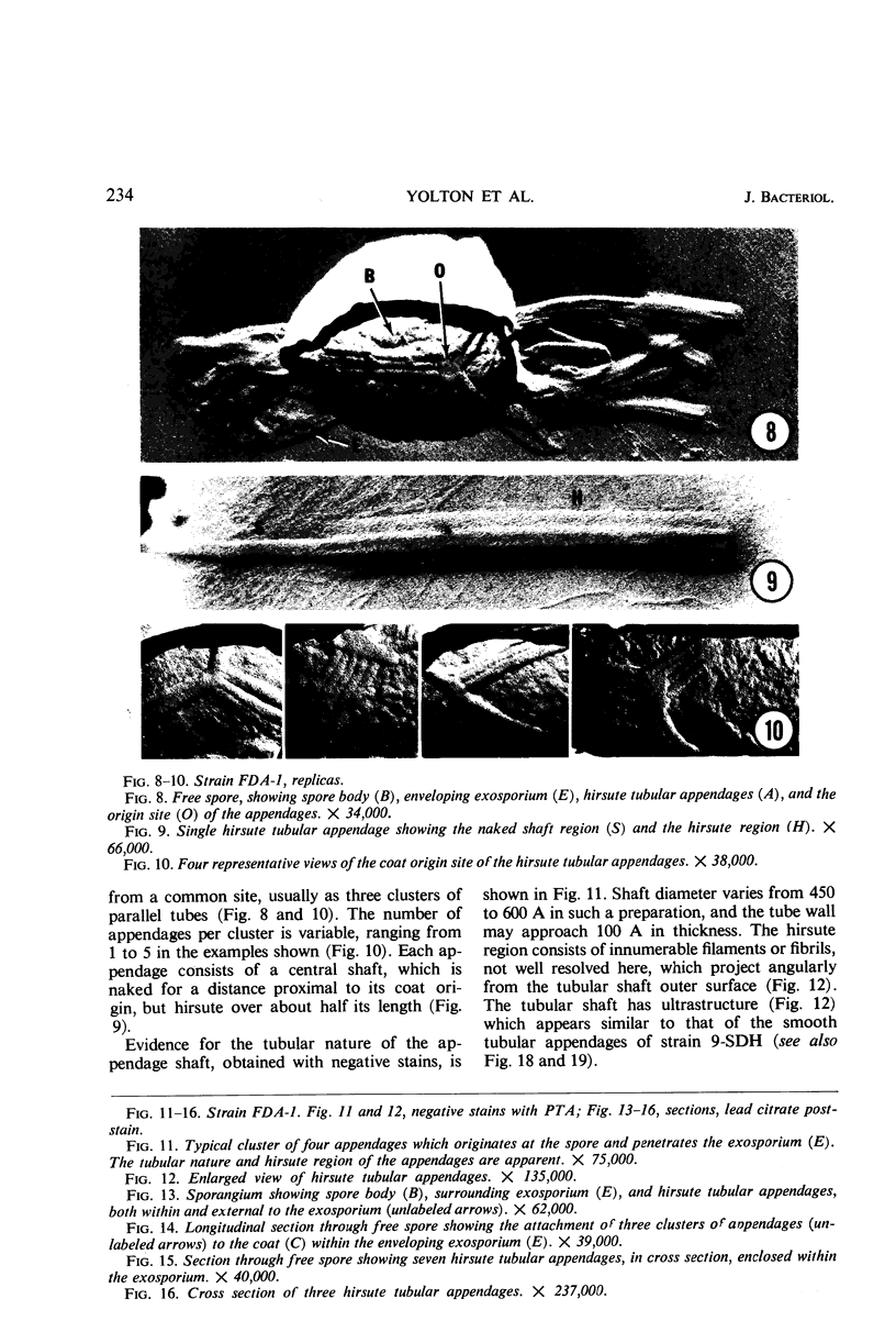
Full text
PDF
Images in this article
Selected References
These references are in PubMed. This may not be the complete list of references from this article.
- GERHARDT P., RIBI E. ULTRASTRUCTURE OF THE EXOSPORIUM ENVELOPING SPORES OF BACILLUS CEREUS. J Bacteriol. 1964 Dec;88:1774–1789. doi: 10.1128/jb.88.6.1774-1789.1964. [DOI] [PMC free article] [PubMed] [Google Scholar]
- Hodgkiss W., Ordal Z. J., Cann D. C. The comparative morphology of the spores of Clostridium botulinum type E and the spores of the "OS mutant". Can J Microbiol. 1966 Dec;12(6):1283–1284. doi: 10.1139/m66-170. [DOI] [PubMed] [Google Scholar]
- Hodgkiss W., Ordal Z. J., Cann D. C. The morphology and ultrastructure of the spore and exosporium of some Clostridium species. J Gen Microbiol. 1967 May;47(2):213–225. doi: 10.1099/00221287-47-2-213. [DOI] [PubMed] [Google Scholar]
- Hodgkiss W., Ordal Z. J. Morphology of the spore of some strains of Clostridium botulinum type E. J Bacteriol. 1966 May;91(5):2031–2036. doi: 10.1128/jb.91.5.2031-2036.1966. [DOI] [PMC free article] [PubMed] [Google Scholar]
- KELLENBERGER E., RYTER A., SECHAUD J. Electron microscope study of DNA-containing plasms. II. Vegetative and mature phage DNA as compared with normal bacterial nucleoids in different physiological states. J Biophys Biochem Cytol. 1958 Nov 25;4(6):671–678. doi: 10.1083/jcb.4.6.671. [DOI] [PMC free article] [PubMed] [Google Scholar]
- Pope L., Yolton D. P., Rode L. J. Appendages of Clostridium bifermentans spores. J Bacteriol. 1967 Oct;94(4):1206–1215. doi: 10.1128/jb.94.4.1206-1215.1967. [DOI] [PMC free article] [PubMed] [Google Scholar]
- REYNOLDS E. S. The use of lead citrate at high pH as an electron-opaque stain in electron microscopy. J Cell Biol. 1963 Apr;17:208–212. doi: 10.1083/jcb.17.1.208. [DOI] [PMC free article] [PubMed] [Google Scholar]
- Rode L. J., Crawford M. A., Williams M. G. Clostridium spores with ribbon-like appendages. J Bacteriol. 1967 Mar;93(3):1160–1173. doi: 10.1128/jb.93.3.1160-1173.1967. [DOI] [PMC free article] [PubMed] [Google Scholar]
- Rode L. J., Williams M. G. Utility of sodium hypochlorite for ultrastructure study of bacterial spore integuments. J Bacteriol. 1966 Dec;92(6):1772–1778. doi: 10.1128/jb.92.6.1772-1778.1966. [DOI] [PMC free article] [PubMed] [Google Scholar]
- SABATINI D. D., MILLER F., BARRNETT R. J. ALDEHYDE FIXATION FOR MORPHOLOGICAL AND ENZYME HISTOCHEMICAL STUDIES WITH THE ELECTRON MICROSCOPE. J Histochem Cytochem. 1964 Feb;12:57–71. doi: 10.1177/12.2.57. [DOI] [PubMed] [Google Scholar]