
Figure 3.
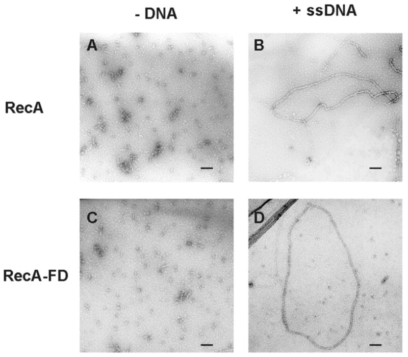
Electron micrographs of RecA and RecA-FD proteins with and without ssDNA. Wild type RecA (A, B) and RecA-FD protein (C, D) were incubated as described in EXPERIMENTAL PROCEDURES in the presence of ATPγS and the absence of φX174 phage ssDNA (A, C) or in the presence of both ATPγS and φX174 phage ssDNA (B, D). Black bar in each panel equals 0.1 μM.