

Abstract
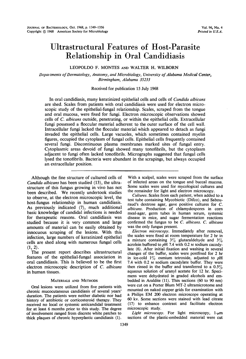
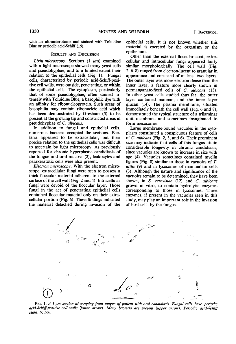
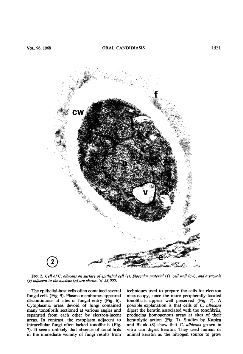
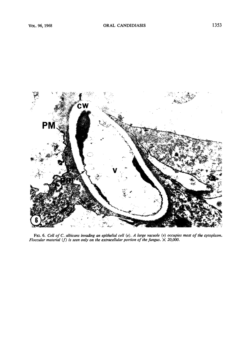
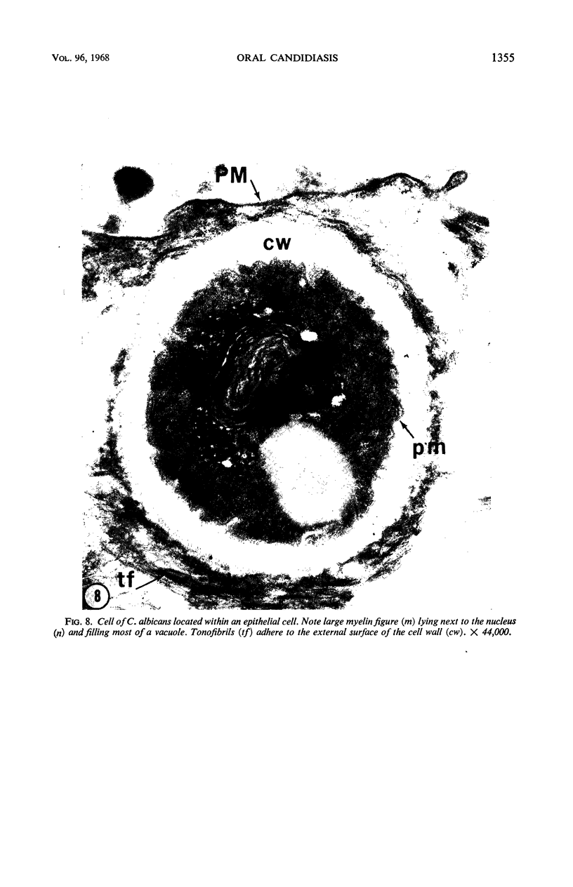
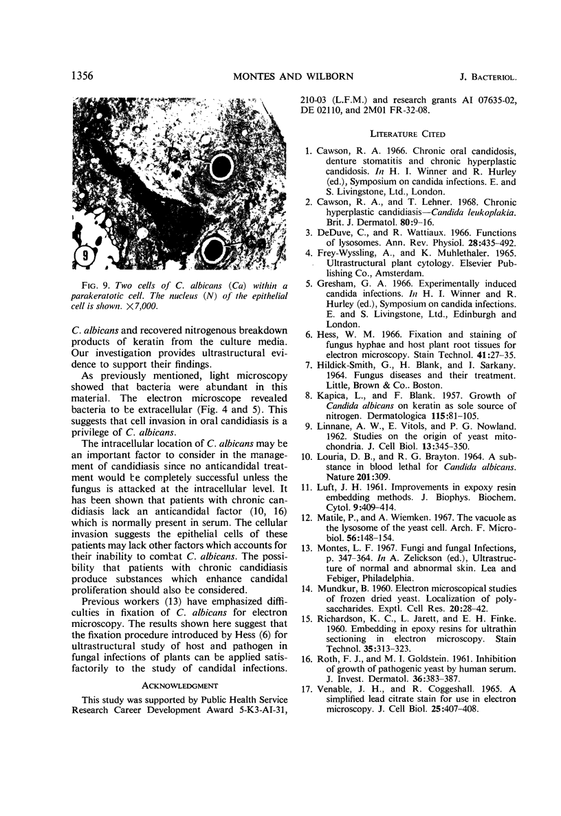
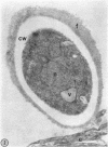
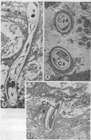
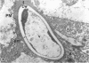
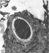
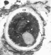
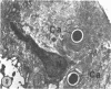
In oral candidiasis, many keratinized epithelial cells and cells of Candida albicans are shed. Scales from patients with oral candidiasis were used for electron microscopic study of the epithelial-fungal relationship. Scales, scraped from the tongue and oral mucosa, were fixed for fungi. Electron microscopic observations showed cells of C. albicans outside, penetrating, or within the epithelial cells. Extracellular fungi possessed a floccular material adherent to the outer surface of the cell wall. Intracellular fungi lacked the floccular material which appeared to detach as fungi invaded the epithelial cells. Large vacuoles, which sometimes contained myelin figures, occupied the cytoplasm of fungal cells. Epithelial cells frequently contained several fungi. Discontinuous plasma membranes marked sites of fungal entry. Cytoplasmic areas devoid of fungi showed many tonofibrils, but the cytoplasm adjacent to fungi often lacked tonofibrils. Micrographs suggested that fungal cells lysed the tonofibrils. Bacteria were abundant in the scrapings, but always occupied an extracellular position.
Full text
PDF

Images in this article
Selected References
These references are in PubMed. This may not be the complete list of references from this article.
- Cawson R. A., Lehner T. Chronic hyperplastic candidiasis--candidal leukoplakia. Br J Dermatol. 1968 Jan;80(1):9–16. doi: 10.1111/j.1365-2133.1968.tb11899.x. [DOI] [PubMed] [Google Scholar]
- De Duve C., Wattiaux R. Functions of lysosomes. Annu Rev Physiol. 1966;28:435–492. doi: 10.1146/annurev.ph.28.030166.002251. [DOI] [PubMed] [Google Scholar]
- Hess W. M. Fixation and staining of fungus hyphae and host plant root tissues for electron microscopy. Stain Technol. 1966 Jan;41(1):27–35. doi: 10.3109/10520296609116276. [DOI] [PubMed] [Google Scholar]
- KAPICA L., BLANK F. Growth of Candida albicans on keratin as sole source of nitrogen. Dermatologica. 1957 Aug;115(2):81–105. doi: 10.1159/000255990. [DOI] [PubMed] [Google Scholar]
- LINNANE A. W., VITOLS E., NOWLAND P. G. Studies on the origin of yeast mitochondria. J Cell Biol. 1962 May;13:345–350. doi: 10.1083/jcb.13.2.345. [DOI] [PMC free article] [PubMed] [Google Scholar]
- LOURIA D. B., BRAYTON R. G. A SUBSTANCE IN BLOOD LETHAL FOR CANDIDA ALBICANS. Nature. 1964 Jan 18;201:309–309. doi: 10.1038/201309a0. [DOI] [PubMed] [Google Scholar]
- LUFT J. H. Improvements in epoxy resin embedding methods. J Biophys Biochem Cytol. 1961 Feb;9:409–414. doi: 10.1083/jcb.9.2.409. [DOI] [PMC free article] [PubMed] [Google Scholar]
- MUNDKUR B. Electron microscopical studies of frozen-dried yeast. I. Localization of polysaccharides. Exp Cell Res. 1960 Jun;20:28–42. doi: 10.1016/0014-4827(60)90219-6. [DOI] [PubMed] [Google Scholar]
- Matile P., Wiemken A. The vacuole as the lysosome of the yeast cell. Arch Mikrobiol. 1967 Feb 20;56(2):148–155. doi: 10.1007/BF00408765. [DOI] [PubMed] [Google Scholar]
- RICHARDSON K. C., JARETT L., FINKE E. H. Embedding in epoxy resins for ultrathin sectioning in electron microscopy. Stain Technol. 1960 Nov;35:313–323. doi: 10.3109/10520296009114754. [DOI] [PubMed] [Google Scholar]
- ROTH F. J., Jr, GOLDSTEIN M. I. Inhibition of growth of pathogenic yeasts by human serum. J Invest Dermatol. 1961 May;36:383–387. doi: 10.1038/jid.1961.59. [DOI] [PubMed] [Google Scholar]
- VENABLE J. H., COGGESHALL R. A SIMPLIFIED LEAD CITRATE STAIN FOR USE IN ELECTRON MICROSCOPY. J Cell Biol. 1965 May;25:407–408. doi: 10.1083/jcb.25.2.407. [DOI] [PMC free article] [PubMed] [Google Scholar]