Abstract
This article describes a modified technique that combines percutaneous pinning and casting. A prospective study was conducted on 54 patients with distal radius fracture who were treated with percutaneous Kirschner wire (K-wire) fixation and pin-in-plaster technique. The surgical indications of this technique included displaced extra-articular fracture, intra-articular fracture without significant comminution, and articular step-off less than 2 mm. The average radial height was 10.96 mm, and the volar tilt was 3.97° on immediate postoperative radiographs. Upon removal of pin-in-plaster and percutaneous K-wires, the average radial height was 9.92 mm, and the volar tilt was 3.93°. Bony union was achieved in all patients; the satisfaction rate was 90.7%. Pin-in-plaster technique is effective for maintaining reduction during bone healing. The procedure provides the ability to achieve anatomic reduction and then maintains this reduction through an adequate method of immobilization.
Keywords: Distal radius fracture, Percutaneous pinning, Pin-in-plaster
Introduction
Distal radius fractures are the most common type of orthopedic fracture. Some surgeons advocate treatment by manipulation and plaster immobilization [19]. Many recommend operative intervention as the only methods to obtain anatomical reduction, and some have proposed that the best functional result will only be achieved by obtaining as near an anatomical radiographic result as possible [12]. Although a study by Young and Rayan [23] found favorable outcomes in low-demand older-aged patients despite deformity, most authors agreed that radial shortening more than 4 mm and radial dorsal angulation of more than 11° would reduce range of motion of the wrist. Furthermore, wrist pain was the most complaint among those patients [2, 7, 12].
In most displaced fractures of the radius, loss of reduction is likely to occur unless accurate management is provided to prevent repeat displacement. Inadequate fixation might result in gradual shortening at the fracture site during the healing process, even with excellent reduction. Percutaneous pinning and casting are simple procedures familiar to most surgeons. This article describes a technique that combines percutaneous Kirschner wire (K-wire) fixation, casting, and external fixation with presentation of treatment results.
Materials and Methods
Fifty-four patients with distal radius fractures who were treated with percutaneous K-wire fixation and pin-in-plaster technique were included in the study. The patients received treatment at our institution during the period of September 2004 to August 2005. Surgical indications for this technique included displaced extra-articular fracture, intra-articular fracture without significant comminution, and articular step-off less than 2 mm. The surgery excluded oblique volar fractures, die-punch fractures, significant dorsal comminution involving more than one third of the anteroposterior diameters of the radius, open fracture, bilateral fractures, and multiple fractures. The patients with multiple injuries were also excluded. All patients sustained closed distal radius fractures. Twenty-two patients were men, and 32 were women. The average age at the time of surgery was 62 years (range = 41–93 years). Eighteen cases were caused by a traffic accident, and thirty-six fractures resulted from falls. The left wrist was affected in 28 patients and the right wrist in 26 others.
Preoperative radiographs were assessed for fracture pattern, degree of comminution, and articular fragmentation. All of the cases were evaluated by one senior physician (Chen) using the Arbeitsgemeinschaft für Osteosynthesefragen (AO) classification [13]. According to the AO classification, there were 16 A2 fractures, 7 A3 fractures, 3 C1 fractures, 18 C2 fractures, and 10 C3 fractures.
Operative Technique
To be included in this study, the patient was scheduled for operative treatment either the same day or within 48 h of injury. Under axillary block or general anesthesia, the patient was placed in the supine position with the involved limb in traction with a finger trap through the index finger, and provided 8 to 10 lbs of countertraction with a water bottle. An accurate reduction in the fracture was the first step in the treatment plan. A neutral position of the wrist was desirable. The application of hyperextension and flexion maneuvers to break up the impaction was not recommended. With a dorsally displaced fracture, the reduction was performed by pushing the distal fragment distally and palmarly while holding the proximal fragment with the fingers around the forearm. The goal was to convert the dorsal angulation to a neutral position as well as to regain radial height. In our study patients, we found that it should not be necessary to manipulate the wrist with pronation or wrist flexion to obtain or hold the reduction. Image intensification fluoroscopy was used to assist the reduction and to assess the accuracy of the reduction.
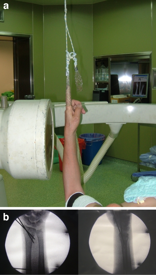
After acceptable reduction of the fracture was achieved, two percutaneous K-wires were inserted through the radial styloid with the wrist in traction to maintain the reduction. Image intensification fluoroscopy was used to assist the insertion of the K-wires throughout the entire procedure (Fig. 1). The wires were drilled proximally through the radial styloid until they penetrated the intact cortex of the shaft. K-wires with a diameter of 1.5 to 2.0 mm were selected for use, with smaller wires for women and larger wires for men. The wire insertion was performed with a power K-wire driver to allow the surgeon to hold part of the reduction with one hand during K-wire insertion. After percutaneous pinning for distal radius fracture was finished, a K-wire was inserted into the base of second metacarpal bone. Another K-wire was inserted into the junction of the distal and middle thirds of the radial shaft. The ends of the wires were bent at a right angle and then cut short outside the skin. Sponge padding with an occlusive dressing was applied to prevent skin irritation. All procedures were carried out under full sterile preparation and draping. A well-fitted pin-in-plaster was applied for external immobilization. Usually a total of four K-wires were used with this technique.
Figure 1.
a Photograph of a study patient’s hand and wrist with percutaneous pinning with pin-in-plaster technique for distal radius fractures. The hand was maintained with a finger strap through the index finger under longitudinal traction and subsequent monitoring with fluoroscopy. b Image intensification fluoroscopy was used to assist the insertion of K-wires through the radial styloid with the wrist in traction to maintain the reduction.
The patients were hospitalized overnight to observe distal circulation of the fingers. The patients underwent follow-up at our outpatient clinic at 2-week intervals following hospital discharge. The healing of the fracture was assessed both clinically and radiographically at each follow-up. Usually, by 6 weeks, clinical and radiographic examination demonstrated progression of fracture healing. The percutaneous wires and the pin-in-plaster were usually removed after 6 weeks of immobilization on an outpatient basis without local anesthesia. Instruction about active-assisted wrist motion was demonstrated to the patient. Physical therapy was arranged because distraction across the wrist possibly could delay the return of motion and strength. A custom-made protective splint was applied after the removal of the cast. The splint could be removed for bathing and exercise. The use of the splint was not needed after 4 to 6 weeks, after which time, the fracture was solidly healed. Most patients favored the splint for protection.
The functional results and radiographic results were evaluated by one senior physician (Chen). The treatment complications were recorded. After bony union, patients underwent further follow-up for 1 year. Wrist function was evaluated at 6 months and 1 year using Solgaard’s modification [17] of the scoring system described by Gartland and Werley [3] (Table 1). The functional outcome was easy to evaluate with simple instruments in this scoring system. The residual deformity and the subjective evaluation were recorded in the same way as the original scoring system. The range of motion was measured using a goniometer to measure dorsal and volar flexion, radial and ulnar deviation, and supination and pronation, and the sum was calculated as the percentage of the unaffected wrist. The grip strength was measured with a gripper, and the result was classified using a nomogram. The final results of the patients with excellent and good functional outcome were considered satisfactory.
Table 1.
Functional scoring system modified after Gartland and Werley.
| Points | ||
|---|---|---|
| Deformity | Prominent ulnar styloid | 1 |
| Radial deviation | 1–2 | |
| Dinner-fork deformity | 1–3 | |
| Maximum | 6 | |
| Subjective evaluation | No pain, no limitation of motion | 0 |
| Slight pain, slight limitation of motion | 2 | |
| Occasional pain, some limitation of motion, weakness | 4 | |
| Pain, limitation of motion, activities restricted | 6 | |
| Maximum | 6 | |
| Range of motion | Limitation of motion <20% | 0 |
| Limitation of motion 20–50% | 2 | |
| Limitation of motion >50% | 6 | |
| Stiffness of wrist | 6 | |
| Maximum | 6 | |
| Grip strength | Normal (within 2 SD) | 0 |
| 2–4 SD | 2 | |
| 4–6 SD | 4 | |
| <6 SD | 6 | |
| Maximum | 6 | |
| Complications | None or minimal | 0 |
| Slight crepitation | 1–2 | |
| Severe crepitation | 3–4 | |
| Median nerve compression | 1–3 | |
| Pulp-palm distance 1 cm | 3 | |
| Pulp-palm distance >2 cm | 5 | |
| Pain in distal radio-ulnar joint | 1–3 | |
| Maximum | 16 | |
| Total score | Excellent | 0–2 |
| Good | 3–7 | |
| Fair | 8–18 | |
| Poor | 19–39 | |
Results
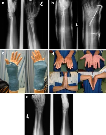
The average follow-up was 15 months (range = 12–24 months). All of the fractures healed in our study group. Excellent results were noted in 27 patients, good in 22, fair in four, and poor in one patient. Most of the patients returned to their preinjury activity level with a 90.7% satisfaction rate (Fig. 2). There were four patients rated as fair, which correlated with radial shortening, especially on the step-off of the radio-ulna joints. There were two cases of pin tract infection in which the removal of the pin-in-plaster in an earlier stage was necessary. Both cases responded well to antibiotic treatment and wound care after the removal of the wires. At the time of last follow-up, there was no recurrent infection, and the functional results were good in both cases. One case had unsightly tethered scars at the site of radial pin insertion, but the functional result was excellent at the time of follow-up. A 93-year-old female patient had radial shaft fracture and metacarpal fracture at the site of pin insertion due to severe osteoporosis. The patient refused further management because of old age and had a poor end result.
Figure 2.
a The anteroposterior and lateral radiographs of the wrist in a 65-year-old man show displaced distal radius fracture. b Radiographs of the wrist show fracture fixation with K-wires. c Photographs of wrist show fixation with pin-in-plaster. d Nearly full forearm pronation, supination, extension, and flexion were regained at 12 weeks. e At 1-year follow-up, the patient’s radiographs show bony union.
Assessment of postoperative radiographs showed that the average radial height was 10.96 mm (range = 7–17 mm) and volar tilt was 3.97° (range = −3.9–12°) on immediate postoperative radiographs. At the time of removal of pin-in-plaster and percutaneous K-wires, the average radial height was 9.92 mm (range = 5–17 mm), and the volar tilt was 3.93° (range = −8.3–12°). The results indicated that pin-in-plaster can provide adequate stability during the time of fracture healing.
Discussion
Distal radius fracture is a common injury. The importance of anatomic reduction has been demonstrated by clinical studies as well as by laboratory assessment of force and stress loading across the radiocarpal joint [8, 20]. In fractures with articular surface displacement greater than 2 mm, radial shortening greater than 5 mm, or dorsal angulation more than 20°, suboptimal results have been reported in previously published studies [8, 11]. Therefore, every effect should be made to restore normal length, alignment, and articular surface congruency of the distal radius.
An accurate reduction in the fracture is the first step in the treatment of the distal radius fracture. After anatomic reduction in the fracture is achieved, many methods are available to maintain alignment and prevent repeat displacement. The methods of immobilization include casting, percutaneous pinning, external fixation, internal fixation with plate, or internal fixation combined with external fixation depending on the different types of fractures. Every method has its advantages and some limitations.
The most common traditional treatment of distal radius fractures in osteoporotic patients is closed reduction and cast immobilization. Three-point fixation with a well-fitted cast is essential for adequate immobilization. However, extreme flexion should be avoided because carpal tunnel pressure will be increased. This is associated with increased wrist flexion and ulnar deviation when the distal fracture was immobilized with a cast. Although cast immobilization alone avoids surgery and complications related to pin placement and pin removal, casts cannot maintain distraction to correct length or control the rotation of the distal fragment when comminution is present [21]. Loss of reduction usually happens after 2 weeks of casting despite a perfect initial anatomic reduction [2]. Gartland and Werley obtained a 68.3% satisfactory result, and Sarmiento et al. reported an 82% satisfactory result treated with the casting technique [15]. Spira and Weigl reported a 51.4% unsatisfactory result with reduction and use of cast in the treatment of comminuted fracture of distal radius with articular involvement [18]. Closed reduction and percutaneous pinning relies on intrafocal manipulation and pinning or manual traction, reduction, and pinning, to hold the fracture in an appropriate anatomic alignment. Clancey reported a 96.4% satisfactory result in 30 patients treated with percutaneous pinning if the articular surface of the radius was not comminuted into more than two fragments [1]. However, the tenting effect is not strong enough in comminuted fracture, which often results in subsiding and dorsal angulation. Anatomic studies reveal the risk of injury to the sensory branch of the radial nerve with percutaneous pinning through the snuffbox [5].
External fixation has been popular for the treatment of displaced fractures of distal radius, and the radial length and dorsal tilt have improved significantly with this method [10, 14, 22]. External fixation can be supplemented with percutaneous wires through the radial styloid for certain intra-articular fractures. Combined internal and external fixation is a technique that attempts to maximize the advantageous features of each of its two components while minimizing their disadvantages. Seitz et al. reported a 92% satisfactory result in 51 patients treated with augmented external fixation using K-wires to reduce and fix unstable fragments [16]. The external fixator could maintain radial length more efficiently than the percutaneous pinning and casting group, but volar tilt was not generally restored [6]. Pin tract infection is another problem that should be concerned.
The pin-in-plaster technique is a combination of percutaneous pinning, casting, and external fixation. The potential biological advantage is that it allows treatment of the fracture with minimal manipulation and devascularization of the bone. Adjacent soft tissue supports structures, including tendons and the joint capsule. After closed reduction in our study patients, the fractures were fixated directly with percutaneous K-wires. Because K-wire fixation seldom provide sufficient stability to allow for early motion and often necessitate use of a cast or splint, the addition of two K-wires incorporated into the pin-in-plaster could increase stability of the fracture fixation. Extreme wrist flexion and ulnar deviation could be avoided with this technique. Extreme wrist flexion might cause potentially uncorrectable wrist stiffness and also is associated with an increased risk for median nerve compression resulting in carpal tunnel syndrome [10]. Pin-in-plaster relies on ligamentotaxis to maintain the reduction. Yet, excessive traction across the wrist ligaments, which leads to stiffness, can be avoided. Postoperatively, the pin-in-plaster functioned as a neutralization device, preventing fracture collapse and decreasing the biomechanical demands on the internal fixation hardware. This procedure can be performed for both intra-articular and extra-articular fractures. Green reported an 86% satisfactory result with this technique used in the treatment of 75 patients with severely comminuted intra-articular fractures [4]. The technique is so easy that most surgeons become familiarized with this procedure in a relatively short time. The wires usually can be withdrawn in the outpatient clinic with relative ease when healing is sufficient.
Despite the above advantages, there were some disadvantages of the pin-in-plaster technique for the treatment of distal radius fracture. There was the risk of injury to the sensory branch of the radial nerve while percutaneous pinning through the snuffbox. Limited incision should be used to protect the distal radial sensory nerve branches from injury. Problems in peripheral circulation might occur if the cast is too tight. Pin tract infection can be an occasional problem because pin tract care is impossible in the pin-in-plaster. Frequent follow-up to assess any sign of pin tract infection is mandatory. The occurrence of pin tract infection can be controlled successfully after removal of the K-wires and initiation of treatment with oral antibiotics and pin tract care. Another problem of pin-in-plaster was poor tolerability in a few patients in our study. The fracture might lose reduction if removal of the pin-in-plaster was performed in an earlier stage before callus formation was strong enough to allow early range of motion of the wrist. A short-arm splint was necessary if the pin-in-plaster was removed in an earlier stage for any reason. Rehabilitation was usually necessary, since wrist stiffness was common immediately following cast and pin removal. However, almost all of the patients could achieve good range of motion of the wrist after a period of physical therapy.
One of the most frequently stated complications associated with most intra-articular fracture is the development of posttraumatic arthritis. In the radio-carpal and distal radio-ulnar joints, the reported incidence of arthritis was variable. However, radiographic findings correlated poorly with symptomatic complaints [9]. There was no incidence of posttraumatic arthritis reported in our patient population because long-term follow-up is not available. Additionally, the need for secondary salvage is unknown. At least, in the short term, the technique of combined fixation seems capable of restoring reasonable joint function for extra- and intra-articular fracture of the distal radius.
In our study, the average radial height was 10.96 mm postoperatively and 9.92 mm after removal of the pin-in-plaster. The average union state of the volar tilt angle was 3.93° compared with 3.97° in the immediate postreduction state. We consider that the pin-in-plaster technique can efficiently stop radial shortening and dorsal tilting during the time of bone healing. Most of our study patients achieved excellent to good functional results 6 months after removal of the pin-in-plaster.
In conclusion, percutaneous pinning incorporated with pin-in-plaster fixation is an excellent technique for both extra-articular and intra-articular fractures in cases without severe comminution of the distal radius. The technique involves a minimal procedure that provides anatomic reduction, fracture fixation, and maintenance of reduction with an adequate method of immobilization.
References
- 1.Clancey GJ. Percutaneous Kirschner-wire fixation of Colles fractures—a prospective study of thirty cases. J Bone Jt Surg Am. 1984;66:1008–14. [PubMed]
- 2.Fu YC, Chien SH, Huang PJ, et al. Use of an external fixation combined with the buttress-maintain pinning method in treating comminuted distal radius fractures in osteoporotic patients. J Trauma. 2006;60:330–3. [DOI] [PubMed]
- 3.Gartland JJ Jr, Werley CW. Evaluation of healed Colles’ fractures. J Bone Jt Surg Am. 1951;33:895–907. [PubMed]
- 4.Green DP. Pins and plaster treatment of comminuted fractures of the distal end of the radius. J Bone Jt Surg Am. 1975;57:304–10. [PubMed]
- 5.Hochwald NL, Levine R, Tornetta P. The risks of Kirschner wire placement in the distal radius: a comparison of techniques. J Hand Surg Am. 1997;22:580–4. [DOI] [PubMed]
- 6.Hutchinson DT, Strenz GO, Cautilli RA. Pins and plaster vs external fixation in the treatment of unstable distal radial fractures. A randomized prospective study. J Hand Surg Br. 1995;20:365–72. [DOI] [PubMed]
- 7.Jenkins NH, Mintowt-Czyz WJ. Mal-union and dysfunction in Colles’ fracture. J Hand Surg Br. 1988;13:291–3. [DOI] [PubMed]
- 8.Knirk JL, Jupiter JB. Intra-articular fractures of the distal end of the radius in young adults. J Bone Jt Surg Am. 1986;68:647–59. [PubMed]
- 9.Kopylov P, Johnell O, Redlund-Johnell I, et al. Fractures of the distal end of the radius in young adults: a 30-year follow-up. J Hand Surg Br. 1993;18:45–9. [DOI] [PubMed]
- 10.McAuliffe JA. Combined internal and external fixation of distal radius fractures. Hand Clin. 2005;21:395–406. [DOI] [PubMed]
- 11.McMurtry RY, Axelrod T, Paley D. Distal radial osteotomy. Orthopedics. 1989;12:149–55. [DOI] [PubMed]
- 12.McQueen M, Caspers J. Colles’ fracture: does the anatomical result affect the final function? J Bone Jt Surg Br. 1988;70:649–51. [DOI] [PubMed]
- 13.Ruch DS. Fractures of the distal radius and ulna. Rockwood and Green’s fractures in adult. 6th ed. Philadelphia, PA: Lippincott Williams & Wilkins; 2006. p. 913–8.
- 14.Ruch DS, Papadonikolakis A. Volar versus dorsal plating in the management of intra-articular distal radius fractures. J Hand Surg Am. 2006;31:9–16. [DOI] [PubMed]
- 15.Sarmiento A, Pratt GW, Berry NC, Sinclair WF. Colles’ fractures—functional bracing in supination. J Bone Jt Surg Am. 1975;57:311–7. [PubMed]
- 16.Seitz WH Jr, Froimson AI, Leb R, et al. Augmented external fixation of unstable distal radius fractures. J Hand Surg Am. 1991;16:1010–6. [DOI] [PubMed]
- 17.Solgaard S. Function after distal radius fracture. Acta Orthop Scand. 1988;59:39–42. [DOI] [PubMed]
- 18.Spira E, Weigl K. The comminuted fracture of the distal end of the radius. Reconstr Surg Traumatol. 1968;11:128–38. [PubMed]
- 19.Stewart HD, Innes AR, Burke FD. Factors affecting the outcome of Colles’ fracture: an anatomical and functional stud. Injury. 1985;16:289–95. [DOI] [PubMed]
- 20.Trumble TE, Schmitt SR, Vedder NB. Factors affecting functional outcome of displaced intra-articular distal radius fractures. J Hand Surg Am. 1994;19:325–40. [DOI] [PubMed]
- 21.Weil WM, Trumble TE. Treatment of distal radius fractures with intrafocal pinning and supplemental skeletal stabilization. Hand Clin. 2005;21:317–28. [DOI] [PubMed]
- 22.Werber KD, Raeder F, Brauer RB, et al. External fixation of distal radial fractures: four compared with five pins: a randomized prospective study. J Bone Jt Surg Am. 2003;85:660–6. [DOI] [PubMed]
- 23.Young BT, Rayan GM. Outcome following nonoperative treatment of displaced distal radius fractures in low-demand patients older than 60 years. J Hand Surg Am. 2000;25:19–28. [DOI] [PubMed]


