Abstract
Pedicle screw instrumentation has become increasingly popular during the past 20 years and a vast selection of products is available on the market. With rising implantation rates, reports about specific complications also have increased. The main reason for these complications is the fact that the course of the pedicle and in turn the positioning of the pedicle screw cannot be adequately controlled visually. Based on the anatomy of the surrounding structures, complications caused by malpositioning can be divided into three main groups: mechanical, neurological and vascular. Beyond mechanical limitations of spinal motion, nerve injury can lead to neurological problems while injuries to vascular structures usually cause hemorrhage. These typical problems in general become apparent intraoperatively or in the immediate postoperative course. We report on a rare delayed complication and analyze the factors that led to it. In addition, we outline our treatment strategy. The goal has to be to avoid such problems in the future by using suitable navigational aids.
Keywords: Complication, Aortic perforation, Thoracic spine, Pedicle screw instrumentation
Material
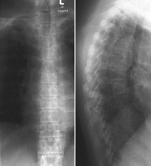
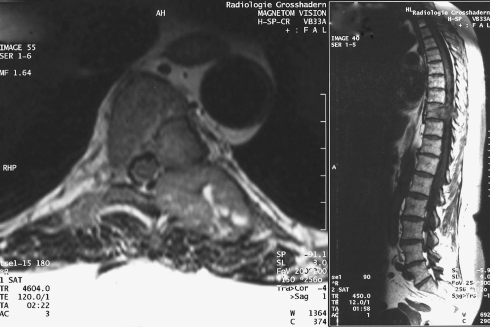
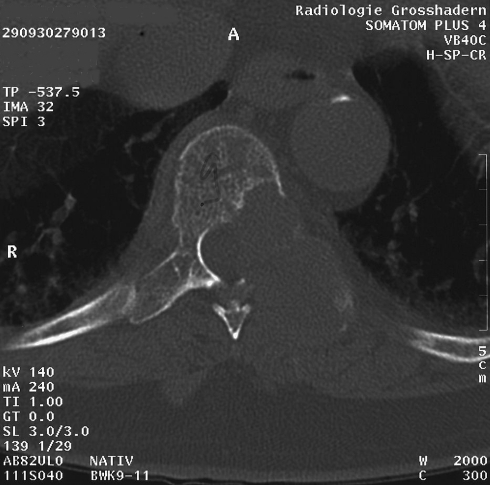
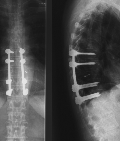
A 69-year-old female patient was first seen by her general practitioner for back pain, which had been present for a period of 4 weeks. She was referred to our department, where clinical examination showed strong pain at the thoracolumbar junction without any peripheral neurological deficit. Plain radiographs showed osteolytic destruction of the 9th thoracic vertebra (Fig. 1). The subsequent diagnostic work-up consisted of a CT of the chest and abdomen, an MRI of the spine, and a 3-phase technetium bone scan and resulted in the diagnosis of a renal cell carcinoma with a solitary metastasis to the 9th thoracic vertebra. The metastasis had penetrated into the spinal canal with beginning compression of the spinal cord (Figs 2, 3). The operative treatment consisted of dorsal decompression by means of an expanded costotransversectomy, bilateral laminectomy, tumor debulking and posterior pedicle screw instrumentation using the USS I-system (Synthes, Umkirch, Germany, Fig. 4) after preoperative tumor embolization. Surgery was followed by local radiation, a nephrectomy and a systemic immunochemotherapy. One year after the procedure, the patient presented again with renewed back pain. She was also experiencing intermittent and pulsating epigastric pain, symptoms which had already been observed immediately after the original surgery, but which had gotten much worse during recent days. In addition, there was left-sided pelvic pain, which could be explained by a new metastasis in the left pubic bone. The epigastric pain did not follow any dermatomal pattern and a CT of the chest and the abdomen was performed. This study showed a suspected penetration of the left-sided T 11-pedicle screw into the descending thoracic aorta (Fig. 5), subsequently confirmed by aortography. We performed a left-sided thoracotomy and the intraoperative findings confirmed screw penetration into the aorta. Brief proximal and distal cross clamping of the aorta allowed for pulling the vessel off the pedicle screw and for oversewing of the defect. The protruding screw tips were subsequently cut flush with the vertebral body cortex using a diamond trephine. After an uneventful recovery, the patient’s epigastric and back pain had completely resolved.
Fig. 1.
Plain radiograph of the thoracic spine in 2 planes. There is an osteolytic destruction of the 9th thoracic vertebra with effacement of the left pedicle root contour and the left costotransverse process in the antero-posterior plane
Fig. 2.
MRI of the thoracic spine in sagittal and axial planes. The osteolytic metastasis has completely destroyed the left pedicle, the left lamina, the left costotransverse process and the base of the rib. The dorsolateral vertebral body is partially destroyed and the spinal canal is being compressed by the tumor that also infiltrates the erector spinae muscles
Fig. 3.
CT of the 9th thoracic vertebra in the axial plane. The same situation as in Fig. 2, visualized by CT for better definition of bony structures
Fig. 4.
Postoperative plain radiographs of the thoracic spine in 2 planes. After decompression, stabilization was achieved by posterior instrumentation with the USS I system from T7 to T11, bridging T9
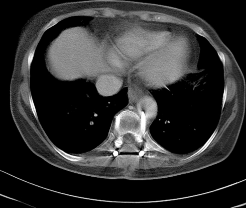
Fig. 5.
CT of the chest in axial planes at the level of T11. The left pedicle screw perforates the aorta with the implant spanning several millimeters into the lumen
Results
The intermittent pulsating pain in this patient had been present since the original surgery, completely resolved after the revision, which indicates that it was caused by the prominent pedicle screw arroding, and eventually perforating into the aorta. The further clinical course in this patient was characterized by further metastases and progression of the underlying disease.
Discussion
Complications caused by malpositioning of pedicle screws are not rare and a host of reasons is being discussed in the literature. The correct choice of entry point into the pedicle and a correctly angulated trajectory trough the pedicle are of great importance. There are several studies characterizing the pedicle angulations for individual vertebrae. When a pedicle screw is placed more or less in the sagittal plane according to the technique described by Roy-Camille, there is a specific risk to penetrate the lateral vertebral cortex [7]. This type of malpositioning is difficult to avoid, since even with biplanar fluoroscopy during surgery, the lateral vertebral cortex can often not be visualized adequately. Weinstein [12] examined the reliability and validity of fluoroscopy-based pedicle screw placement in relation to the entry point into the pedicle. He was able to demonstrate that the technique according to Roy-Camille leads to a significantly higher number of malpositioned screws than his own technique with a more lateral entry point. With decreasing pedicle diameters at higher levels, the issue of safe pedicle screw placement is even more challenging in the thoracic spine than in the lumbar spine [10, 11]. A biomechanical study by Morgenstern et al. [6] finds that extrapedicular pedicle screw placement in the thoracic spine leads to instrumentation with a stability that is equal to transpedicular screw placement while reducing the risk of malpositioned screws.
Acute injuries of the aorta caused by malpositioned pedicle screws have been described in the literature. Jendrisak [3] reports on a 25-year-old patient with a fracture of the 2nd lumbar vertebra, who suffered an acute hemorrhage 6 days after surgery from an aortic laceration caused by a pedicle screw. Stewart et al. [9] describe the rupture of a lumbar artery with retroperitoneal hemorrhage and the formation of a pseudoaneurysm. Such acute hemorrhages usually lead to hypovolemia and hence rapidly become apparent. The paper by Anda [1] describes vascular complications after discectomy, which essentially are based on the same mechanism, the anterior penetration of a surgical instrument. A delayed but massive hemorrhage, which would have been likely in our case, carries specific risks that might have had a lethal effect. The patient might not have been in hospital at the time of the hemorrhage and the association with the original spine surgery might not have been obvious, causing considerable delay in establishing a correct diagnosis and performing the life-saving procedure. Other authors report perforations of the lung, the ureter, the gut and the esophagus in their analyses [2]. There clearly is a necessity for methods that would allow for a more controlled positioning of pedicle screws. Probing of the pedicle canal using a straight or a bent K-wire, as it was first described by Matsuzaki [5] does improve safety to some extent and special pedicle probes have since been developed. In recent years, 3-dimensional image intensifiers, intraoperative CT and navigation systems have been established and several authors have reported reduced rates of malpositioned pedicle screws when employing these systems. While these technologies are disputed, they appear to be the method of choice in our opinion, at least in the long-term perspective [4, 8].
Conclusion
Complications caused by pedicle screws can usually be observed perioperatively or in the short-term postoperative course. The aortic perforation that we report on in this paper must be considered a rare occurrence. However, with uncharacteristic complaints after pedicle screw instrumentation, such a rare complication should be considered and appropriate imaging studies (CT) should be performed to ascertain the implant position. In the case of an internal organ injury, operative revision should be performed. The strategy chosen by us represents one possible way of correcting the problem. Alternatively, exchanging the implants would have been an option, but this would have required a simultaneous anterior and posterior approach in order to repair the vascular injury. Improved intraoperative imaging and navigation are promising technologies that will increasingly be available to assist with correct implant positioning in the years to come.
Acknowledgments
Conflict of interest statement None of the authors has any potential conflict of interest.
References
- 1.Anda S, Akhus S, Skaanes KO, Sande E, Schrader H. Anterior perforations in lumbar discectomies—a report four cases of vascular complications and a CT study of the prevertrebral lumbar anatomy. Spine. 1991;16:54–60. doi: 10.1097/00007632-199101000-00011. [DOI] [PubMed] [Google Scholar]
- 2.Fujita T, Kostuik JP, Huckell CB, Sieber AN. Complications of spinal fusion in adult patients more than 60 years of age. Orthop Clin North Am. 1998;29(4):669–678. doi: 10.1016/S0030-5898(05)70040-7. [DOI] [PubMed] [Google Scholar]
- 3.Jendrisak MD. Spontaneous abdominal aortic rupture from erosion by a lumbar spine fixation device: a case report. Surgery. 1986;99(5):631–633. [PubMed] [Google Scholar]
- 4.Kosmopoulos V, Schizas C. Pedicle screw placement accuracy. Spine. 2007;32:E111–E120. doi: 10.1097/01.brs.0000254048.79024.8b. [DOI] [PubMed] [Google Scholar]
- 5.Matsuzaki H, Tokuhashi Y, Matsumoto F, Hoshino M, Kinchi T, Toriyama S. Problems and solutions of pedicle screw plate fixation of lumbal spine. Spine. 1990;15:1159–1165. doi: 10.1097/00007632-199011010-00014. [DOI] [PubMed] [Google Scholar]
- 6.Morgenstern W, Ferguson SJ, Berey S, Orr TE, Nolte LP. Posterior thoracic extrapedicular fixation: a biomechanical study. Spine. 2003;28:1829–1835. doi: 10.1097/01.BRS.0000083280.72978.D1. [DOI] [PubMed] [Google Scholar]
- 7.Roy-Camile R, Saillant G, Mazel C. Internal fixation of the lumbar spine with pdicle srew plating. Clin Orthop. 1986;203:7–17. [PubMed] [Google Scholar]
- 8.Schröder J, Wassermann H. Spinal navigation: an accepted standard of care? Current situation and literature review. Zentralbl Neurochir. 2006;67:123–128. doi: 10.1055/s-2006-942146. [DOI] [PubMed] [Google Scholar]
- 9.Stewart JR, Barth KH, Williams GM. Ruptured lumbar artery pseudoaneyrysm: an unusual cause of retroperitoneal haemorrhage. Surgery. 1983;94:592–4. [PubMed] [Google Scholar]
- 10.Vaccaro AR, Rizzolo SJ, Balderston RA, Allardyce TJ, Garfin SR, Dolinskas C, An HS. Placement of pedicle screws in the thoracic spine, part I: morphometric analysis of the thoracic vertebrae. J Bone Joint Surg. 1995;77-A:1193–1199. doi: 10.2106/00004623-199508000-00008. [DOI] [PubMed] [Google Scholar]
- 11.Vaccaro AR, Rizzolo SJ, Balderston RA, Allardyce TJ, Garfin SR, Dolinskas C, An HS. Placeent of pedicle screws in the thoracic spine, Part II: An anatomical and radiographic assessment. J Bone Joint Surg. 1995;77-A:1200–1206. doi: 10.2106/00004623-199508000-00009. [DOI] [PubMed] [Google Scholar]
- 12.Weinstein JN, Spratt KF, Sprengler D, Brick C, Reid S. Spinal pedicle fixation: reliability and validity of roentgenogram-based assessment and surgical factors on screw placement. Spine. 1988;13:1012–1018. doi: 10.1097/00007632-198809000-00008. [DOI] [PubMed] [Google Scholar]


