
Figure 1.
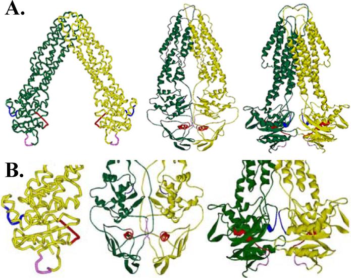
A. Crystal structure of MsbA from E. coli, V. cholera, and S. typhimurium (left to right). B. Enlarged NBD domains from each of the crystal structures are shown with the Walker A (red), LSGGQ (blue), and H-motif (pink) highlighted to demonstrate the differences in these conserved residues between the three structures.