Abstract
Aflatoxin B1 (AFB1) is a potent carcinogen, which can significantly increase the risk of hepatocellular carcinoma development through food contamination. In past decades, chemopreventive agents such as oltipraz and chlorophyllins have demonstrated that chemo-intervention is an effective approach to reduce the hepatotoxicity by AFB1. However, due to the potential adverse effects of these agents, alternative novel mechanism-based chemopreventive agents are needed. We report here that novel cis-terpenones 1-3, which were synthesized as the precursors of natural product analogues in our laboratory, showed promising protective effects against AFB1 induced cytotoxicity in HepG2 cells. Chemo-protection was observed with increasing concentrations of cis-terpenones in the co-treatment of AFB1, and no cytotoxicity was observed with cis-terpenones alone. In addition, cis-terpenones 1-3 at 10 μM effectively inhibited induced cytochrome P450 1A/1B activity by 50% in HepG2 cells, as indicated by an EROD assay. P450 1A/B is involved in the activation of many pre-carcinogens and is highly inducible in liver cells. These results suggested that novel terpenones 1-3 are candidates for the development of novel mechanism-based chemopreventive agents against AFB1 and other carcinogenic stimuli.
Introduction
Aflatoxin B1 (AFB1)1 is a major mycotoxin produced by the fungus A. flavus and is commonly found as a crop contaminant (1). The potent cytotoxicity of AFB1 is attributed to the covalent DNA modification by the exo-epoxide metabolite (2,3), resulting in liver damage and possibly hepatocellular carcinoma (4-8). In past decades, several effective chemoprevention agents, including oltipraz and chlorophyllins, have been developed and showed promising results in clinical trials (9-12). Unfortunately, adverse effects were the major concern for oltipraz (9,10), while more mechanistic studies are required for chlorophyllins (12). Recently, screening of natural products, such as extracts from algae (13), onion bulbs (14), and kola seeds (15), has identified several alternatives as potential candidates against AFB1. However, it is difficult to predict at this time whether these agents will be effective drugs. Thus, novel mechanism-based chemoprevention agents against AFB1 are needed.
cis-Terpenones 1-3 were designed as precursors of analogues of the natural product tingenone (16,17), which could be metabolically converted by Phase I enzymes through hydroxylation and oxidation, based on the metabolism of similar compounds, such as estrogens (18,19). On the other hand, cis-terpenones 1-3 differ from estrogens due to the unique bent conformation (17). The hypothesized hydroxylated cis-terpenone metabolite resembles partially the structure of the chemopreventive agent resveratrol, which is a potent inhibitor of P450 1A/B activity (20,21). Because P450 1A/B is highly inducible in liver cells and is involved in the activation of many pre-carcinogens, including benzopyrenes, estrogens and even AFB1 (20-24); it is important to evaluate the inhibitory potential of these cis-terpenones on P450 1A/B.
We report in this paper that novel cis-terpenones 1-3 (Scheme 1) showed promising protective effects against AFB1 induced cytotoxicity in HepG2 cells, while no cytotoxicity was observed by cis-terpenones alone. The chemo-protective effect of cis-terpenones 1-3 was observed with both cell viability and apoptosis assays when co-treated with AFB1. In addition, cis-terpenones 1-3 at 10 μM effectively inhibited the induced activity of Phase I P450 1A/B in HepG2 cells as indicated by an EROD assay. These results suggest that cis-terpenones 1-3 are potential candidates for novel mechanism-based preventive agents against both AFB1 and other carcinogenic stimuli.
Scheme 1.
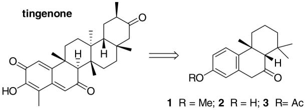
cis-Terpenones 1-3 as Precursors of Analogues of the Natural Bioactive Tingenone.
Experimental Procedures
All chemicals were purchased from Fisher Scientific (Pittsburg, PA) or Sigma-Aldrich (Milwaukee, WI) and used without further purification. NMR spectra of the synthesized compounds were obtained by Variant NMR spectrometers. Electrospray ionization mass spectroscopy (ESI-MS) analysis was carried out with Q-TOF2 from Micromass (Manchester, UK).
2-(3-Methoxybenzyl)-2-(2,6-dimethylhepta-1,5-dienyl)-1,3-dithiane (5)
To a solution of thiol-protected citral as a 1:1 cis/trans mixture (1.10 g, 4.54 mmol) in dry THF (20 mL) at -40 °C (acetonitrile/dry ice bath) was slowly added a solution of 1.05 equiv n-BuLi (1.6 M in hexanes, 2.84 mL) under N2. The resulting reaction solution was stirred at -40 °C for 1 h, and then a solution of 3-methoxybenzyl chloride (650 mg, 4.16 mmol) in dry THF (10 mL) was added. The reaction mixture was maintained at -40 °C for 8 h and then slowly warmed to room temperature over 16 h under N2. The reaction solution was quenched with brine (100 mL) and extracted with ether (100 mL × 2). The organic layers were collected, dried with MgSO4 and concentrated. A flash column separation (0-2% EtOAc in hexanes) afforded the desired product as a cis/trans diastereoisomeric mixture (0.60 g) in 40% yield. Further separation of the isomers was not carried out because the subsequent cyclization step afforded diastereoisomerically pure cis-compounds. 1H NMR (CDCl3, 300 MHz) for the cis/trans diastereomeric mixture: δ 7.23-7.14 (m, 1H), 6.95-6.72 (m, 3H), 5.40 (s, 1H), 5.19-5.05 (m, 1H), 3.83-3.76 (m, 3H), 3.37-3.25 (m, 2H), 3.00-2.74 (m, 4H), 2.54-2.44 (m, 1H), 2.17-1.90 (m, 5H), 1.84-1.52 (m, 9H). 13C NMR (CDCl3, 75 MHz) observed: δ 159.1, 143.2, 142.5, 137.8, 137.5, 132.0, 128.8, 128.7, 127.9, 127.4, 124.5, 124.3, 123.7, 117.0, 116.9, 112.5, 112.4, 55.3, 54.4, 47.0, 46.8, 41.8, 32.8, 28.2, 28.0, 26.9, 26.4, 26.0, 25.6, 24.7, 18.0, 17.9, 17.3. ESI-MS calcd for C21H31OS2 (M - H+), 361.17; found, 361.07.
1-(3-Methoxyphenyl)-4,8-dimethylnona-3,7-dienyl-2-one (6)
To a solution of 5 (300 mg, 0.83 mmol) in MeOH/H2O (9:1, 25 mL) was added 1.1 equiv HgO and HgCl2. The resulting reaction solution was stirred at room temperature for 4 h. The reaction solution was diluted with CH2Cl2 and the precipitation was filtered through a 5 μm Acrodisc filter. The solution was washed with brine, dried with MgSO4 and concentrated. The desired product was purified by a flash column separation (0-10 % EtOAc in hexanes) as a cis/trans diastereomeric mixture (160 mg) in 71% yield. 1H NMR (CDCl3, 300 MHz) for the cis/trans diastereomeric mixture: δ 7.24-7.14 (m, 1H), 6.84-6.74 (m, 2H), 6.09 (s, 1H), 5.05-4.98 (m, 1H), 3.79 (s, 3H), 3.74-3.67 (m, 2H), 2.18-2.09 (m, 7H), 1.17-1.56 (m, 6H). 13C NMR (CDCl3, 75 MHz) observed: δ 198.1, 160.3, 160.0, 136.7, 132.7, 129.7, 129.5, 123.2, 123.1, 122.6, 122.1, 121.9, 119.7, 115.3, 115.2, 114.1, 113.0, 112.5, 64.1, 55.5, 55.3, 51.7, 46.7, 41.5, 34.2, 26.9, 26.3, 26.0, 25.9, 19.7, 17.9. ESI-MS calcd for C18H25O2 (M + H+), 273.19; found, 273.07.
4b,5,6,7,8,8a-cis-Hexahydro-2-methoxy-4b,8,8-trimethylphenanthren-9(10H)-one (1)
To a solution of 6 (130 mg, 0.48 mmol) in dry CH3NO2 (10 mL) at room temperature was added 10 equiv BF3·Et2O, and the resulting reaction solution was stirred under N2 for 2 h. The reaction solution was diluted with saturated NaHCO3 (150 mL) and extracted with CH2Cl2 (150 mL × 3). The organic layers were collected, dried with MgSO4 and concentrated. The desired compound was purified by a flash column separation (0-5% EtOAc in hexanes) as a diastereoisomeric pure product (50 mg) as an oil in 38% yield. 1H NMR (CDCl3, 300 MHz): δ 7.23 (d, J = 8.7 Hz, 1H), 6.80 (dd, J1 = 8.7 Hz, J2 = 2.7 Hz, 1H), 6.61 (d, J = 2.7 Hz, 1H), 3.80 (s, 3H), 3.69 (d, J = 22.8 Hz, 1H), 3.48 (d, J = 22.8 Hz, 1H), 2.48 (d, J = 14.1 Hz, 1H), 2.10 (s, 1H), 1.54-1.46 (m, 2H), 1.34-1.23 (m, 3H), 1.05 (s, 3H), 0.95 (s, 3H), 0.34 (s, 3H). 13C NMR (CDCl3, 75 MHz): δ 212.5, 158.2, 135.6, 134.0, 125.2, 113.9, 112.7, 66.7, 55.4, 44.4, 42.4, 38.3, 36.4, 34.4, 33.8, 32.4, 22.7, 19.0. ESI-MS calcd for C18H25O2 (M + H+), 273.19; found, 273.12.
4b,5,6,7,8,8a-cis-Hexahydro-2-hydroxy-4b,8,8-trimethylphenanthren-9(10H)-one (2)
To a solution of 1 (15 mg, 0.05 mmol) in CH2Cl2 (2.0 mL) under N2 was added a solution of BBr3 (1.0 M in heptane, 1.0 mL). The resulting solution was stirred at room temperature for 2 h. The reaction solution was quenched with brine (100 mL) and extracted with EtOAc (100 mL × 2). The organic layers were collected, dried with MgSO4 and concentrated. A flash column separation (5-35 % EtOAc in hexanes) afforded the desired product (8 mg) as an oil in 57% yield. 1H NMR (CDCl3, 300 MHz): δ 7.18 (d, J = 8.7 Hz, 1H), 6.73 (dd, J1 = 8.7 Hz, J2 = 2.4 Hz, 1H), 6.57 (d, J = 2.4 Hz, 1H), 4.78 (bs, 1H), 3.67 (d, J = 22.8 Hz, 1H), 3.45 (d, J = 22.8 Hz, 1H), 2.48 (d, J = 12.3 Hz, 1H), 2.10 (s, 1H), 1.56-1.47 (m, 2H), 1.35-1.23 (m, 3H), 1.05 (s, 3H), 0.95 (s, 3H), 0.35 (s, 3H). 13C NMR (CDCl3, 75 MHz): δ 212.6, 154.1, 135.9, 134.0, 125.4, 115.3, 114.2, 66.7, 44.2, 42.4 38.3, 36.4, 34.4, 33.8, 32.4, 22.7, 19.0. ESI-MS calcd for C17H21O2 (M - H+), 257.15; found, 257.33.
4b,5,6,7,8,8a-cis-Hexahydro-2-acetyloxy-4b,8,8-trimethylphenanthren-9(10H)-one (3)
To a solution of 2 (5 mg, 0.02 mmol) in CH3CN (2.0 mL) was added solutions of Ac2O and Et3N (1.0 M, 1.0 mL each). The resulting solution was stirred at room temperature for 4 h. The reaction solution was extracted with Et2O (50 mL × 2). The organic layers were collected, dried with MgSO4 and concentrated. A flash column separation (5-20 % EtOAc in hexanes) afforded the desired product (5 mg) as an oil in 84% yield. 1H NMR (CDCl3, 300 MHz): δ 7.33 (d, J = 8.7 Hz, 1H), 6.99 (dd, J1 = 8.7 Hz, J2 = 2.4 Hz, 1H), 6.83 (d, J = 2.4 Hz, 1H), 3.71 (d, J = 22.8 Hz, 1H), 3.50 (d, J = 22.8 Hz, 1H), 2.50 (d, J = 14.1 Hz, 1H), 2.29 (s, 3H), 2.12 (s, 1H), 1.56-1.48 (m, 2H), 1.37-1.23 (m, 3H), 1.07 (s, 3H), 0.95 (s, 3H), 0.33 (s, 3H). 13C NMR (CDCl3, 75 MHz): δ 211.7, 169.7, 149.2, 139.3, 135.8, 125.2, 121.5, 120.3, 66.5, 44.2, 42.3, 38.7, 36.4, 34.5, 33.5, 32.3, 22.7, 21.4, 18.9. ESI-MS calcd for C19H23O3 (M - H+), 299.16; found, 299.33.
Cell Culture Study
Human HepG2 cells were maintained in the growth medium at 37 °C with 5% CO2. The growth medium contained 90% Minimum Essential Eagle Medium (Invitrogen, CA) and 10% heat inactivated fetal bovine serum, and was enriched with 2 mM L-glutamine, 0.75 g/L sodium bicarbonate, 0.1 mM non-essential amino acids, 1.0 mM sodium pyruvate, 50 U/mL penicillin, and 50 μg/mL streptomycin. Stock solutions of TCDD, AFB1, and cis-terpenones 1-3 were prepared in DMSO and stored at -20 °C. The MTT solution was prepared at 5 mg/mL in PBS solution (pH 7.4), filtered and stored at 4 °C. The MTT lysis buffer was prepared by dissolving 25 g SDS in 100 mL of 50% DMF in water, and the pH was adjusted to 4.7 with a solution of 2.5% HCl in 80% acetic acid.
For the cell viability MTT assay, HepG2 cells were seeded at 30,000 cells/well on a 96 well plate for 4 h before the treatment with AFB1 and cis-terpenones. The treatments included medium only, AFB1 only, AFB1 plus cis-terpenones 1-3, and cis-terpenones only. The final concentration for AFB1 was 2 μM (25), and those for cis-terpenones 1-3 were 10, 20, and 40 μM. The final solution of each well contained 9% serum with a volume of 200 μL and DMSO less than 0.18%. After incubation for 72 h, the MTT assay was carried out as reported (26). Briefly, the MTT solution (10 μL) was added to each well followed by incubation for 10 h. The medium was then removed, and lysis buffer (100 μL each) was added. After 10 h, the absorbance of each well at 570 nm (background correction at 690 nm) was obtained with a plate reader (μQuant, BioTek Instruments, VT). The statistical analyses (one-way ANOVAwith Dunnett’s test) were performed by GraphPad Prism (version 4.00, GraphPad Software, San Diego, CA). Each treatment was performed in quadruplicate, and all of the experiments were repeated at least three times independently.
For the apoptosis assay, HepG2 cells were plated at 50,000 cells/well and treated with AFB1 and/or cis-terpenone 2 as described in the viability assay. Apoptosis induced by AFB1 treatments was determined with Annexin V fluorescein isothiocyanate (FITC) in conjunction with propidium iodide (PI), according to the procedures provided by the manufacturer (BD Biosciences, San Jose, CA). Labeled cells were quantitatively resolved by a Coulter EPICS XL-MCL flow cytometer (Beckman Coulter, Inc., Fullerton, CA). Apoptotic cells (Annexin V-FITC positive and PI negative) were distinguished from cells that were either at the end of apoptosis, undergoing necrosis, or already dead (Annexin V-FITC positive and PI positive) (27,28).
For the EROD assay, HepG2 cells were seeded at 50,000 cells/well on a 96 well plate. After plating for 4 h, various treatments included DMSO only, TCDD only, TCDD plus cis-terpenones 1-3, and cis-terpenones only. The final concentration of TCDD was 1 nM, and the concentrations of cis-terpenones 1-3 were 0.1, 1, 10, and 20 μM. The final volume of each well was 200 μL containing 0.1% DMSO. After 24 h, the EROD assay was performed as reported (23,24). Briefly, fresh medium (175 μL per well) containing 2% serum, 8 μM 7-ethoxyresorufin and 10 μM dicumarol was added to each well, and then incubated for 1 h. The resulting medium (150 μL each) was transferred to a black plate, and ethanol (150 μL each) was added. The fluorescence intensity between 580 and 600 nm of each well was obtained by a Cary Eclipse fluorescence spectrometer (Varian Instruments, CA) with an exciting wavelength at 550 nm. The fluorescent intensity at 590 nm (λmax) was used as the index for the EROD activity, and the statistical analyses (one-way ANOVAwith Dunnett’s test) were performed by GraphPad Prism. All the experiments were repeated at least three times independently. Also, the cell viability MTT assay was carried out similarly as described above. No cytotoxicity in HepG2 cells was observed in the treatment of TCDD and/or cis-terpenones after 24 h.
Results and Discussion
Synthesis of cis-Terpenones
cis-Terpenones 1-3 were synthesized with a modified conjugated polyene cyclization as reported previously (17). For the synthesis, methoxylbenzyl chloride 4 was first coupled with 1,3-dithiol protected citral (1:1 cis/trans isomers) at -40°C in 40% yield (Scheme 2). The thiol groups of the resulting 5 were removed with HgCl2 and HgO in 71% yield. The obtained polyene 6 was cyclized in the presence of BF3·OEt2, affording cis-terpenone 1 diastereomeric selectively. The cis-conformation of terpenone 1 was confirmed by 2-D NMR ROESY analysis (see Supporting Information), and was consistent with that of reported cis-conformers (17). Removal of the methyl ether group of 1 was achieved with BBr3 to the desired cis-terpenone 2 in 57% yield, which was acetylated to cis-terpenone 3 in 84% yield.
Scheme 2.
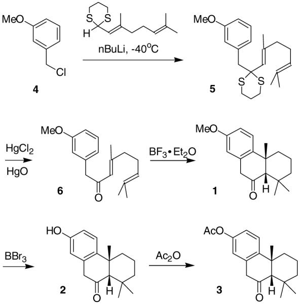
Synthesis of cis-Terpenones 1-3.
Chemo-protection with cis-Terpenones against AFB1 Induced Cytotoxicity
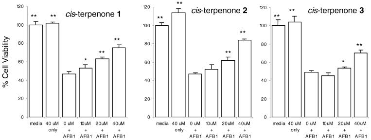
The chemo-protection against AFB1 with cis-terpenones 1-3 was confirmed by a cell viability assay in HepG2 cells, and no cytotoxicity was observed by cis-terpenones alone. The cell viability was determined after the co-treatment with AFB1 (2 μM) and cis-terpenones 1-3 at various concentrations (10-40 μM) for 72 h. As shown in Figure 1, 40 μM cis-terpenones exhibited no cytotoxicity in HepG2 cells, while AFB1 at 2 μM resulted in more than 50% cell death. The co-treatment of AFB1 and cis-terpenones showed a gradual increase of cell viability with increasing concentrations of cis-terpenones (up to 80% at 40 μM), and all of three cis-terpenones showed similar chemo-protection at the concentrations investigated. These results indicated that novel cis-terpenones 1-3 effectively reduce the AFB1-induced cytotoxicity in HepG2 cells, and suggested that cis-terpenones could be a promising candidate as a chemopreventive agent against AFB1.
Figure 1.
Chemo-protection with cis-terpenones against AFB1 induced cytotoxicity. HepG2 cells were co-treated with AFB1 (2 μM) and cis-terpenones 1-3 at various concentrations (10-40 μM) and incubated for 72 h. Cell viability was measured with the MTT assay. The percentage of viable cells was based on cells treated with medium only. Each bar represents the mean ± SD of 4 replicates. The data are representative of three independent experiments. * P<0.05, ** P<0.001 compared to treatment with AFB1 by one way ANOVA and Dunnett’s test.
Chemo-protection with cis-Terpenones against AFB1 Induced Apoptosis
The chemo-protection with cis-terpenone 2 at various concentrations (10-40 μM) against AFB1 was further validated with an Annexin V-apoptosis assay by flow cytometry. Annexin V has a high affinity for the membrane phospholipid phosphatidylserine, an early marker when cells start to undergo apoptosis, and thus is a sensitive probe for identifying apoptotic cells (27,28). When cell death eventually occurs, apoptotic cells will lose the membrane integrity and expose the nucleus to the vital dye PI, besides being stained positive for Annexin V-FITC. As shown in Figure 2, each graph (Figure 2a-d) is divided into four boxes, which are PI positive (at top left), both PI and annexin V positive (at top right), both PI and annexin V negative (at lower left), and annexin V positive (at lower right). The numbers within each box indicate the percent distribution of the cell population. The treatment with AFB1 alone (Figure 2b) resulted in only 41.7% viable HepG2 cells, while 11.8% were at early stage apoptosis and 41.8% at late stage apoptosis or dead. In contrast, co-treatment with 40 μM cis-terpenone 2 (Figure 2d) protected 84% of cells from the induced apoptosis by AFB1. Collectively, with the results from the cell viability assay, these data fully confirmed the chemo-protective role of cis-terpenones against AFB1 induced toxicity in HepG2 cells.
Figure 2.
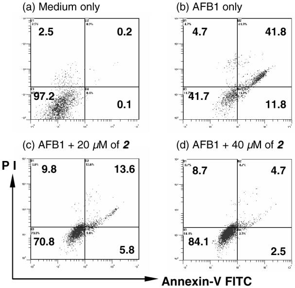
Chemo-protection with cis-terpenone 2 against AFB1 induced apoptosis. HepG2 cells were co-treated with AFB1 (2 μM) and cis-terpenone 2 at various concentrations (10-40 μM) and incubated for 72 h. The cells were stained with PI (shown as the Y-axis of each graph) and Annexin-V FITC (shown as the X-axis of each graph), and analyzed with a flow cytometer. The numbers in each divided box represent the percent distribution of HepG2 cells within the gated areas. PI positive (at top left), both PI and annexin V positive - late apoptosis or dead (at top right), both PI and annexin V negative - viable (at lower left), and annexin V positive - early apoptosis (at lower right). The data are representative of three independent experiments.
Chemoprevention of Induced P450 1A/B Activity in HepG2 Cells with cis-Terpenones
The chemopreventive agent resveratrol is a potent inhibitor of P450 1A/B activity, which is involved in the activation of pro-carcinogens including benzopyrenes, estrogens and even AFB1 (20,21). In liver cells, the levels of P450 1A/B are low yet highly inducible by poly aromatic compounds, such as TCDD and benzopyrenes (20-24). Because the hypothesized hydroxylated cis-terpenone metabolite resembles the structure of resveratrol partially, it is important to evaluate potential effects of the cis-terpenones on P450 1A/B activity, which would demonstrate chemopreventive actions in addition to anti-AFB1 activity.
Indeed, we found that cis-terpenones effectively inhibited TCDD-induced P450 1A/B activity in HepG2 cells (23,24). The activity of P450 1A/B was determined by the EROD assay with either TCDD alone (1 nM) or the co-treatment of TCDD and cis-terpenones after 24 h (Figure 3). As shown in Figure 3, the basal level of P450 1A/B activity in the absence of the inducer TCDD is negligible, while TCDD (1 nM) significantly induced the enzymatic activity. In the co-treatment of cis-terpenones and TCDD, the EROD activity showed a decreasing trend in a concentration-dependent manner. At 10 μM concentration, cis-terpenones 1-3 were able to reduce the induced enzyme activity by approximately 50%. In addition, no cytotoxicity in HepG2 cells under these conditions was observed with the MTT assay. These results implied that cis-terpenones 1-3 could be used as a generic chemopreventive agent that suppresses activity of P450 1A/B, besides the protective effect against AFB1. The inhibitory effect with cis-terpenones may be due to either the inhibition of enzyme activity or the suppression of the expression level of enzyme. Therefore, the detailed study on the inhibitory mechanism with cis-terpenones 1-3 is currently under investigation.
Figure 3.
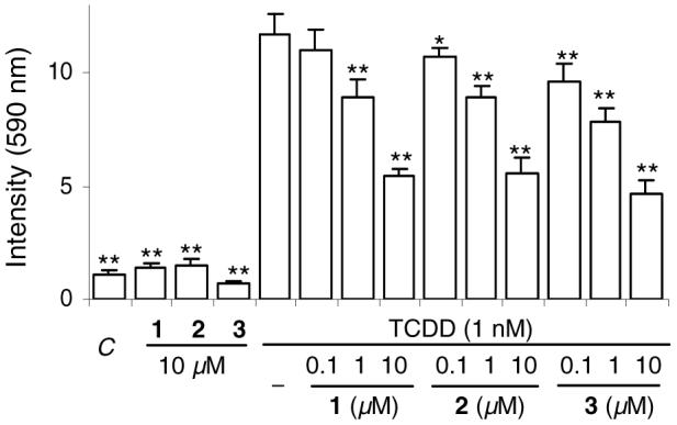
Inhibition of TCDD-induced P450 1A/B activity with cis-terpenones. HepG2 cells were co-treated with TCDD (1 nM) and cis-terpenones 1-3 and incubated for 24 h. The activity of P450 1A/B was determined with the EROD assay. Bars represent the mean ± SD of EROD activity at 590 nm in 4 replicates. The data are representative of three independent experiments. C: DMSO only. * P<0.05, ** P<0.001 compared to TCDD only with one way ANOVA and Dunnett’s test.
In conclusion, we have demonstrated that cis-terpenones 1-3 have effective chemoprevention against both AFB1 and carcinogenic stimuli TCDD in liver cells, and thus are promising candidates for novel mechanism-based preventive agents. Future research efforts are focusing on the detailed mechanism by which cis-terpenones effectively prevent the cytotoxicity induced by AFB1 and potentially impact other metabolic P450 enzymes.
Supplementary Material
Acknowledgement
We thank John B. Mangrum for his help on the mass analysis. This study was supported partially by NSF grant MCB-0131419.
Footnotes
- AFB1
- aflatoxin B1
- ANOVA
- analysis of variance
- EROD
- ethoxyresorufin-O-deethylase
- ESI-MS
- electrospray ionization mass spectroscopy
- FITC
- fluorescein isothiocyanate
- MTT
- methylthiazolyldiphenyl-tetrazolium bromide
- ROESY
- rotating-frame Overhauser spectroscopy
- PI
- propidium iodine
- TCDD
- 2,3,7,8-tetrachlorodibenzo-p-dioxin
References
- (1).Wilson DM, Payne GA. Factors affecting Aspergillus flavus group infection and aflatoxin contamination of crops. In: Eaton DL, Groopman JD, editors. Toxicology of Aflatoxin: Human Health, Veterinary, and Agricultural Significance. 1994. pp. 309–326. [Google Scholar]
- (2).Guengerich FP. Cytochrome P450 oxidations in the generation of reactive electrophiles: epoxidation and related reactions. Arch. Biochem. Biophys. 2003;409:59–71. doi: 10.1016/s0003-9861(02)00415-0. [DOI] [PubMed] [Google Scholar]
- (3).Guengerich FP, Johnson WW, Shimada T, Ueng YF, Yamazaki H, Langouët S. Activation and detoxication of aflatoxin B1. Mut. Res. 1998;402:121–128. doi: 10.1016/s0027-5107(97)00289-3. [DOI] [PubMed] [Google Scholar]
- (4).Thomas MB, Zhu AX. Hepatocellular Carcinoma: The need for progress. J. Clin. Oncology. 2005;23:2892–2899. doi: 10.1200/JCO.2005.03.196. [DOI] [PubMed] [Google Scholar]
- (5).Gong YY, Egal S, Hounsa A, Turner PC, Hall AJ, Cardwell KF, Wild CP. Determinants of aflatoxin exposure in young children from Benin and Togo, West Africa: the critical role of weaning. Int. J. Epidemiol. 2003;32:556–562. doi: 10.1093/ije/dyg109. [DOI] [PubMed] [Google Scholar]
- (6).Ming L, Thorgeirsson SS, Gail MH, Lu P, Harris CC, Wang N, Shao Y, Wu Z, Liu G, Wang X, Sun Z. Dominant role of hepatitis B virus and cofactor role of aflatoxin in hepatocarcinogenesis in Qidong, China. Hepatology. 2002;36:1214–1220. doi: 10.1053/jhep.2002.36366. [DOI] [PubMed] [Google Scholar]
- (7).Wang JS, Huang T, Su J, Liang F, Wei Z, Liang Y, Luo H, Kuang SY, Qian GS, Sun G, He X, Kensler TW, Groopman JD. Hepatocellular carcinoma and aflatoxin exposure in Zhuqing Village, Fusui County, People’s Republic of China. Cancer Epidemiol. Biomarkers Prev. 2001;10:143–146. [PubMed] [Google Scholar]
- (8).Li FQ, Yoshizawa T, Kawamura O, Luo XY, Li YW. Aflatoxins and Fumonisins in Corn from the High-Incidence Area for Human Hepatocellular Carcinoma in Guangxi, China. J. Agric. Food Chem. 2001;49:4122–4126. doi: 10.1021/jf010143k. [DOI] [PubMed] [Google Scholar]
- (9).Zhang BC, Zhu YR, Wang JB, Wu Y, Zhang QN, Qian GS, Kuang SY, Li YF, Fang X, Yu LY, De Flora S, Jacobson LP, Zarba A, Egner PA, He X, Wang JS, Chen B, Enger CL, Davidson NE, Gordon GB, Gorman MB, Prochaska HJ, Groopman JD, Munoz A, Kensler TW. Oltipraz chemoprevention trial in Qidong, Jiangsu Province, People’s Republic of China. J. Cell Biochem. Suppl. 1997;28-29:166–173. [PubMed] [Google Scholar]
- (10).Jacobson LP, Zhang BC, Zhu YR, Wang JB, Wu Y, Zhang QN, Yu LY, Qian GS, Kuang SY, Li YF, Fang X, Zarba A, Chen B, Enger CL, Davidson NE, Gorman MB, Gordon GB, Prochaska HJ, Egner PA, Groopman JD, Munoz A, Helzlsouer KJ, Kensler TW. Oltipraz chemoprevention trial in Qidong, People’s Republic of China: study design and clinical outcomes. Cancer Epidemiol. Biomarkers Prev. 1997;6:257–265. [PubMed] [Google Scholar]
- (11).Egner PA, Wang JB, Zhu YR, Zhang BC, Wu Y, Zhang QN, Qian GS, Kuang SY, Gange SJ, Jacobson LP, Helzlsouer KJ, Bailey GS, Groopman JD, Kensler TW. Chlorophyllin intervention reduces aflatoxin-DNA adducts in individuals at high risk for liver cancer. Proc. Natl. Acad. Sci. USA. 2001;98:14601–14606. doi: 10.1073/pnas.251536898. [DOI] [PMC free article] [PubMed] [Google Scholar]
- (12).Egner PA, Stansbury KH, Snyder EP, Rogers ME, Hintz PA, Kensler TW. Identification and characterization of chlorin e(4) ethyl ester in sera of individuals participating in the chlorophyllin chemoprevention trial. Chem. Res. Toxicol. 2000;13:900–906. doi: 10.1021/tx000069k. [DOI] [PubMed] [Google Scholar]
- (13).Abdel-Wahhab MA, Ahmed HH, Hagazi MM. Prevention of aflatoxin B1-initiated hepatotoxicity in rat by marine algae extracts. J. Appl. Toxicol. 2006;26:229–238. doi: 10.1002/jat.1127. [DOI] [PubMed] [Google Scholar]
- (14).Lee JK, Choi EH, Lee KG, Chun HS. Alleviation of aflatoxin B1-induced oxidative stress in HepG2 cells by volatile extract from Allii Fistulosi Bulbus. Life Sci. 2005;77:2896–2910. doi: 10.1016/j.lfs.2005.03.028. [DOI] [PubMed] [Google Scholar]
- (15).Nwankwo JO, Tahnteng JG, Emerole GO. Inhibition of aflatoxin B1 genotoxicity in human liver-derived HepG2 cells by kolaviron biflavonoids and molecular mechanisms of action. Eur. J. Cancer Prev. 2000;9:351–361. doi: 10.1097/00008469-200010000-00010. [DOI] [PubMed] [Google Scholar]
- (16).Zhou Q, Zuniga MA. Quinone Methides Formations in the Cu2+-induced Oxidation of a Diterpenone Catechol and Concurrent Damage on DNA. Chem. Res. Toxicol. 2005;18:382–388. doi: 10.1021/tx049703a. [DOI] [PubMed] [Google Scholar]
- (17).Zuniga MA, Dai J, Wehunt MP, Zhou Q. DNA Oxidative Damage by Terpene Catechols as Analogs of Natural Terpene Quinone Methide Precursors in the Presence of Cu (II) and/or NADH. Chem. Res. Toxicol. 2006;19:828–836. doi: 10.1021/tx060021s. [DOI] [PubMed] [Google Scholar]
- (18).Bolton JL, Pisha E, Zhang F, Qiu S. Role of quinoids in estrogen carcinogenesis. Chem. Res. Toxicol. 1998;10:1113–1127. doi: 10.1021/tx9801007. [DOI] [PubMed] [Google Scholar]
- (19).Hayes CL, Spink DC, Spink BC, Cao JQ, Walker NJ, Sutter TR. 17-β-Estradiol hydroxylation catalyzed by human cytochrome P450 1B1. Proc. Natl. Acad. Sci. USA. 1996;93:9776–9781. doi: 10.1073/pnas.93.18.9776. [DOI] [PMC free article] [PubMed] [Google Scholar]
- (20).Guengerich FP, Chun YJ, Kim D, Gillman EMJ, Shimada T. Cytochrome P450 1B1: A target for in inhibition in anticarcinogenesis strategies. Mut. Res. 2003;523-524:173–182. doi: 10.1016/s0027-5107(02)00333-0. [DOI] [PubMed] [Google Scholar]
- (21).Chun YJ, Kim S. Discovery of cytochrome P450 inhibitors as new promising anti-cancer agents. Med. Res. Rev. 2003;23:657–668. doi: 10.1002/med.10050. [DOI] [PubMed] [Google Scholar]
- (22).Kamdem LK, Meineke I, Gödtel-Armbrust U, Brockmöller J, Wojnowski L. Dominant Contribution of P450 3A4 to the Hepatic Carcinogenic Activation of Aflatoxin B1. Chem. Res. Toxicol. 2006;19:577–586. doi: 10.1021/tx050358e. [DOI] [PubMed] [Google Scholar]
- (23).Iwanari M, Nakajima M, Kizu R, Hayakawa K, Yokoi T. Induction of CYP1A1, CYP1A2, and CYP1B1 mRNAs by nitropolycyclic aromatic hydrocarbons in various human tissue-derived cells: chemical-, cytochrome P450 isoform-, and cell-specific differences. Arch. Toxicol. 2002;76:287–298. doi: 10.1007/s00204-002-0340-z. [DOI] [PubMed] [Google Scholar]
- (24).Lai KP, Wong MH, Wong CKC. Modulation of Ahr-mediated CYP1A1 mRNA and EROD activities by 17β-estradiol and dexamethasone in TCDD-induced H411E cells. Toxicol. Sci. 2004;78:41–49. doi: 10.1093/toxsci/kfh045. [DOI] [PubMed] [Google Scholar]
- (25).Renzulli C, Galvano L, Pierdomenico L, Speroni E, Guerra MC. Effects of rosmarinic acid against aflatoxin B1 and ochratoxin-A induced cell damage in a human hepatoma cell line (HepG2) J. Appl. Toxicol. 2004;24:289–296. doi: 10.1002/jat.982. [DOI] [PubMed] [Google Scholar]
- (26).Xie H, Rath NC, Huff GR, Huff WE, Balog JM. Effects of Salmonella typhimurium lipopolysaccharide on broiler chickens. Poult. Sci. 2000;79:33–40. doi: 10.1093/ps/79.1.33. [DOI] [PubMed] [Google Scholar]
- (27).van Engeland M, Ramaekers FC, Schutte B, Reutelingsperger CP. A novel assay to measure loss of plasma membrane asymmetry during apoptosis of adherent cells in culture. Cytometr. 1996;24:131–139. doi: 10.1002/(SICI)1097-0320(19960601)24:2<131::AID-CYTO5>3.0.CO;2-M. [DOI] [PubMed] [Google Scholar]
- (28).Koopman G, Reutelingsperger CPM, Kuijten GAM, Keehnen RMJ, Pals ST, van Oers MHJ. Annexin V for flow cytometric detection of phosphatidylserine expression on B cells undergoing apoptosis. Blood. 1994;84:1415–1420. [PubMed] [Google Scholar]
Associated Data
This section collects any data citations, data availability statements, or supplementary materials included in this article.


