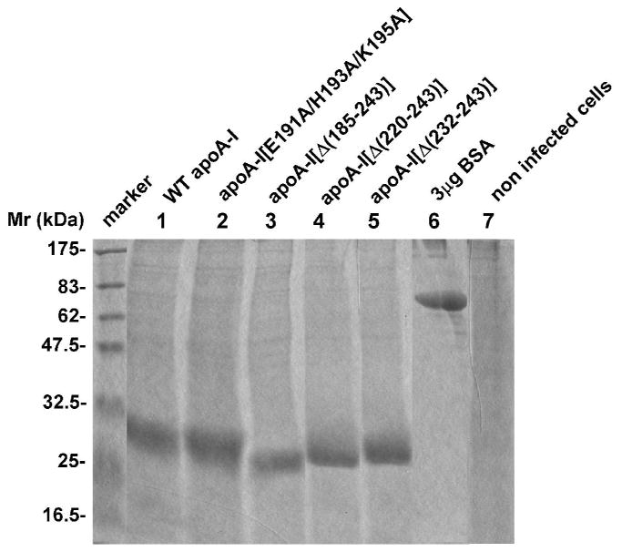
FIGURE 2. Expression of WT and mutant apoA-I forms in cultures of HTB-13 cells following infection with the corresponding recombinant adenoviruses.

SDS-PAGE analysis of medium obtained from HTB-13 cells in 100 mm dishes infected with adenoviruses expressing the WT and mutant apoA-I forms as described in the Experimental Procedures. An aliquot of 30 μL of serum-free culture medium was analyzed. “Marker” indicates protein markers of different molecular mass, as shown in the figure. Lane 6 contains 3 μg BSA. It was estimated that the infected cultures (5×10⁶ cells) secreted approximately 60-100 μg/ml WT and mutant apoA-I forms over 24 hours of incubation.
