Abstract
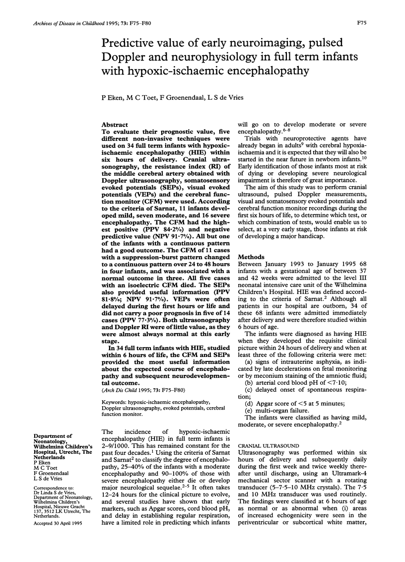
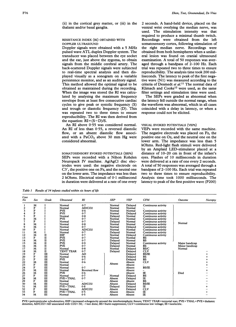
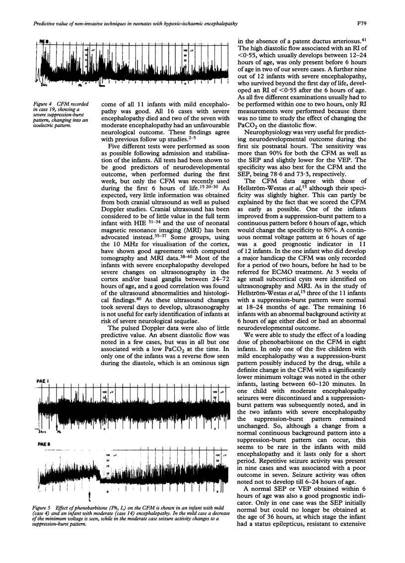
To evaluate their prognostic value, five different non-invasive techniques were used on 34 full term infants with hypoxic-ischaemic encephalopathy (HIE) within six hours of delivery. Cranial ultrasonography, the resistance index (RI) of the middle cerebral artery obtained with Doppler ultrasonography, somatosensory evoked potentials (SEPs), visual evoked potentials (VEPs) and the cerebral function monitor (CFM) were used. According to the criteria of Sarnat, 11 infants developed mild, seven moderate, and 16 severe encephalopathy. The CFM had the highest positive (PPV 84.2%) and negative predictive value (NPV 91.7%). All but one of the infants with a continuous pattern had a good outcome. The CFM of 11 cases with a suppression-burst pattern changed to a continuous pattern over 24 to 48 hours in four infants, and was associated with a normal outcome in three. All five cases with an isoelectric CFM died. The SEPs also provided useful information (PPV 81.8%; NPV 91.7%). VEPs were often delayed during the first hours or life and did not carry a poor prognosis in five of 14 cases (PPV 77.3%). Both ultrasonography and Doppler RI were of little value, as they were almost always normal at this early stage. In 34 full term infants with HIE, studied within 6 hours of life, the CFM and SEPs provided the most useful information about the expected course of encephalopathy and subsequent neurodevelopmental outcome.
Full text
PDF


Images in this article
Selected References
These references are in PubMed. This may not be the complete list of references from this article.
- Archer L. N., Levene M. I., Evans D. H. Cerebral artery Doppler ultrasonography for prediction of outcome after perinatal asphyxia. Lancet. 1986 Nov 15;2(8516):1116–1118. doi: 10.1016/s0140-6736(86)90528-3. [DOI] [PubMed] [Google Scholar]
- Babcock D. S., Ball W., Jr Postasphyxial encephalopathy in full-term infants: ultrasound diagnosis. Radiology. 1983 Aug;148(2):417–423. doi: 10.1148/radiology.148.2.6867334. [DOI] [PubMed] [Google Scholar]
- Baenziger O., Martin E., Steinlin M., Good M., Largo R., Burger R., Fanconi S., Duc G., Buchli R., Rumpel H. Early pattern recognition in severe perinatal asphyxia: a prospective MRI study. Neuroradiology. 1993;35(6):437–442. doi: 10.1007/BF00602824. [DOI] [PubMed] [Google Scholar]
- Bjerre I., Hellström-Westas L., Rosén I., Svenningsen N. Monitoring of cerebral function after severe asphyxia in infancy. Arch Dis Child. 1983 Dec;58(12):997–1002. doi: 10.1136/adc.58.12.997. [DOI] [PMC free article] [PubMed] [Google Scholar]
- Bode H., Sauer M., Pringsheim W. Diagnosis of brain death by transcranial Doppler sonography. Arch Dis Child. 1988 Dec;63(12):1474–1478. doi: 10.1136/adc.63.12.1474. [DOI] [PMC free article] [PubMed] [Google Scholar]
- Couture A., Veyrac C., Baud C., Leboucq N., Montoya F. New imaging of cerebral ischaemic lesions. High frequency probes and pulsed Doppler. Ann Radiol (Paris) 1987;30(7):452–461. [PubMed] [Google Scholar]
- Desmedt J. E., Brunko E., Debecker J. Maturation of the somatosensory evoked potentials in normal infants and children, with special reference to the early N1 component. Electroencephalogr Clin Neurophysiol. 1976 Jan;40(1):43–58. doi: 10.1016/0013-4694(76)90178-4. [DOI] [PubMed] [Google Scholar]
- Dijxhoorn M. J., Visser G. H., Huisjes H. J., Fidler V., Touwen B. C. The relation between umbilical pH values and neonatal neurological morbidity in full term appropriate-for-dates infants. Early Hum Dev. 1985 May;11(1):33–42. doi: 10.1016/0378-3782(85)90117-3. [DOI] [PubMed] [Google Scholar]
- Eken P., Jansen G. H., Groenendaal F., Rademaker K. J., de Vries L. S. Intracranial lesions in the fullterm infant with hypoxic ischaemic encephalopathy: ultrasound and autopsy correlation. Neuropediatrics. 1994 Dec;25(6):301–307. doi: 10.1055/s-2008-1073044. [DOI] [PubMed] [Google Scholar]
- Ergander U., Eriksson M., Zetterström R. Severe neonatal asphyxia. Incidence and prediction of outcome in the Stockholm area. Acta Paediatr Scand. 1983 May;72(3):321–325. doi: 10.1111/j.1651-2227.1983.tb09722.x. [DOI] [PubMed] [Google Scholar]
- Finer N. N., Robertson C. M., Richards R. T., Pinnell L. E., Peters K. L. Hypoxic-ischemic encephalopathy in term neonates: perinatal factors and outcome. J Pediatr. 1981 Jan;98(1):112–117. doi: 10.1016/s0022-3476(81)80555-0. [DOI] [PubMed] [Google Scholar]
- Gibson N. A., Graham M., Levene M. I. Somatosensory evoked potentials and outcome in perinatal asphyxia. Arch Dis Child. 1992 Apr;67(4 Spec No):393–398. doi: 10.1136/adc.67.4_spec_no.393. [DOI] [PMC free article] [PubMed] [Google Scholar]
- Gray P. H., Tudehope D. I., Masel J. P., Burns Y. R., Mohay H. A., O'Callaghan M. J., Williams G. M. Perinatal hypoxic-ischaemic brain injury: prediction of outcome. Dev Med Child Neurol. 1993 Nov;35(11):965–973. doi: 10.1111/j.1469-8749.1993.tb11578.x. [DOI] [PubMed] [Google Scholar]
- Hagberg B., Hagberg G., Olow I. The changing panorama of cerebral palsy in Sweden 1954-1970. I. Analysis of the general changes. Acta Paediatr Scand. 1975 Mar;64(2):187–192. doi: 10.1111/j.1651-2227.1975.tb03820.x. [DOI] [PubMed] [Google Scholar]
- Hagberg B., Hagberg G., Olow I. The changing panorama of cerebral palsy in Sweden. VI. Prevalence and origin during the birth year period 1983-1986. Acta Paediatr. 1993 Apr;82(4):387–393. doi: 10.1111/j.1651-2227.1993.tb12704.x. [DOI] [PubMed] [Google Scholar]
- Klimach V. J., Cooke R. W. Maturation of the neonatal somatosensory evoked response in preterm infants. Dev Med Child Neurol. 1988 Apr;30(2):208–214. doi: 10.1111/j.1469-8749.1988.tb04752.x. [DOI] [PubMed] [Google Scholar]
- Kuenzle C., Baenziger O., Martin E., Thun-Hohenstein L., Steinlin M., Good M., Fanconi S., Boltshauser E., Largo R. H. Prognostic value of early MR imaging in term infants with severe perinatal asphyxia. Neuropediatrics. 1994 Aug;25(4):191–200. doi: 10.1055/s-2008-1073021. [DOI] [PubMed] [Google Scholar]
- Levene M. I., Fenton A. C., Evans D. H., Archer L. N., Shortland D. B., Gibson N. A. Severe birth asphyxia and abnormal cerebral blood-flow velocity. Dev Med Child Neurol. 1989 Aug;31(4):427–434. doi: 10.1111/j.1469-8749.1989.tb04020.x. [DOI] [PubMed] [Google Scholar]
- Levene M. I., Gibson N. A., Fenton A. C., Papathoma E., Barnett D. The use of a calcium-channel blocker, nicardipine, for severely asphyxiated newborn infants. Dev Med Child Neurol. 1990 Jul;32(7):567–574. doi: 10.1111/j.1469-8749.1990.tb08540.x. [DOI] [PubMed] [Google Scholar]
- Levene M. L., Kornberg J., Williams T. H. The incidence and severity of post-asphyxial encephalopathy in full-term infants. Early Hum Dev. 1985 May;11(1):21–26. doi: 10.1016/0378-3782(85)90115-x. [DOI] [PubMed] [Google Scholar]
- Martin D. J., Hill A., Fitz C. R., Daneman A., Havill D. A., Becker L. E. Hypoxic/ischaemic cerebral injury in the neonatal brain. A report of sonographic features with computed tomographic correlation. Pediatr Radiol. 1983;13(6):307–312. doi: 10.1007/BF01625955. [DOI] [PubMed] [Google Scholar]
- Martin E., Barkovich A. J. Magnetic resonance imaging in perinatal asphyxia. Arch Dis Child Fetal Neonatal Ed. 1995 Jan;72(1):F62–F70. doi: 10.1136/fn.72.1.f62. [DOI] [PMC free article] [PubMed] [Google Scholar]
- McCulloch D. L., Taylor M. J., Whyte H. E. Visual evoked potentials and visual prognosis following perinatal asphyxia. Arch Ophthalmol. 1991 Feb;109(2):229–233. doi: 10.1001/archopht.1991.01080020075047. [DOI] [PubMed] [Google Scholar]
- Muttitt S. C., Taylor M. J., Kobayashi J. S., MacMillan L., Whyte H. E. Serial visual evoked potentials and outcome in term birth asphyxia. Pediatr Neurol. 1991 Mar-Apr;7(2):86–90. doi: 10.1016/0887-8994(91)90002-3. [DOI] [PubMed] [Google Scholar]
- Pryds O., Greisen G., Lou H., Friis-Hansen B. Vasoparalysis associated with brain damage in asphyxiated term infants. J Pediatr. 1990 Jul;117(1 Pt 1):119–125. doi: 10.1016/s0022-3476(05)72459-8. [DOI] [PubMed] [Google Scholar]
- Robertson C., Finer N. Term infants with hypoxic-ischemic encephalopathy: outcome at 3.5 years. Dev Med Child Neurol. 1985 Aug;27(4):473–484. doi: 10.1111/j.1469-8749.1985.tb04571.x. [DOI] [PubMed] [Google Scholar]
- Rutherford M. A., Pennock J. M., Dubowitz L. M. Cranial ultrasound and magnetic resonance imaging in hypoxic-ischaemic encephalopathy: a comparison with outcome. Dev Med Child Neurol. 1994 Sep;36(9):813–825. doi: 10.1111/j.1469-8749.1994.tb08191.x. [DOI] [PubMed] [Google Scholar]
- Sarnat H. B., Sarnat M. S. Neonatal encephalopathy following fetal distress. A clinical and electroencephalographic study. Arch Neurol. 1976 Oct;33(10):696–705. doi: 10.1001/archneur.1976.00500100030012. [DOI] [PubMed] [Google Scholar]
- Siegel M. J., Shackelford G. D., Perlman J. M., Fulling K. H. Hypoxic-ischemic encephalopathy in term infants: diagnosis and prognosis evaluated by ultrasound. Radiology. 1984 Aug;152(2):395–399. doi: 10.1148/radiology.152.2.6739805. [DOI] [PubMed] [Google Scholar]
- Sykes G. S., Molloy P. M., Johnson P., Gu W., Ashworth F., Stirrat G. M., Turnbull A. C. Do Apgar scores indicate asphyxia? Lancet. 1982 Feb 27;1(8270):494–496. doi: 10.1016/s0140-6736(82)91462-3. [DOI] [PubMed] [Google Scholar]
- Taylor M. J., Murphy W. J., Whyte H. E. Prognostic reliability of somatosensory and visual evoked potentials of asphyxiated term infants. Dev Med Child Neurol. 1992 Jun;34(6):507–515. doi: 10.1111/j.1469-8749.1992.tb11471.x. [DOI] [PubMed] [Google Scholar]
- Thornberg E., Ekström-Jodal B. Cerebral function monitoring: a method of predicting outcome in term neonates after severe perinatal asphyxia. Acta Paediatr. 1994 Jun;83(6):596–601. doi: 10.1111/j.1651-2227.1994.tb13088.x. [DOI] [PubMed] [Google Scholar]
- Whyte H. E., Taylor M. J., Menzies R., Chin K. C., MacMillan L. J. Prognostic utility of visual evoked potentials in term asphyxiated neonates. Pediatr Neurol. 1986 Jul-Aug;2(4):220–223. doi: 10.1016/0887-8994(86)90051-2. [DOI] [PubMed] [Google Scholar]
- Willis J., Duncan C., Bell R. Short-latency somatosensory evoked potentials in perinatal asphyxia. Pediatr Neurol. 1987 Jul-Aug;3(4):203–207. doi: 10.1016/0887-8994(87)90017-8. [DOI] [PubMed] [Google Scholar]
- de Vries L. S. Somatosensory-evoked potentials in term neonates with postasphyxial encephalopathy. Clin Perinatol. 1993 Jun;20(2):463–482. [PubMed] [Google Scholar]


