
Figure 4.
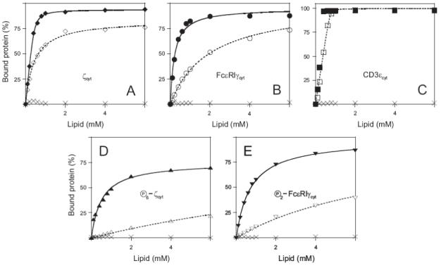
The membrane binding of the MIRR cytoplasmic domains. (A-E) Partitioning of ζcyt (A), FcεRIγcyt (B), CD3εcyt (C) as well as fully phosphorylated ζcyt (D) and FcεRIγcyt (E) onto large (100 nm diameter) unilamellar phospholipid vesicles (LUV). The results were obtained with a sucrose-loaded vesicle assay using LUV formed from POPG (A-E, filled symbols, solid lines) and 1:1 POPG/POPC (A-E, empty symbols, dotted lines), and POPC (A-E, crossings) in 100 mM KCl buffered to pH 7.0 with 1 mM MOPS. Each plotted point is the average of at least two samples at that total lipid concentration; the errors associated with the points are <10%. The solid and dotted lines correspond to theoretical binding curves that were obtained as described under Materials and Methods. Cytoplasmic domains of CD3γ (net charge of 0), CD3δ (net charge of 0), Igα (net charge of -9) and Igβ (net charge of -10) did not bind to phospholipid vesicles of any of the compositions tested (data not shown).