Abstract
Subungual metastasis as a presenting feature of lung adenocarcinoma is rare and may be confused with a benign inflammatory disorder of the nail unit with subsequent diagnostic and management delay. We discuss the case of a 46-year-old man with a subungual metastasis as a presenting feature of lung adenocarcinoma.
Keywords: Metastasis, Primary digital adenocarcinoma, Primary lung adenocarcinoma, Paronychia
Introduction
Visceral malignancy that manifests primarily as a metastatic deposit in the nailfold is extremely rare. There are very few documented cases in the literature. We report on a patient who presented with a chronic paronychial infection, which proved to be an undiagnosed lung adenocarcinoma.
Case Report
A 46-year-old refuse collector was referred with a 3-month history of an overgranulating wound on the ulnar aspect of his ring finger. This had been treated over the last 3 months, before referral to our unit, as a paronychial infection and managed with oral antibiotics. He had originally noted a swelling on the ulnar aspect of the dorsum of his finger, at the paronychial fold. This had progressively increased in size and intermittently discharged pus. He had no previous history of trauma or hospital admissions and was well apart from smoking 400-pack years.
An area of overgranulation at the paronychial fold was noted on examination (Fig. 1). This area was excised with a narrow margin (Fig. 2), and specimens were sent for microbiological and histological examination.
Figure 1.
Palmar view of lesion.
Figure 2.
Dorsal view of lesion post incision biopsy.
Histopathology reported an invasive adenocarcinoma; most probably a secondary deposit. Immunochemistry stains were TTF-1-positive (thyroid transcription factor-1), suggesting a primary in the lung or thyroid [7]. Amputation of the ring finger was performed at the level of the PIPJ. The tumor was found to infiltrate the distal phalanx.
Subsequent examination revealed three mobile firm lymph nodes in the right axilla and bilateral multiple small groin nodes. There were no signs of anemia or clubbing. Respiratory and abdominal examinations were unremarkable.
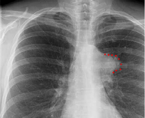
The chest radiograph revealed a diffuse opacity in the left upper lobe of the lung (Fig. 3). Computed tomography scan confirmed a large left hilar mass and pleural effusion. The patient was referred to the oncology service for management of his primary and metastatic lung adenocarcinoma. He died 4 months later.
Figure 3.
Chest radiograph showing lesion. Red dotted line indicates lesion.
Discussion
The incidence of cutaneous metastasis from all types of lung cancer is reported to be 1.5–12%. Cutaneous metastasis of a lung cancer generally reflects the progression or recurrence of cancer. The average survival after diagnosis of skin metastases from lung cancer ranges from 3–5 months. An isolated cutaneous metastasis is rarely the presenting feature of an occult lung tumor.
Although subungual metastasis from a malignant lung tumor is rare [9], in 44% of these patients, this lesion has been reported as the presenting feature of an otherwise occult tumor [3]. The diagnosis of subungual metastasis is rarely entertained and typically misdiagnosed as a benign lesion. Confirmation of malignancy is obtained by tissue biopsy. Radiography may demonstrate adjacent bone erosion [10].
Clinical features of subungual metastasis of a malignant lung tumor are variable. An erythematous tender swelling of the distal digit involving the nail folds and subungual area has often been reported. Subungual metastatic lesions can mimic other skin lesions including pyogenic granulomas, acute paronychial infections, erysipelas, and herpes zoster [6, 8].
The surgical management of metastases is complete surgical excision, often resulting in amputation. Complex reconstruction is not indicated.
Visceral malignancies rarely present with skin cancer. Metastatic malignancy or atypical organisms should be suspected in wounds that persist or worsen in spite of ordinary antibacterial medications and local wound. Biopsy and routine histopathology will identify primary or metastatic tumors. Special stains and/or culture techniques usually identify atypical organisms. Although there are documented cases of metastatic lung lesions of the nail bed [1, 4], there are no documented cases in the literature of lung adenocarcinoma presenting in this way. Keramidas and Brotherston [5] reported a case of extensive metastasis to the hand from an undiagnosed adenocarcinoma, and Chang et al. [2] and Vanhooteghem et al. [11] reported on subungual metastases secondary to squamous cell carcinoma of the lung. When the diagnosis of a metastasis has been confirmed, a complete evaluation should be performed to identify the origin of the primary tumor, and the patient should be referred for oncological management.
Summary
This case highlights the importance of fully investigating (chronic infections) of the hand that are not being successfully treated with antibiotics. Metastatic tumor may present as a subungual lesion in asymptomatic patients or those already diagnosed with cancer. However, the diagnosis of a subungual metastasis is often not initially considered, as the symptoms and appearance of the subungual tumor may mimic other conditions.
References
- 1.Bueno RA, Neumeister MW, Wilhemi BJ. Aggressive digital papillary adenocarcinoma presenting as a finger infection. Ann Plast Surg 2002;49(3):326–7 (Sep). [DOI] [PubMed]
- 2.Chang SE, Choi JH, Sung KJ, Moon KC, Koh JK. Metastatic squamous cell carcinoma of the nail bed: a presenting sign of lung cancer. Br J Dermatol 1999;141:939–40. [DOI] [PubMed]
- 3.Cohen PR. Metastatic tumors to the nail unit: subungal metastases. Dermatol Surg 2001;27(3):280–93. [DOI] [PubMed]
- 4.Gorva AD, Mohil R, Srinivasan MS. Aggressive digital papillary adenocarcinoma presenting as a paronychial of the finger. J Hand Surg (Br) 2005;30(5):534 (Oct). [DOI] [PubMed]
- 5.Keramidas E, Brotherston M. Extensive metastasis to the hand from undiagnosed adenocarcinoma of the lung. Scand J Plast Reconstr Surg Hand Surg 2005;39:113–5. [DOI] [PubMed]
- 6.Keyser JJ, Littler JW, Eaton RG. Surgical treatment of infections and lesions. Hand Clin 1990;6:113–34. [PubMed]
- 7.Miller RT. Focus on Immunohistochemistry. Thyroid transcription factor-1. http://www.ihcworld.com/_newsletter/2001/focus_apr_2001.pdf.
- 8.Moran GJ, Talan, DA. Hand infections. Emerg Med Clin North Am 1993;11:603. [PubMed]
- 9.Rye JS, Cho JW, Moon TH, Lee HL, Han HS, Choi GS. Squamous cell lung cancer with solitary subungal metastasis. Yonsei Med J 2000;41(5):666–8. [DOI] [PubMed]
- 10.Sommer N, Neumeister MW. Tumors of the perionychium. Hand Clin 2002;18(4):673–89, vii; discussion 691 (Nov). [DOI] [PubMed]
- 11.Vanhooteghem O, Dumont M, Andre J, Leempoel M, de la Brassinne M. Bilateral subungual metastasis from squamous cell carcinoma of the lung: a diagnostic trap! Rev Med Liege 1999;54(8):653–4 (Aug). [PubMed]


