Abstract
Dorsally angulated malunions of the distal radius have historically been corrected with an opening wedge osteotomy fixed with a dorsal plate. Volar locking plates may facilitate a less morbid approach to corrective osteotomies of the wrist. Eight consecutive patients with an average age of 40 years (range, 15–52 years) underwent correction of a distal radius deformity through a volar approach. Clinical follow-up averaged 17.4 months (range, 7–41 months). Preoperative radiographs revealed an average of 24° of dorsal tilt in patients with dorsal deformity. Postoperatively, their average measurement was <3° of volar tilt. Patients were initially ulnar-positive with an average of 4 mm ulnar-positive variance (range, 2–7 mm). This corrected to less than 1 mm postoperatively. Postoperative disabilities of the arm, shoulder, and hand (DASH), SF-12, and Mayo Wrist scores averaged 10.8, 40.5, and 82.5, respectively. There were no nonunions, and no plates required removal. Distal radius deformity can be effectively addressed through a volar approach with the use of a locking plate.
Keywords: Wrist, Hand, Osteotomy, Bone trauma, Fracture
Introduction
Malunion remains one of the most common complications after extra-articular distal radius fractures. Although the severity of the deformity does not often warrant surgical intervention [22, 51], patients who have lost normal volar tilt, radial inclination, and radial length relative to the ulna will complain of pain, limited motion, decreased grip strength, and cosmetic deformity [19, 44, 45]. Once angulation of the distal articular surface of the radius becomes greater than 25 to 30° in the sagittal plane, Fernandez recommended corrective osteotomy [8, 7]. However, no strict guidelines for surgical intervention exist, and patients with even 20°of true deformity may be symptomatic and benefit from corrective osteotomy [44]. In Bacorn and Kurtzke’s review of 2,132 fractures, increasing dysfunction correlated with increasing deformity [2], and other investigators have noted decreased grip strength when dorsal angulation was greater than 20° [29].
Several biomechanical studies have demonstrated abnormal wrist contact pressures with extra-articular deformity that may pre-dispose to arthritis [23, 28, 30]. Loss of radial height, particularly with dorsal angulation, results in increased contact pressures across the ulna. Whereas load transmitted across the wrist is dependent on both the direction of the force and wrist position [21], in the normal wrist, approximately 82% of the axial load transmitted across the wrist is born by the distal radius [54, 55]. Increasing ulnar positivity by 2.5 mm increases the load across the ulna to 42% of the total, and dorsal tilt rather than the normal volar tilt of the distal radius leads to further increases in load across the ulna [54, 55]. The increased load across the ulnar side of the wrist may lead to arthrosis or problems with the triangular fibrocartilage complex [35].
Malunion of the distal radius is particularly problematic at the distal radioulnar joint [1, 17, 16]. As radial height is lost, radio-ulnar length mismatch may result in distal radioulnar joint incongruity, causing instability, decreased motion, and arthrosis [1, 50]. Distal radius malunions frequently have an unappreciated rotational component that may affect distal radioulnar joint (DRUJ) motion and stability. In a study that examined computed tomography (CT) scans of both affected and normal wrists, Prommesberger et al. [33] reported a rotational component to the malunion in 23 of 37 patients.
Distal radius malunion may also lead to compensatory motion of the carpus [46]. Dorsally angulated distal radius fractures may lead to an adaptive carpal instability with a dorsal intercalary segment instability (DISI)-type deformity [27]. Patients with excessively volar malunited distal radius fractures or Madelung’s type deformity may also exhibit adaptive carpal instability [38].
Because of the long-term problems associated with a distal radius malunion, multiple techniques for corrective osteotomy have been developed [8–9, 31, 47, 49, 53]. For severely malunited distal radius fractures, these techniques have been shown to improve radiographic parameters and, more importantly, to improve motion, pain, and grip strength [19, 39]. Fernandez described the traditional treatment of osteotomy and dorsal plating with bone graft for dorsally angulated malunions [8]. This technique effectively restores anatomy, improves function, and relieves pain, but frequent morbidity has been associated with the use of dorsal plates [4, 14, 34]. The most common problems are hardware prominence and irritation or rupture of extensor tendons.
Volar-fixed angled plates have added a new dimension to the treatment of distal radius fractures [25, 26]. The low morbidity of the surgical approach and strength of final construct allow early mobilization and return to function [25, 26]. While volar locking plates are not indicated for all fractures, their advent has made the use of external fixation or dorsal plating far less common. Because of the success of using volar locked plating for acute distal radius fractures, the senior author began using this technique for corrective osteotomy of the distal radius.
The purpose of this study is to report the outcomes of a cohort of patients who underwent corrective osteotomy of the distal radius using a volar locking plate. Outcome measures for this study were the results of physical and radiographic examination, Short Form (SF)-12, and the Upper Limb Disabilities of the Arm, Shoulder, and Hand (DASH), and the Mayo Wrist score.
Materials and Methods
Study Subjects
The potential study group for this analysis included 14 patients who were identified by medical record search by Current Procedural Terminology (CPT) code during the study period of 2002–2005. Inclusion criteria consisted of a patient with a symptomatic deformity of the distal radius who underwent distal radius corrective osteotomy at this institution. Patients with pre-existing intracarpal arthrosis and without at least 6 months of follow-up were excluded. Examination of the medical records revealed that 8 of the 14 patients met the inclusion criteria. After Institutional Review Board approval, patients were contacted and invited to participate in this study. This search yielded six of eight patients who were available for current clinical review. The information from the medical record on the remaining two patients was combined with the data from the patients who were available for review.
Surgical Technique
A volar approach to the distal radius was performed [25]. A 5-cm longitudinal incision was made over the flexor carpi radialis tendon proximal to the wrist flexion crease. The radial artery was identified, and its superficial branch at the distal portion of the incision was preserved. The pronator quadratus was released along its radial and distal borders, and the brachioradialis and radial septum were released. After exposure, the distal portion of the small five-hole plate from DePuy (Warsaw, IN) was provisionally held to the distal radius with K-wires. Fluoroscopy images were taken to assure appropriate position of the plate and for planning of the osteotomy. The position of the plate and osteotomy were marked but the distal holes were not predrilled. The plate was then removed and an osteotomy was performed. It was attempted to place the osteotomy at the site of deformity and roughly perpendicular to the shaft of the radius. After the osteotomy was completed, the dorsal periosteum was further released, and the fragments were distracted initially using a small osteotome as a lever followed by a small lamina spreader to allow creep of the soft tissues. The plate was then fixed to the distal fragment to allow appropriate correction of volar tilt and radial inclination. Once the plate was fixed to the distal fragment in appropriate position, the lamina spreader was reinserted for distraction, and the proximal portion of the plate was held to the radial shaft using a tenaculum. Fluoroscopy was then used to assess plate position, and adjustments to radial inclination and height were made. The oval hole in the proximal portion of the plate was filled with a 3.5-mm cortical screw. Next, corticocancellous bone graft from the patient’s inner table of iliac crest was harvested and placed in the gap. The remaining proximal 3.5-mm screws were then placed. The pronator quadratus was sutured back into place covering the plate, and the skin closed with a running absorbable subcuticular suture. All patients were placed into a well-padded, above-elbow splint and were discharged to home on the same day.
Outcome Measures
Each patient in the study group underwent clinical evaluation consisting of history, physical examination of both extremities, and radiographic examination with PA, lateral and ulnar variance views [36, 48, 56]. Each patient was asked to fill out a combined SF-12 and DASH outcome measurement instrument. Additionally, functional outcomes were calculated with the Mayo Wrist score [12].
The SF-12 is a commonly used measure of health-related quality of life. The SF-12 consists of 12 questions relating to physical functioning, limitations of physical health, pain, general health, emotional well-being, and social functioning. Scores range from 0 to 100 with the higher scores defining a more favorable state of health. The mean score of the general US population is 50 with a SD of 10 [12].
The DASH instrument measures the effects of upper extremity condition on general functioning through 30 items that measure patient ability to perform daily care routines. All items are scored from 1 to 5, with 1 indicative of no disability and 5 indicating severe disability. The raw score is subsequently converted to a 0-to-100 scale in which a score of 0 indicates no disability and a score of 100 indicates maximum disability. There is no normative data for the DASH instrument.
The Mayo Wrist scoring system allows for a total count of 100 points in four categories. Range of motion, grip strength, and pain were measured. Range of motion was reported as both the absolute measurement and that compared with the contra lateral side. Maximal grip strength on the operated side was measured with a Jamar™ (Chicago, IL) dynamometer and was reported as a percentage of maximal strength of the opposite side. The pain scale and satisfaction score were self-reported and graded with the use of a questionnaire.
Results
After retrospective medical record review, the study group consisted of eight patients, six of whom were available for complete evaluation (SF-12, DASH, and Mayo Wrist score), physical examination, and radiographs. Two of the eight patients could not be contacted despite extensive calls and Internet search for availability, so their medical records were reviewed to complete the analysis.
Eight consecutive patients (three females and five males), with an average age at surgery of 40 years (range, 15–52) formed the study population (Table 1). Clinical follow-up averaged 17.4 months (range, 7–41 months). Four patients had left-sided operations, and four patients had right-sided operations. All patients had wrist deformities with limited range of motion and pain preoperatively.
Table 1.
Results of the clinical follow-up.
| Patient | Age | Gender | Side | Follow-up (Months) | Flexion | Extension | Supination | Pronation | Radial Deviation | Ulnar Deviation | Grip Strength |
|---|---|---|---|---|---|---|---|---|---|---|---|
| 1 | 54 | F | R | 41 | 65 | 75 | 80 | 70 | 20 | 24 | 64/62 |
| 2 | 35 | M | L | 15 | 50 | 55 | 70 | 60 | 20 | 25 | 100/80 |
| 3 | 45 | F | L | 13 | 45 | 60 | 75 | 70 | 10 | 15 | 60/45 |
| 4 | 52 | M | L | 29 | 65 | 65 | 75 | 70 | 15 | 20 | 100/85 |
| 5 | 51 | F | R | 17 | 35 | 40 | 40 | 60 | 15 | 15 | 45/30 |
| 6 | 37 | M | R | 9 | 75 | 60 | 80 | 80 | 18 | 40 | 125/125 |
| 7 | 15 | M | L | 8 | 65 | 70 | 85 | 75 | 20 | 25 | 90/80 |
| 8 | 29 | M | R | 7 | 65 | 60 | 75 | 70 | 20 | 30 | 100/100 |
| Mean | 40 | N/A | N/A | 17 | 58 | 61 | 73 | 69 | 17 | 24.3 | 86/76 |
Radiographs
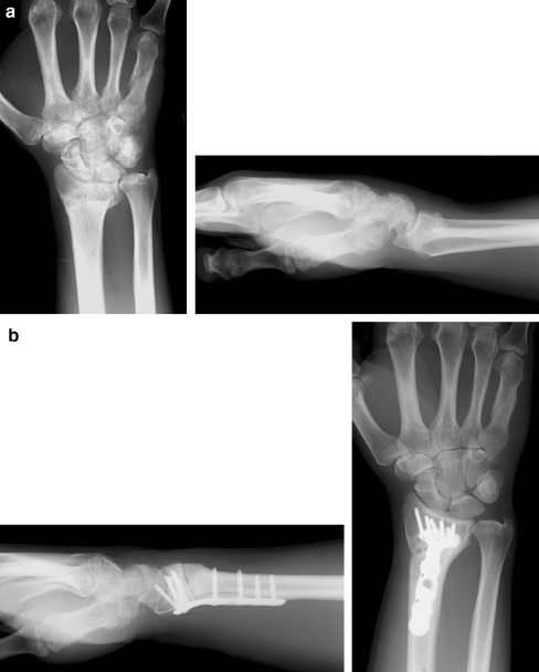
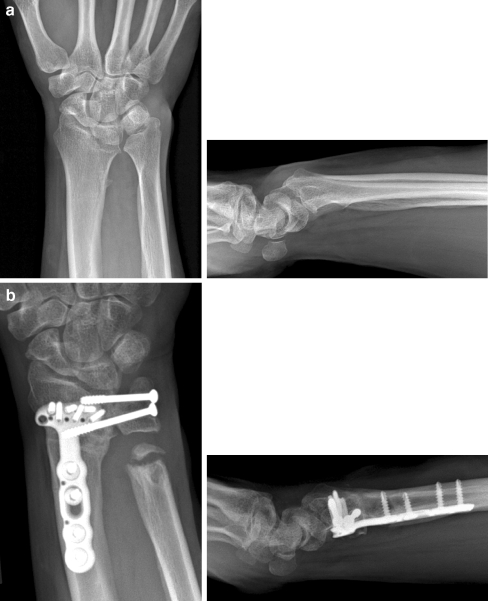
There were six patients with dorsally angulated malunions. Preoperative radiographs revealed an average of 24.3° (range, 5–49°) of dorsal tilt. Postoperative radiographs revealed that their average measurement was 2.5° volar tilt (Fig. 1). Two patients had excessive volar tilt preoperatively. One patient suffered a malunion of a Smith-type fracture and had a volar tilt of 24°; the other patient had a severe Madelung’s deformity with 50° of volar tilt (Fig. 2). The volar tilt in these two patients was corrected to 13 and 25°, respectively. All patients in this study were initially ulnar positive; the average was 4.1 mm of positive ulnar variance (range, 2–7 mm). Postoperatively, the average positive ulnar variance was less than 1 mm. In addition, radial inclination was corrected (Table 2). Preoperatively, radial inclination averaged 18.5°; postoperatively, radial inclination measurements averaged 23.6° (Table 2).
Figure 1.
a Preoperative radiographs of a patient with a dorsally angulated malunion of the distal radius. b Postoperative radiographs. At latest follow-up, the patient had near-normal grip strength and range of motion and no pain.
Figure 2.
a Preoperative images of a 51-year-old woman with bilateral severe Madelung’s deformity. The patient was a smoker, and after her osteotomy, she was treated with an external bone stimulator. The patient had a Sauve–Kapandji for her DRUJ symptoms as well. b The patient healed her osteotomy and elected to have the same procedure done on her opposite wrist.
Table 2.
Results of the radiography.
| Patient | Age | Gender | Side | Preoperative Volar Tilt | Postoperative Volar Tilt | Preoperative Ulnar Variance | Postoperative Ulnar Variance | Preoperative Radial Inclination | Postoperative Radial Inclination |
|---|---|---|---|---|---|---|---|---|---|
| 1 | 54 | F | R | 22 dorsal | 5 volar | +4 | +1 | 15 | 24 |
| 2 | 35 | M | L | 21 dorsal | 0 | +2 | 0 | 18 | 26 |
| 3 | 45 | F | L | 5 dorsal/volar translocation | 5 volar | +5 | +1 | 26 | 26 |
| 4 | 52 | M | L | 24 volar | 13 volar | +3 | −1 | 18 | 28 |
| 5 | 51 | F | R | 50 volar | 25 volar | +7 | 0 | 31 | 31 |
| 6 | 37 | M | R | 49 dorsal | 2 dorsal | +5 | 0 | 15 | 19 |
| 7 | 15 | M | L | 22 dorsal | 0 | +3 | −2 | 14 | 19 |
| 8 | 29 | M | R | 27 dorsal | 3 volar | +3.5 | +1 | 11 | 16 |
| Mean | 39.8 | N/A | N/A | 24 dorsal/37 volara | 3 dorsal/19 volara | +4 | 0 | 19 | 24 |
aAverage preoperative tilt in patients with dorsal deformity was 24.3°, and this corrected to 2.5° of dorsal tilt postoperatively. Average preoperative tilt in patients with volar deformity was 37°, and this corrected to 19° degrees of volar tilt postoperatively.
Clinical Examination
Preoperative examination revealed patients’ range of motion to be 20° of wrist flexion and 42° of wrist extension. Pronation and supination averaged 45 and 55°, respectively. The latest postoperative examination revealed an average of 60.6° of wrist extension, 58.1° of wrist flexion, 69.4° of pronation, and 72.5° of supination. Radial and ulnar deviation averaged 17.3° and 24.3°. Grip strengths measured improved from an average of 42 to 80.7 lb (Table 1).
Functional Outcomes
Subjective outcome measurements are reported in Table 3. DASH, SF-12, and Mayo Wrist scores averaged 10.8, 81, and 82.5, respectively. Postoperatively, the Mayo wrist scores of all patients in the study were above 80 and, therefore, classified as “good” under the Mayo Wrist scoring system.
Table 3.
Outcomes.
| Patient | Age | Gender | Side | Follow-up (Months) | DASH | SF12 | Mayo | Additional Radiographic Findings |
|---|---|---|---|---|---|---|---|---|
| 1 | 54 | F | R | 41 | 12.5 | 82 (83%) | 85 | |
| 2 | 35 | M | L | 15 | Ulnar styloid fx | |||
| 3 | 45 | F | L | 13 | Ulnar styloid fx | |||
| 4 | 52 | M | L | 29 | 9 | 80 | 80 | Ulnar styloid fx |
| 5 | 51 | F | R | 17 | 24 | 72 (69%) | 80 | Madelung’s, also underwent Sauve–Kapandji |
| 6 | 37 | M | R | 9 | 6 | 82 | 80 | |
| 7 | 15 | M | L | 8 | 8 | 84 | 85 | Open ulnar physis, ulnar styloid fx |
| 8 | 29 | M | R | 7 | 5 | 86 | 85 | Ulnar styloid fx |
| Mean | 40 | N/A | N/A | 17 | 10.8 | 81 | 83 |
Complications
There were no nonunions, tendon ruptures, or tendon irritations; additionally, plate removal was not required in any of the patients. All patients reported improvements from their preoperative function. One patient with a severe Madelung deformity had an additional Sauve–Kapandji procedure for unrelieved DRUJ pain, but she was so pleased with her final results that she elected to have the same procedures (osteotomy and Sauve–Kapandji) done on her other wrist as well.
Discussion
Malunion of the distal radius may result in biomechanical abnormalities in the radio-ulnar, radiocarpal, and midcarpal articulations [6, 23, 27, 28]. For the distal radius, normal radiographic values are typically cited as 11° of volar tilt, 22° of radial inclination, neutral ulnar variance, and a congruent radiocarpal articulation [3, 10]. The acceptable ranges of these parameters are typically cited as up to 15° of dorsal radial tilt or 20° of volar tilt, a 15° change in radial inclination, 4 mm of ulnar variance, and 2 mm of articular step-off. When the deformity exceeds these parameters, wrist dysfunction follows certain patterns. With increasing dorsal angulation, biomechanical studies have demonstrated increasing force concentration on the radioscaphoid, radiolunate, and ulnocarpal articulations [3]. Clinically, patients may develop dorsal carpal subluxation or an adaptive DISI pattern [27]. Both abnormal dorsal angulation of the distal radius, as well as ulnar variance, may affect the distal radioulnar joint and wrist pronation/supination [11, 35, 48]. In addition, increasing ulnar positivity results in ulnocarpal impaction and degenerative changes on the ulnar side of the wrist [52, 55].
For dorsally angulated fractures, techniques involving a dorsal approach and fixation may improve radiographic parameters as well as pain and function, but there are well-documented complications associated with the use of plates on the dorsal surface of the wrist [14, 34, 37, 41, 43]. Recently, Keller et al. reported on a series of 49 patients who underwent dorsal plating of the distal radius. At a mean follow-up of 32 months, patients had an average DASH score of 14.4 with good motion and grip strength. However, 37 of the 49 patients had undergone plate removal, and of the 12 patients who did not undergo plate removal, one patient suffered a rupture of the extensor indicis proprius [15]. Other authors have argued that extensor tendon complications are the result of the profile of the dorsal plate. In 2006, Kamath et al. [13] reported on a series of 30 patients who underwent dorsal plating with a low profile plate. At a mean follow-up of 18 months, patients had an average DASH score of 15 without need for plate removal, although two patients had undergone screw removal and one patient underwent extensor pollicis tenolysis. Simic et al. [41] reported on 60 patients who underwent dorsal plating with a low-profile plate. At a mean follow-up of 2 years, the average DASH score was 11.9 and only one patient underwent removal of hardware. Low profile dorsal plates may reduce some of the extensor tendon morbidity; however, studies in both canine and rabbit models indicate a reactive, inflammatory response to both stainless steel and titanium plates, which increases with time [5, 24].
The volar approach is potentially less morbid, but traditionally, volar plating has been performed on malunions with excess volar angulation [32, 40, 47]. Locking plates offer mechanical advantages for treating acute fractures of the distal radius [18, 42], and these properties make them attractive for corrective osteotomy as well. To our knowledge, this is the largest series of corrective osteotomy and volar locked plating to treat dorsally angulated and complex deformities of the distal radius. Malone et al. described four cases of dorsally malunited fractures in which they used a volar fixed angle plate. They used autogenous iliac crest bone graft in only 50% (two) of their cases, but the severity of the deformity that they addressed was less than that reported in this paper [20]. Prommersberger and Lanz [30] have published on a radiovolar approach to treat distal radius malunions [32]. They began using a fixed angled device in 2000 and have reported their technique [31], which differs from that reported in this paper. In both Malone’s and Prommersberger’s reports, low morbidity associated with the volar approach were documented.
In our series of eight patients, a single design of volar locking distal radius plate was used. The contour of this plate simplified the restoration of normal or near-normal distal radius anatomy. In addition, the strength of the plate led to a stable construct that did not require structural bone graft. In all of our cases, the deformity to be corrected was severe in that there was no cortical apposition between the distal and proximal fragments once the plate was in its final position. The resulting gap from the osteotomy was filled with iliac crest autograft in this series. We used autograft for its osteoinductive potential. There was no need for structural support (i.e., cortico-cancellous graft) because of the locking nature of the plate. It is possible to use bone graft substitute or recombinant bone morphogenetic protein, but we have not done so in this series.
There were no nonunions in our series. One patient who was a smoker was treated with the Exogen™ (Smith and Nephew, Memphis, TN) device. This patient did go on to have solid union and elected to have the same procedure on the opposite wrist.
All patients reported improved function and pain relief from their preoperative condition. Three patients complained of wrist pain that mildly limited their function at the last follow-up. As mentioned previously, one patient had a severe Madelung’s deformity and underwent a Sauve–Kapandji procedure. One patient was the youngest in our series and had sustained multiple fractures of his distal radius. After his osteotomy healed, he returned to playing football and re-injured his wrist, which resulted in ulnar-sided wrist pain; an MR arthrogram revealed only the previous ulnar styloid fracture without a triangular fibrocartilage complex (TFCC) tear. A second patient who reported that her surgery had helped her return to work complained of occasional ulnar-sided wrist pain. An arthrogram revealed a central TFCC tear. Neither patient elected to undergo any further surgical intervention at the last follow-up. Their Mayo Wrist scores were both 85 at the latest follow-up visit.
In conclusion, this report reviews a series of eight patients with severe deformity of the distal radius who underwent corrective osteotomy. This surgical technique allowed excellent correction of deformity based on radiographic parameters, with low morbidity and no nonunions, hardware failure, or need for hardware removal. Outcome scores as well as pre- and postoperative range of motion and grip strength tests document significant improvements in function. We believe that the volar approach and locking plate is an extremely effective technique for addressing symptomatic and even severe deformities of the distal radius, and it shows promise for restoring the maximum long-term function of the wrist.
Footnotes
Level IV evidence
The authors have no relevant financial interests with any organizations related to commercial products or services.
This study was begun after receiving approval from the UC Davis Medical Center IRB.
References
- 1.Adams BD. Effects of radial deformity on distal radioulnar joint mechanics. J Hand Surg (Am) 1993;18(3):492–8. [DOI] [PubMed]
- 2.Bacorn RW, Kurtzke JF. Colles’ fracture; a study of two thousand cases from the New York State Workmen’s Compensation Board. J Bone Jt Surg Am 1953;35-A(3):643–58. [PubMed]
- 3.Bushnell BD, Bynum DK. Malunion of the distal radius. J Am Acad Orthop Surg 2007;15(1):27–40. [DOI] [PubMed]
- 4.Carter PR, Frederick HA, Laseter GF. Open reduction and internal fixation of unstable distal radius fractures with a low-profile plate: a multicenter study of 73 fractures. J Hand Surg (Am) 1998;23(2):300–7. [DOI] [PubMed]
- 5.Cohen MS, Turner TM, Urban RM. Effects of implant material and plate design on tendon function and morphology. Clin Orthop Relat Res 2006;445:81–90. [DOI] [PubMed]
- 6.Crisco JJ, Moore DC, Marai GE, Laidlaw DH, Akelman E, Weiss AP, Wolfe SW. Effects of distal radius malunion on distal radioulnar joint mechanics—an in vivo study. J Orthop Res 2007;25(4):547–55. [DOI] [PubMed]
- 7.Fernandez DL. Radial osteotomy and Bowers arthroplasty for malunited fractures of the distal end of the radius. J Bone Jt Surg Am 1988;70(10):1538–51. [PubMed]
- 8.Fernandez DL. Malunion of the distal radius: current approach to management. Instr Course Lect 1993;42:99–113. [PubMed]
- 9.Fernandez DL. Reconstructive procedures for malunion and traumatic arthritis. Orthop Clin North Am 1993;24(2):341–63. [PubMed]
- 10.Graham TJ. Surgical correction of malunited fractures of the distal radius. J Am Acad Orthop Surg 1997;5(5):270–81. [DOI] [PubMed]
- 11.Hirahara H, Neale PG, Lin YT, Cooney WP, An KN. Kinematic and torque-related effects of dorsally angulated distal radius fractures and the distal radial ulnar joint. J Hand Surg (Am) 2003;28(4):614–21. [DOI] [PubMed]
- 12.Jansen JC, Adams BD. Long-term outcome of nonsurgically treated patients with wrist pain and a normal arthrogram. J Hand Surg (Am) 2002;27(1):26–30. [DOI] [PubMed]
- 13.Kamath AF, Zurakowski D, Day CS. Low-profile dorsal plating for dorsally angulated distal radius fractures: an outcomes study. J Hand Surg (Am) 2006;31(7):1061–7. [DOI] [PubMed]
- 14.Kambouroglou GK, Axelrod TS. Complications of the AO/ASIF titanium distal radius plate system (pi plate) in internal fixation of the distal radius: a brief report. J Hand Surg (Am) 1998;23(4):737–41. [DOI] [PubMed]
- 15.Keller M, Steiger R. Open reduction and internal fixation of distal radius extension fractures in women over 60 years of age with the dorsal radius plate (pi-plate). Handchir Mikrochir Plast Chir 2006;38(2):82–9. [DOI] [PubMed]
- 16.Kihara H, Short WH, Werner FW, Fortino MD, Palmer AK. The stabilizing mechanism of the distal radioulnar joint during pronation and supination. J Hand Surg (Am) 1995;20(6):930–6. [DOI] [PubMed]
- 17.Kihara H, Palmer AK, Werner FW, Short WH, Fortino MD. The effect of dorsally angulated distal radius fractures on distal radioulnar joint congruency and forearm rotation. J Hand Surg (Am) 1996;21(1):40–7. [DOI] [PubMed]
- 18.Koh S, Morris RP, Patterson RM, Kearney JP, Buford WL Jr, Viegas SF. Volar fixation for dorsally angulated extra-articular fractures of the distal radius: a biomechanical study. J Hand Surg (Am) 2006;31(5):771–9. [DOI] [PubMed]
- 19.Ladd AL, Huene DS. Reconstructive osteotomy for malunion of the distal radius. Clin Orthop Relat Res 1996;(327):158–71. [DOI] [PubMed]
- 20.Malone KJ, Magnell TD, Freeman DC, Boyer MI, Placzek JD. Surgical correction of dorsally angulated distal radius malunions with fixed angle volar plating: a case series. J Hand Surg (Am) 2006;31(3):366–72. [DOI] [PubMed]
- 21.Markolf KL, Lamey D, Yang S, Meals R, Hotchkiss R. Radioulnar load-sharing in the forearm. A study in cadavera. J Bone Jt Surg Am 1998;80(6):879–88. [DOI] [PubMed]
- 22.McKay SD, MacDermid JC, Roth JH, Richards RS. Assessment of complications of distal radius fractures and development of a complication checklist. J Hand Surg (Am) 2001;26(5):916–22. [DOI] [PubMed]
- 23.Miyake T, Hashizume H, Inoue H, Shi Q, Nagayama N. Malunited Colles’ fracture. Analysis of stress distribution. J Hand Surg (Br) 1994;19(6):737–42. [DOI] [PubMed]
- 24.Nazzal A, Lozano-Calderon S, Jupiter JB, Rosenzweig JS, Randolph MA, Lee SG. A histologic analysis of the effects of stainless steel and titanium implants adjacent to tendons: an experimental rabbit study. J Hand Surg (Am) 2006;31(7):1123–30. [DOI] [PubMed]
- 25.Orbay J. Volar plate fixation of distal radius fractures. Hand Clin 2005;21(3):347–54. [DOI] [PubMed]
- 26.Orbay JL, Badia A, Indriago IR, Infante A, Khouri RK, Gonzalez E, Fernandez DL. The extended flexor carpi radialis approach: a new perspective for the distal radius fracture. Tech Hand Up Extrem Surg 2001;5(4):204–11. [DOI] [PubMed]
- 27.Park MJ, Cooney WP III, Hahn ME, Looi KP, An KN. The effects of dorsally angulated distal radius fractures on carpal kinematics. J Hand Surg (Am) 2002;27(2):223–32. [DOI] [PubMed]
- 28.Pogue DJ, Viegas SF, Patterson RM, Peterson PD, Jenkins DK, Sweo TD, Hokanson JA. Effects of distal radius fracture malunion on wrist joint mechanics. J Hand Surg (Am) 1990;15(5):721–7. [DOI] [PubMed]
- 29.Porter M, Stockley I. Fractures of the distal radius. Intermediate and end results in relation to radiologic parameters. Clin Orthop Relat Res 1987;(220):241–52. [PubMed]
- 30.Prommersberger KJ, Lanz U. Biomechanical aspects of malunited distal radius fracture. A review of the literature. Handchir Mikrochir Plast Chir 1999;31(4):221–6. [DOI] [PubMed]
- 31.Prommersberger KJ, Lanz UB. Corrective osteotomy of the distal radius through volar approach. Tech Hand Up Extrem Surg 2004;8(2):70–7. [DOI] [PubMed]
- 32.Prommersberger KJ, Moossavi S, Lanz U. Results of corrective osteotomy of malunited extension fractures of the radius at the usual site. Handchir Mikrochir Plast Chir 1999;31(4):234–40. [DOI] [PubMed]
- 33.Prommersberger KJ, Froehner SC, Schmitt RR, Lanz UB. Rotational deformity in malunited fractures of the distal radius. J Hand Surg (Am) 2004;29(1):110–5. [DOI] [PubMed]
- 34.Sanchez T, Jakubietz M, Jakubietz R, Mayer J, Beutel FK, Grunert J. Complications after Pi Plate osteosynthesis. Plast Reconstr Surg 2004;116(1):153–8. [DOI] [PubMed]
- 35.Sato S. Load transmission through the wrist joint: a biomechanical study comparing the normal and pathological wrist. Nippon Seikei Geka Gakkai Zasshi 1995;69(7):470–83. [PubMed]
- 36.Sauerbier M, Kluge S, Bickert B, Germann G. Subjective and objective outcomes after total wrist arthrodesis in patients with radiocarpal arthrosis or Kienbock’s disease. Chir Main 2000;19(4):223–31. [DOI] [PubMed]
- 37.Schnur DP, Chang B. Extensor tendon rupture after internal fixation of a distal radius fracture using a dorsally placed AO/ASIF titanium pi plate. Arbeitsgemeinschaft fur Osteosynthesefragen/Association for the Study of Internal Fixation. Ann Plast Surg 2000;44(5):564–6. [DOI] [PubMed]
- 38.Schroven I, De Smet L, Zachee B, Steenwerckx A, Fabry G. Radial osteotomy and Sauve–Kapandji procedure for deformities of the distal radius. Acta Orthop Belg 1995;61(1):1–5. [PubMed]
- 39.Sharpe F, Stevanovic M. Extra-articular distal radial fracture malunion. Hand Clin 2005;21(3):469–87. [DOI] [PubMed]
- 40.Shea K, Fernandez DL, Jupiter JB, Martin C Jr. Corrective osteotomy for malunited, volarly displaced fractures of the distal end of the radius. J Bone Jt Surg Am 1997;79(12):1816–26. [DOI] [PubMed]
- 41.Simic PM, Robison J, Gardner MJ, Gelberman RH, Weiland AJ, Boyer MI. Treatment of distal radius fractures with a low-profile dorsal plating system: an outcomes assessment. J Hand Surg (Am) 2006;31(3):382–6. [DOI] [PubMed]
- 42.Smith DW, Henry MH. Volar fixed-angle plating of the distal radius. J Am Acad Orthop Surg 2005;13(1):28–36. [DOI] [PubMed]
- 43.Suckel A, Spies S, Munst P. Dorsal (AO/ASIF) pi-Plate osteosynthesis in the treatment of distal intraarticular radius fractures. J Hand Surg (Br) 2006;31(6):673–9. [DOI] [PubMed]
- 44.Szabo RM. Extra-articular fractures of the distal radius. Orthop Clin North Am 1993 24(2):229–37. [PubMed]
- 45.Szabo RM, Hotchkiss RN, Slater RR Jr. The use of frozen-allograft radial head replacement for treatment of established symptomatic proximal translation of the radius: preliminary experience in five cases. J Hand Surg (Am) 1997;22(2):269–78. [DOI] [PubMed]
- 46.Taleisnik J, Watson HK. Midcarpal instability caused by malunited fractures of the distal radius. J Hand Surg (Am) 1984;9(3):350–7. [DOI] [PubMed]
- 47.Thivaios GC, McKee MD. Sliding osteotomy for deformity correction following malunion of volarly displaced distal radial fractures. J Orthop Trauma 2003;17(5):326–33. [DOI] [PubMed]
- 48.Tomaino MM. The importance of the pronated grip X-ray view in evaluating ulnar variance. J Hand Surg (Am) 2000;25(2):352–7. [DOI] [PubMed]
- 49.Van Cauwelaert de Wyels J, De Smet L. Corrective osteotomy for malunion of the distal radius in young and middle-aged patients: an outcome study. Chir Main 2003;22(2):84–9. [DOI] [PubMed]
- 50.Van Schoonhoven J, Lanz U. Salvage operations and their differential indication for the distal radioulnar joint. Orthopade 2004;33(6):704–14. [DOI] [PubMed]
- 51.Voche P, Merle M, Dautel G. Non-articular malunions of the distal radius: evaluation and techniques of correction. Rev Chir Orthop Repar Appar Mot 2001;87(3):263–75. [PubMed]
- 52.Wada T, Tsuji H, Iba K, Aoki M, Yamashita T. Simultaneous radial closing wedge and ulnar shortening osteotomy for distal radius malunion. Tech Hand Up Extrem Surg 2004;9(4):188–94. [DOI] [PubMed]
- 53.Watson HK, Castle TH Jr. Trapezoidal osteotomy of the distal radius for unacceptable articular angulation after Colles’ fracture. J Hand Surg (Am) 1988;13(6):837–43. [DOI] [PubMed]
- 54.Werner FW, Glisson RR, Murphy DJ, Palmer AK. Force transmission through the distal radioulnar carpal joint: effect of ulnar lengthening and shortening. Handchir Mikrochir Plast Chir 1986;18(5):304–8. [PubMed]
- 55.Werner FW, Palmer AK, Fortino MD, Short WH. Force transmission through the distal ulna: effect of ulnar variance, lunate fossa angulation, and radial and palmar tilt of the distal radius. J Hand Surg (Am) 1992;17(3):423–8. [DOI] [PubMed]
- 56.Yeh GL, Beredjiklian PK, Katz MA, Steinberg DR, Bozentka DJ. Effects of forearm rotation on the clinical evaluation of ulnar variance. J Hand Surg (Am) 2001;26(6):1042–6. [DOI] [PubMed]


