Abstract
Idiopathic interstitial pneumonias (IIPs) have a progressive and often fatal course, and their enigmatic etiology has complicated approaches to effective therapies. Idiopathic pulmonary fibrosis (IPF) is the most common of IIPs and shares with IIPs an increased incidence with age and unexplained scarring in the lung. Short telomeres limit tissue renewal capacity in the lung and germ-line mutations in telomerase components, hTERT and hTR, underlie inheritance in a subset of families with IPF. To examine the hypothesis that short telomeres contribute to disease risk in sporadic IIPs, we recruited patients who have no family history and examined telomere length in leukocytes and in alveolar cells. To screen for mutations, we sequenced hTERT and hTR. We also reviewed the cases for features of a telomere syndrome. IIP patients had shorter leukocyte telomeres than age-matched controls (P < 0.0001). In a subset (10%), IIP patients had telomere lengths below the first percentile for their age. Similar to familial cases with mutations, IPF patients had short telomeres in alveolar epithelial cells (P < 0.0001). Although telomerase mutations were rare, detected in 1 of 100 patients, we identified a cluster of individuals (3%) with IPF and cryptogenic liver cirrhosis, another feature of a telomere syndrome. Short telomeres are thus a signature in IIPs and likely play a role in their age-related onset. The clustering of cryptogenic liver cirrhosis with IPF suggests that the telomere shortening we identify has consequences and can contribute to what appears clinically as idiopathic progressive organ failure in the lung and the liver.
Keywords: interstitial lung disease, liver fibrosis, telomerase, aplastic anemia, dyskeratosis congenita
Idiopathic interstitial pneumonias (IIPs) have a predictable, progressive course that often leads to respiratory failure. As their name indicates, the etiology of IIPs is unknown, and this has hampered progress in the development of therapies for patients with this disease. Idiopathic pulmonary fibrosis (IPF) is the most common of IIPs and accounts for greater than 70% of all cases (1). It has a characteristic radiographic appearance associated with the pathologic lesion of usual interstitial pneumonia. Age is the biggest risk factor for the development of IIPs, with the majority of cases diagnosed after the sixth decade, yet the factors that contribute to the age-related onset of IIPs are not known (1). As many as one in five patients with IPF reports a family history of the disease, establishing genetic factors as a critical contributor to disease risk (2). Histological features of IPF and other IIP subtypes are often present in the same individual and in individuals from a single family, indicating that IIPs could share a common etiology (3, 4). Mutations in telomerase components, hTERT and hTR, underlie inheritance of IPF in 8–15% of individuals who have a documented family history (5, 6). In these families, affected individuals have a clinical presentation indistinguishable from sporadic forms of the disease (5). Telomere length, not telomerase mutations, predicts disease onset in syndromes of telomere shortening (7–10). Here, we examine the role of telomere shortening and mutations in telomerase components in the pathogenesis of nonfamilial forms of idiopathic interstitial lung disease.
Telomeres are DNA–protein structures that protect chromosome ends. Telomeres shorten successively with each cell division, and short telomeres ultimately activate a DNA damage response that leads to cell death or permanent cell cycle arrest (11–13). This biology has implicated telomere shortening in degenerative age-related disease. Telomerase is a specialized polymerase responsible for telomere elongation (14–16). Mutations in either of the essential components of telomerase, hTERT, the catalytic reverse transcriptase, or hTR, telomerase RNA, lead to haploinsufficiency, a decrease in telomerase dose that leads to accelerated telomere shortening and ultimately to organ failure (5, 7, 17, 18). Mutations in telomerase components were initially identified in the setting of dyskeratosis congenita, a severe form of a syndrome of telomere shortening characterized by abnormal skin manifestations and premature mortality due to bone marrow failure and interstitial lung disease (19–22). In IPF families with telomerase mutations, cases of aplastic anemia can be hidden and are uncovered after a thorough family history, suggesting that mutations in telomerase have heterogeneous manifestations in an individual patient and within families, and that a subset of families with IPF falls on the same spectrum of telomere disorders as dyskeratosis congenita (5).
Recently, we described a pedigree with autosomal dominant dyskeratosis congenita that carried a mutation in hTERT that abolished catalytic activity (7). In this kindred, both pulmonary and liver fibrosis displayed anticipation, an earlier more severe onset of disease with successive generations. The anticipation of the fibrosis phenotypes, along with aplastic anemia, correlated with inheritance of the shortest telomeres across generations and suggested that telomere shortening underlies the predisposition to fibrosis in parenchymal organs and that the fibrosis, similar to aplasia in the marrow, may represent a loss of regenerative capacity (7). Telomere length, and not mutations in telomerases themselves, predicts disease onset and severity in models of aplastic anemia and dyskeratosis congenita (9). In these animal models, wild-type mice who inherit short telomeres display phenotypes similar to heterozygous mice. Thus, even when telomerase is wild type, short telomeres limit tissue renewal capacity (9). Here, we examine the hypothesis that telomere shortening, in the presence or absence of telomerase mutations, contributes to disease risk in IIP patients who have no family history. We show that, similar to familial IPF patients with telomerase mutations, individuals with idiopathic interstitial lung disease have short telomeres in both peripheral blood and in the lung. In this group of patients, there is an increased incidence of other features of dyskeratosis congenita, specifically of cryptogenic liver cirrhosis. Our findings establish a role for telomere shortening in IPF pathogenesis beyond a subset of families with telomerase mutations and suggest that telomere shortening may be an important contributor to the genetic susceptibility to this age-related disease.
Results
IIP Patients Have Short Telomeres in Peripheral Blood Leukocytes.
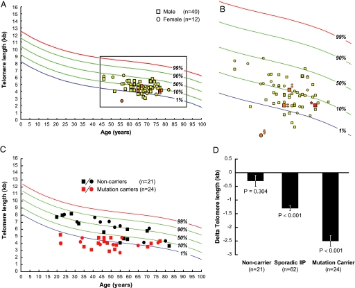
To examine whether individuals with IIP have short telomeres, we measured telomeres in peripheral blood lymphocytes using flow cytometry and FISH. We found that, compared with healthy age-matched controls (n = 400), IIP patients had shorter telomeres (P < 0.0001, Wilcoxon signed-rank test; Fig. 1 A and B). Specifically, 97% (60 of 62) of IIP patients had shorter telomeres than the median for their age (mean delta telomere length −1.3 kb, range −0.3 to −2.7; probability of random event P < 0.0001). To determine whether this effect was cell-type specific, we examined telomere length in granulocytes from the same patients and found a similar trend. We did not detect differences in telomere length within our cohort by gender (P = 0.30, multivariate regression analysis adjusting for age), smoking status (P = 0.50), or by diagnosis of IPF compared with non-IPF IIP (P = 0.58). These data suggested that individuals with IIP have shorter telomeres in peripheral blood cells than healthy age-matched controls.
Fig. 1.
Telomere length in lymphocytes from IIP patients and families with known telomerase mutations compared with healthy controls. (A) IIP patients, in yellow, have shorter telomeres than age-matched controls (P < 0.0001, Wilcoxon signed rank). IIP patients [60 of 62 (97%)] have telomeres shorter than the median of healthy controls (P < 0.0001). Of 62 IIP patients, 50 (81%) carried the diagnosis of IPF. (B) Detailed view of A with individuals with features of a telomere syndrome highlighted in red: *, a 77-year-old IPF patient with hTR 325G→T mutation; §, a patient with very short telomeres who had chronic unexplained thrombocytopenia, a feature of subclinical aplastic anemia; ‡, two individuals with both IPF and cryptogenic liver cirrhosis who have short telomeres. Ten percent of IIP patients (6 of 62) have short telomeres below the first percentile; a range predictive of the presence of a telomerase mutation. (C) Telomere length from 45 individuals from 10 families with known mutations in hTERT (n = 17), hTR (n = 3), and DKC1 (n = 4). (D) Bar graph illustrates the mean difference in telomere length from the median of age-matched healthy controls. Compared with noncarriers whose telomere length was similar to controls (P = 0.304, Wilcoxon signed rank), both sporadic IIP patients and known telomerase mutation carriers had shorter telomeres (P < 0.0001 for both).
Telomere Length Is a Surrogate for Mutation Status in Families with Telomerase Mutations.
Short telomeres are associated with telomerase mutations in familial IPF and dyskeratosis congenita. To determine whether telomere length can be a surrogate for mutation status, we examined 45 individuals from 10 families with known mutations in telomerase components and compared mutation carriers with their first-degree relatives who did not carry mutations. We found that individuals with mutations in telomerase components had significantly shorter telomeres than noncarriers (P < 0.0001, regression analysis adjusting for age; Fig. 1C). Furthermore, mutation carriers had shorter telomeres compared with the median telomere length of age-matched controls (n = 24, P < 0.0001, Wilcoxon signed-rank test). In contrast, noncarriers had telomere lengths that were not different from the median (n = 21, P = 0.304; Fig. 1D). Individuals with mutations in telomerase components also had short telomeres in granulocytes. These data suggest that telomere length in peripheral blood can be a useful surrogate for mutation status in relatives of individuals with known telomerase mutations. Specifically, individuals with lymphocyte telomere length greater than the 50th percentile for age never had mutations (100% predictive value). In contrast, individuals with very short lymphocyte telomeres (less than the first percentile for age) who also had short telomeres in granulocytes (less than the 10th percentile), independent of disease status, had a 95% likelihood of carrying the same mutation as the proband in their family. Similar patterns were also recently reported in a cohort of children with dyskeratosis congenita and their families (23). Thus, within these parameters, telomere length can be a useful surrogate for predicting mutation status in relatives of probands with known telomerase mutations.
A Subset of IIP Patients Has Short Telomeres Similar to Mutation Carriers.
To examine the significance and magnitude of telomere shortening in sporadic IIP patients, we compared their telomere length with known mutation carriers and their relatives. We found that, similar to mutation carriers, IIP patients had shorter telomeres than noncarriers (P = 0.007, multivariate regression analysis adjusting for age). We then examined the proportion of IIP patients with short leukocyte telomeres and found that in 10% (6 of 62), telomeres were very short in both lymphocytes (below the first percentile) and granulocytes (below the 10th percentile). Thus, similar to telomerase mutation carriers, a subset of sporadic IIP patients has very short telomeres in a range similar to mutation carriers.
Detectable Telomerase Mutations Are Rare in Patients with Sporadic IIP.
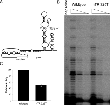
To examine the hypothesis that telomerase mutations underlie the telomere shortening in sporadic IPF, we sequenced the essential components of telomerase, hTERT and hTR, in 100 consecutive patients from the Vanderbilt Interstitial Lung Disease Clinic, including the 62 individuals where telomere length was available. We identified one individual who carried a mutation in hTR 325G→T, which predicted disruption of a conserved helix in telomerase RNA (Fig. 2A) (24). Younger asymptomatic siblings of this individual also carried the mutation confirming that the hTR 325G→T is germ line. This previously undescribed mutation was absent in a large series of healthy controls (n = 194) (25), was associated with short telomeres, and led to a loss of activity as quantitated by the direct telomerase activity assay (Figs. 1B and 2). We also identified three heterozygous nonsynonymous variants of hTERT: Ala279Thr (n = 8), His412Tyr (n = 1), and Ala1062Thr (n = 5) [supporting information (SI) Table S1]. All three variants had been previously noted in series of healthy controls (18) with the His412Tyr heterozygous allele identified in 1 of 22 healthy controls of European descent (dbSNP rs 34094729). To assess the functional consequences of these alleles, we quantitated telomerase activity. Although we did not detect any compromise in activity for the Ala279Thr and Ala1062Thr alleles, we detected a modest (15%) decrease in activity of the His412Tyr allele in vitro (P = 0.002, two-sample t test) but not in cells (P = 0.359; Fig. S1). These results are in contrast to previous studies where a drastic decrease in telomerase activity of the His412Tyr allele was seen when assayed by the semiquantitative PCR-based telomere repeat amplification protocol (18, 26). The role of this potentially functional polymorphic allele in telomere length variation across populations will need further exploration. Thus, readily detectable mutations in individuals with sporadic idiopathic lung fibrosis are rare. A similar frequency was seen by Tsakiri et al., who identified 1 hTERT mutation in 44 sporadic cases (2%) (6). The presence of individuals with very short telomeres in our cohort suggests that other genetic mechanisms that lead to telomere shortening play a role.
Fig. 2.
Germ-line hTR mutation in an IPF patient with no family history. (A) Secondary structure of hTR. hTR 325 G→T predicts disrupting the integrity of the conserved P5 helix and is thus expected to compromise function. (B) Gel of in vitro reconstituted telomerase with 5-fold dilutions as indicated. Telomerase activity of mutant hTR is compromised as shown by the decreased intensity of the repeat ladder compared with wild type. Quantitation of three independent experiments shown in C indicates that this allele is hypomorphic. Hypomorphic alleles of hTERT and hTR have been previously described in both aplastic anemia and familial IPF patients (5, 6, 18).
IPF Patients Have Short Telomeres in Alveolar Epithelium.
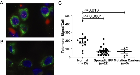
Peripheral blood telomeres may reflect a germ-line telomere length but are also susceptible to states of high turnover in leukocytes. To examine whether telomere shortening occurs in the IPF lung and reflects a genetic predisposition to having short telomeres, we examined telomere length in alveolar epithelium using quantitative FISH. We compared telomere length from age-matched individuals with normal lungs, sporadic IPF, and IPF patients with known telomerase mutations. We found that alveolar epithelium from individuals with IPF with known telomerase mutations had shorter telomeres than normal controls (P = 0.013, two-sample t test; Fig. 3). Additionally, individuals with sporadic IPF also had shorter telomeres than healthy controls (P < 0.0001). When we compared alveolar and lymphocyte telomere lengths from the same individuals (n = 9), we found a positive correlation (P = 0.045, Pearson's correlation coefficient; Fig. S2). These data indicate that, similar to peripheral blood leukocytes, telomeres in alveolar epithelium are shorter in IPF, and the IPF phenotype, even in the absence of a family history and a detectable mutation in telomerase, is associated with short telomeres in the lung.
Fig. 3.
Telomere length quantitation in alveolar epithelium by FISH. Lung cells from patients with usual interstitial pneumonia have short telomeres. (A) Representative images of nuclei of surfactant positive C cells (cytoplasmic staining in green) from an individual with no known lung disease showing bright telomere signals after hybridizing with a fluorescent telomere probe (pink). In contrast, alveolar cells from a patient with IPF have significantly shorter telomeres as seen by the dim or absent telomere signal in B. (C) Telomere signal per alveolar cell from age-matched individuals with normal lungs, sporadic IPF, and known hTERT (n = 4) or hTR mutation (n = 1) carriers. Mean age per group was 61, 59, and 64 years, respectively. IPF patients, with and without a family history, have shorter telomeres than controls with P values, as shown.
Cryptogenic Cirrhosis in Patients with Idiopathic Pulmonary Fibrosis.
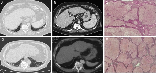
To probe the clinical relevance of short telomeres in IIP patients, we examined medical records for additional features of a syndrome of telomere shortening. None of the 100 patients had diagnosed aplastic anemia, although the patient with the shortest telomeres in our cohort had chronic unexplained thrombocytopenia, a feature of subclinical aplastic anemia (Fig. 1B). Ten percent of the patients in our cohort had platelet counts less than the normal range. In the absence of a formal work-up, it is difficult to discern, but it is interesting to consider the possibility that subclinical aplastic anemia may be another manifestation of the short telomeres in some IIP patients. Unexplained liver fibrosis is associated with IPF in individuals with dyskeratosis congenita (7, 19); we therefore queried our cohort for cases of cryptogenic liver disease. We identified two Vanderbilt patients who were diagnosed with cryptogenic liver cirrhosis after a thorough workup for an etiology (Table 1). To probe this observation further, we independently reviewed the records of 50 consecutive IPF patients seen in the Johns Hopkins Interstitial Lung Disease Clinic for the diagnosis of cryptogenic liver cirrhosis. We identified two additional patients who underwent liver transplant for decompensated liver cirrhosis and who carried the diagnosis of cryptogenic liver disease (Fig. 4). In total, in this series, we identified 4 of 150 IIP patients (3%) with a history of unexplained liver cirrhosis. None of these patients had detectable telomerase mutations, although they had telomeres in the lowest percentiles of the population (Fig. 1B and data not shown). Based on a prevalence rate of 100/100,000, the likelihood of cryptogenic liver cirrhosis and IPF coexisting in the same individual by chance alone is rare (P < 10−22). This association will need to be verified in larger studies. In the meantime, it is intriguing to consider the possibility that telomere shortening, even in the absence of readily detectable mutations, is genetically relevant and underlies an increased predisposition to both pulmonary and liver failure that manifest as progressive idiopathic-cryptogenic disease in the same patient.
Table 1.
Clinical features of patients with both idiopathic pulmonary fibrosis and cryptogenic liver cirrhosis
| Gender | IIP diagnosis | Age at diagnosis, presenting symptom | Evidence of cirrhosis | Age at diagnosis |
|---|---|---|---|---|
| M | IPF | 60, cough | Liver transplant | 58 |
| M | IPF | 72, cough | Liver transplant | 65 |
| M | IPF | 65, dyspnea | Portal hypertension | 65 (died 66) |
| M | IPF | 67, cough | Compensated cirrhosis | 59 |
M, male.
Fig. 4.
Imaging and pathology from patients with both idiopathic pulmonary fibrosis and cryptogenic liver cirrhosis. Computed tomography A and D shows honeycomb changes of IPF in the lung bases. B and E show representative abnormalities in the same patients with evidence of decompensated cirrhosis with splenomegaly and portal hypertension in B and nodular and abnormal liver contour in E associated with intraoperative description of a cirrhotic liver. (C and F) Reticulin stains of liver explants from a patient with scans in A and B and of a second patient who underwent liver transplant 3 years before his IPF diagnosis. The fibrosis on the background of cirrhotic lobules is prominent in the interstitial and perivascular space.
Discussion
Telomere Length as a Risk Factor for IIPs.
The factors that contribute to the age-related predisposition of IIPs are not known. Based on the finding in IPF families that mutations in telomerase exert their effect through telomere shortening, we examined the incidence of short telomeres in individuals with sporadic IIP. We found that compared with age-matched controls, individuals with IIP have shorter telomeres both in peripheral blood and in the lung. Moreover, a subset of patients (10%) with no family history had telomere lengths in the range of known mutation carriers even when mutations were not detected. Mutations in hTERT and hTR were readily detectable in only 1% of individuals suggesting that other genetic mechanisms that lead to telomere shortening underlie the prominent differences seen in this cross-sectional study. A homozygous mutant allele in NOP10, a component of the dyskerin complex, has been reported in one autosomal recessive dyskeratosis congenita family (27); however, we examined and did not identify coding sequence variants in 73 familial IPF probands (unpublished data). Additionally, mutant alleles in exon 6 of the telomere-binding protein TIN2 were recently identified in cases of dyskeratosis congenita (28); however, we did not identify exon 6 sequence variants in a screen of the same IPF probands (unpublished data).
Even when telomerase is wild type, telomere-mediated degenerative disease can occur, and it is possible that the IPF-IIP phenotype enriches for individuals with the shortest telomeres in the population. Our findings support the idea that individuals with the shortest telomeres across the population may be at increased risk for developing idiopathic interstitial lung disease compared with individuals with long telomeres and suggest that the genetics of telomere shortening underlie at least a component of the age-related predisposition to what appears as unprovoked or idiopathic progressive processes in the lung. This idea is further supported by clinical observations of asymptomatic IPF in the elderly and suggests that the IPF phenotype may be a clinically important manifestation of aging in the lung. Considering telomere length in future studies examining the risk factors that underlie IPF will be important in fully uncovering its genetic epidemiology.
Implications for Treatment.
Approaches to therapy in IPF have been hindered by the poorly understood pathophysiology that underlies the progressive nature of alveolar destruction and the accumulation of fibrosis. Because of the end replication problem, telomere shortening inevitably occurs in cells over time, and short telomeres ultimately activate a DNA damage response that manifests clinically as aplasia in the bone marrow and fibrosis in the lung and liver. As such, we have proposed that the fibrosis phenotype represents an irreversible loss of tissue renewal capacity as a result of the loss of replicative potential of local progenitors in parenchymal organs (5, 7). Interstitial lung and liver disease are the most common causes of mortality in dyskeratosis congenita patients who are exposed to cytotoxic chemotherapy in the setting of bone marrow transplant for aplastic anemia (29). Additionally, mice with short telomeres are at increased risk for developing fibrotic liver disease when exposed to toxins compared with wild-type mice (30). Short telomeres are sufficient to induce the IPF phenotype in familial IPF (5). The presence of short telomeres in both peripheral blood and the lung of sporadic IPF patients, beyond the subset of patients who have readily detected telomerase mutations, suggests that telomere shortening, rather than being a secondary effect, plays a primary role in both familial and sporadic disease pathogenesis, and that strategies aimed at preventing cell death or local responses to it may have an impact in attenuating the course of this disease.
A Subset of IPF Cases Falls on the Spectrum of a Telomere Syndrome.
Finally, our data suggest that some individuals with IPF may harbor subtle features of a syndrome of telomere shortening. Our observation of an increased incidence of unexplained liver failure in IPF patients underscores the clinical relevance of short telomeres in this context. IPF and cryptogenic cirrhosis, to our knowledge, have been described only in the setting of dyskeratosis congenita. It will be interesting to examine whether patients with cryptogenic cirrhosis similarly have an increased incidence of interstitial lung disease. Systematic studies of personal and family history for aplastic anemia in patients with unexplained fibrosis in the lung or liver will also further define the prevalence of a syndrome of telomere shortening.
Methods
Patients.
Patients were eligible for the study if they had a diagnosis of IIP as defined by the 2002 consensus classification in the absence of a family history of IIP (1). From 2006–2007, subjects were recruited from the Vanderbilt Interstitial Lung Disease and the Johns Hopkins Hematology and Interstitial Lung Disease Clinics. Patient characteristics are summarized in Table S1. The majority of patients carried the diagnosis of IPF (84 of 100). The study was approved by the local institutional review boards, and written informed consent was obtained from all subjects. We systematically reviewed the medical records of IIP patients for features of dyskeratosis congenita by focusing on the diagnosis of aplastic anemia and liver cirrhosis and examining complete blood counts and liver function tests. Paraffin-embedded lung tissue specimens were retrieved from patients with IPF/usual interstitial pneumonia who had a surgical lung biopsy obtained during clinical management. Normal lung tissue was obtained from individuals who died with no recognized lung disorders via the National Disease Research Interchange and the Vanderbilt Autopsy program.
Telomere Length and Sequencing Studies.
The average length of telomeres was measured in peripheral blood leukocytes by flow cytometry and FISH, as described (5, 18, 23, 31). Telomere length was measured in paraffin-embedded tissues in alveolar type 2 cells using quantitative FISH, as described (32). Quantitation of telomere length was specific to surfactant protein C-positive cells (i.e., alveolar type 2 cells) identified by immunostaining with rabbit anti-human SPC antibodies (Chemicon) followed by detection with goat anti-rabbit Alexa Fluor-488 conjugated antibody (Invitrogen). We obtained four images per slide at a fixed exposure time and three to five nuclei were analyzed per high power field (×100). We analyzed the raw images and obtained data on 15 nuclei for each sample using Telometer, an ImageJ plugin available at http://bui2.win.ad.jhu.edu/telometer kindly provided by Alan Meeker (Johns Hopkins School of Medicine, Baltimore). We measured telomere length in each cell by dividing the total Cy3 signal (telomere signal) in the nucleus by the DAPI area to account for the assessable nuclear area in cross section. We manually amplified and sequenced hTERT and hTR from genomic DNA prepared from peripheral blood as described (5). For hTERT, we analyzed the 16 coding exons and at least 200 nucleotides within introns from splice junction boundaries. hTERT variants are listed in Table S2. Statistical analyses were performed by using Stata 10.0 for Windows (Stata Corporation). All P values are two-sided, and error bars represent standard error of the mean. All of the telomere length and sequencing analyses were performed blind.
Telomerase Activity.
To assess the functional significance of suspected mutants, point mutations were generated, and the telomerase complex was reconstituted in vitro (5, 33). Briefly, recombinant hTERT protein was synthesized in 10 μl of TnT quick-coupled rabbit reticulocyte lysate (Promega) at 30°C for 60 min following the manufacturer's instructions. Specifically, 10 μM methionine and 4 μM 35S methionine (1,175 Ci/mmole, 10 mCi/ml, Perkin-Elmer) were supplied together in the 10 μI reaction. In vitro synthesized full-length hTR was added to a near-saturated concentration of 1 μM to the TnT reaction of hTERT synthesis and incubated at 30°C for 30 min. Telomerase activity was assayed without amplification by using the direct assay (33, 34). Briefly, a 10-μl reaction was carried out with 3 μl of in vitro reconstituted telomerase sample in the presence of 1× PE buffer (50 mM Tris·HCl, pH 8.3, 50 mM KCl, 2 mM DTT, 3 mM MgCl2, and 1 mM spermidine); 1 mM dATP, 2 μM dGTP, 1 mM dTTP, 1 μM (TTAGGG)3 telomere primer; and 1.25 μM [α-32P] dGTP (800 Ci/mmol, 10 mCi/ml, Perkin–Elmer) at 30°C for 1 h. The reactions were mixed with 32P end-labeled 15-mer oligonucleotide, phenol-chloroform-extracted, and ethanol-precipitated. The products were resolved by 10% denaturing polyacrylamide gel electrophoresis and analyzed using a Bio-Rad FX Pro Imager. Activity was determined by measuring the total intensity of telomere product, correcting for background, and normalizing against the 35S labeled hTERT expresson in the rabbit reticulocyte lysate and the loading controls (32P end-labeled oligonucleotide). For activity assays in cells, we transfected 293FT cells (Invitrogen) with plasmids overexpressing hTERT and hTR genes kind gift from Joachim Lingner (Swiss Institute for Experimental Cancer Research, Lausonne, Switzerland), as described (35) and quantitated telomerase activity using the direct assay.
Supplementary Material
Acknowledgments.
We thank the patients and families who participated and their clinicians. We are grateful to Laura Kasch-Semenza and Roxann Ingersoll of the Johns Hopkins Genetics Resources Core Facility for assistance with the DNA sequencing. This work was supported by a grant from the National Institutes of Health National Cancer Institute K08 118416 and funding from the Doris Duke Charitable Foundation (M.Y.A.). J.K.A. received support from an National Institutes of Health/National Cancer Institute Training Grant (T32 60441).
Footnotes
Conflict of interest statement: P.M.L. is a founding shareholder in Repeat Diagnostics, a company that specializes in length measurement of leukocyte telomeres with the use of flow FISH.
This article is a PNAS Direct Submission.
This article contains supporting information online at www.pnas.org/cgi/content/full/0804280105/DCSupplemental.
References
- 1.American Thoracic Society/European Respiratory Society International Multidisciplinary Consensus Classification of the Idiopathic Interstitial Pneumonias. This joint statement of the American Thoracic Society (ATS), and the European Respiratory Society (ERS) was adopted by the ATS board of directors, June 2001 and by the ERS Executive Committee, June 2001. Am J Respir Crit Care Med. 2002;165:277–304. doi: 10.1164/ajrccm.165.2.ats01. [DOI] [PubMed] [Google Scholar]
- 2.Loyd JE. Pulmonary fibrosis in families. Am J Respir cell Mol Biol. 2003;29:S47–S50. [PubMed] [Google Scholar]
- 3.Steele MP, et al. Clinical and pathologic features of familial interstitial pneumonia. Am J Respir Crit Care Med. 2005;172:1146–1152. doi: 10.1164/rccm.200408-1104OC. [DOI] [PMC free article] [PubMed] [Google Scholar]
- 4.Thomas AQ, et al. Heterozygosity for a surfactant protein C gene mutation associated with usual interstitial pneumonitis and cellular nonspecific interstitial pneumonitis in one kindred. Am J Respir Crit Care Med. 2002;165:1322–1328. doi: 10.1164/rccm.200112-123OC. [DOI] [PubMed] [Google Scholar]
- 5.Armanios MY, et al. Telomerase mutations in families with idiopathic pulmonary fibrosis. N Engl J Med. 2007;356:1317–1326. doi: 10.1056/NEJMoa066157. [DOI] [PubMed] [Google Scholar]
- 6.Tsakiri KD, et al. Adult-onset pulmonary fibrosis caused by mutations in telomerase. Proc Natl Acad Sci USA. 2007;104:7552–7557. doi: 10.1073/pnas.0701009104. [DOI] [PMC free article] [PubMed] [Google Scholar]
- 7.Armanios M, et al. Haploinsufficiency of telomerase reverse transcriptase leads to anticipation in autosomal dominant dyskeratosis congenita. Proc Natl Acad Sci USA. 2005;102:15960–15964. doi: 10.1073/pnas.0508124102. [DOI] [PMC free article] [PubMed] [Google Scholar]
- 8.Blasco MA, et al. Telomere shortening and tumor formation by mouse cells lacking telomerase RNA. Cell. 1997;91:25–34. doi: 10.1016/s0092-8674(01)80006-4. [DOI] [PubMed] [Google Scholar]
- 9.Hao LY, et al. Short telomeres, even in the presence of telomerase, limit tissue renewal capacity. Cell. 2005;123:1121–1131. doi: 10.1016/j.cell.2005.11.020. [DOI] [PubMed] [Google Scholar]
- 10.Vulliamy T, et al. Disease anticipation is associated with progressive telomere shortening in families with dyskeratosis congenita due to mutations in TERC. Nat Genet. 2004;36:447–449. doi: 10.1038/ng1346. [DOI] [PubMed] [Google Scholar]
- 11.d'Adda di Fagagna F, et al. A DNA damage checkpoint response in telomere-initiated senescence. Nature. 2003;426:194–198. doi: 10.1038/nature02118. [DOI] [PubMed] [Google Scholar]
- 12.Harley CB, Futcher AB, Greider CW. Telomeres shorten during ageing of human fibroblasts. Nature. 1990;345:458–460. doi: 10.1038/345458a0. [DOI] [PubMed] [Google Scholar]
- 13.Lee HW, et al. Essential role of mouse telomerase in highly proliferative organs. Nature. 1998;392:569–574. doi: 10.1038/33345. [DOI] [PubMed] [Google Scholar]
- 14.Greider CW, Blackburn EH. Identification of a specific telomere terminal transferase activity in Tetrahymena extracts. Cell. 1985;43:405–413. doi: 10.1016/0092-8674(85)90170-9. [DOI] [PubMed] [Google Scholar]
- 15.Greider CW, Blackburn EH. The telomere terminal transferase of Tetrahymena is a ribonucleoprotein enzyme with two kinds of primer specificity. Cell. 1987;51:887–898. doi: 10.1016/0092-8674(87)90576-9. [DOI] [PubMed] [Google Scholar]
- 16.Nakamura TM, et al. Telomerase catalytic subunit homologs from fission yeast and human. Science. 1997;277:955–959. doi: 10.1126/science.277.5328.955. [DOI] [PubMed] [Google Scholar]
- 17.Vulliamy T, et al. The RNA component of telomerase is mutated in autosomal dominant dyskeratosis congenita. Nature. 2001;413:432–435. doi: 10.1038/35096585. [DOI] [PubMed] [Google Scholar]
- 18.Yamaguchi H, et al. Mutations in TERT, the gene for telomerase reverse transcriptase, in aplastic anemia. N Engl J Med. 2005;352:1413–1424. doi: 10.1056/NEJMoa042980. [DOI] [PubMed] [Google Scholar]
- 19.Dokal I. Dyskeratosis congenita in all its forms. Br J Haematol. 2000;110:768–779. doi: 10.1046/j.1365-2141.2000.02109.x. [DOI] [PubMed] [Google Scholar]
- 20.Dokal I. Dyskeratosis congenita. A disease of premature ageing. Lancet. 2001;358(Suppl):S27. doi: 10.1016/s0140-6736(01)07040-4. [DOI] [PubMed] [Google Scholar]
- 21.Heiss NS, et al. X-linked dyskeratosis congenita is caused by mutations in a highly conserved gene with putative nucleolar functions. Nat Genet. 1998;19:32–38. doi: 10.1038/ng0598-32. [DOI] [PubMed] [Google Scholar]
- 22.Mitchell JR, Wood E, Collins K. A telomerase component is defective in the human disease dyskeratosis congenita. Nature. 1999;402:551–555. doi: 10.1038/990141. [DOI] [PubMed] [Google Scholar]
- 23.Alter BP, et al. Very short telomere length by flow fluorescence in situ hybridization identifies patients with dyskeratosis congenita. Blood. 2007;110:1439–1447. doi: 10.1182/blood-2007-02-075598. [DOI] [PMC free article] [PubMed] [Google Scholar]
- 24.Chen JL, Blasco MA, Greider CW. Secondary structure of vertebrate telomerase RNA. Cell. 2000;100:503–514. doi: 10.1016/s0092-8674(00)80687-x. [DOI] [PubMed] [Google Scholar]
- 25.Yamaguchi H, et al. Mutations of the human telomerase RNA gene (TERC) in aplastic anemia and myelodysplastic syndrome. Blood. 2003;102:916–918. doi: 10.1182/blood-2003-01-0335. [DOI] [PubMed] [Google Scholar]
- 26.Du HY, et al. Complex inheritance pattern of dyskeratosis congenita in two families with 2 different mutations in the telomerase reverse transcriptase gene. Blood. 2008;111:1128–1130. doi: 10.1182/blood-2007-10-120907. [DOI] [PMC free article] [PubMed] [Google Scholar]
- 27.Walne AJ, et al. Genetic heterogeneity in autosomal recessive dyskeratosis congenita with one subtype due to mutations in the telomerase-associated protein NOP10. Hum Mol Genet. 2007;16:1619–1629. doi: 10.1093/hmg/ddm111. [DOI] [PMC free article] [PubMed] [Google Scholar]
- 28.Savage SA, et al. TINF2, a component of the shelterin telomere protection complex, is mutated in dyskeratosis congenita. Am J Hum Genet. 2008;82:501–509. doi: 10.1016/j.ajhg.2007.10.004. [DOI] [PMC free article] [PubMed] [Google Scholar]
- 29.de la Fuente J, Dokal I. Dyskeratosis congenita: Advances in the understanding of the telomerase defect and the role of stem cell transplantation. Pediatr Transplant. 2007;11:584–594. doi: 10.1111/j.1399-3046.2007.00721.x. [DOI] [PubMed] [Google Scholar]
- 30.Rudolph KL, Chang S, Millard M, Schreiber-Agus N, DePinho RA. Inhibition of experimental liver cirrhosis in mice by telomerase gene delivery. Science. 2000;287:1253–1258. doi: 10.1126/science.287.5456.1253. [DOI] [PubMed] [Google Scholar]
- 31.Baerlocher GM, Vulto I, de Jong G, Lansdorp PM. Flow cytometry and FISH to measure the average length of telomeres (flow FISH) Nat Protoc. 2006;1:2365–2376. doi: 10.1038/nprot.2006.263. [DOI] [PubMed] [Google Scholar]
- 32.Meeker AK, et al. Telomere length assessment in human archival tissues: Combined telomere fluorescence in situ hybridization and immunostaining. Am J Pathol. 2002;160:1259–1268. doi: 10.1016/S0002-9440(10)62553-9. [DOI] [PMC free article] [PubMed] [Google Scholar]
- 33.Xie M, et al. Structure and function of the smallest vertebrate telomerase RNA from teleost fish. J Biol Chem. 2008;283:2049–2059. doi: 10.1074/jbc.M708032200. [DOI] [PubMed] [Google Scholar]
- 34.Drosopoulos WC, Direnzo R, Prasad VR. Human telomerase RNA template sequence is a determinant of telomere repeat extension rate. J Biol Chem. 2005;280:32801–32810. doi: 10.1074/jbc.M506319200. [DOI] [PubMed] [Google Scholar]
- 35.Cristofari G, Lingner J. Telomere length homeostasis requires that telomerase levels are limiting. EMBO J. 2006;25:565–574. doi: 10.1038/sj.emboj.7600952. [DOI] [PMC free article] [PubMed] [Google Scholar]
Associated Data
This section collects any data citations, data availability statements, or supplementary materials included in this article.


