Abstract
Ten cases of histologically proven chondromyxoid fibroma (CMF) of the foot and ankle with a mean follow-up of 6.1 years were retrospectively reviewed using the Scottish Bone Tumour Registry. The patients' mean age was 19 years; there were six males and four females. The anatomical locations were five phalangeal, three metatarsal, one tarsal affecting body of os calcis and one distal tibial. The median delay in presentation was 4.5 months. The modes of presentation were pain only (n=4), painful lump (n=4) and painless lump (n=2). The typical radiological finding was an expansile, lobulated, cystic lesion. Cortical erosion was documented in 80% patients. In four cases, curettage alone was carried out, while five patients underwent curettage along with autogenous bone grafting. One patient with distal phalangeal CMF had a primary toe amputation. Two patients had recurrences 9 and 16 months after their initial curettage. Both of them were males with proximal phalangeal CMF, associated with cortical erosion. Foot and phalangeal CMF initially treated with curettage only should be closely followed up, as we observed a 20% recurrence rate within a 2-year period. Cases featuring cortical erosion require thorough curettage and may require autogenous bone grafting to prevent fracture.
Résumé
Nous avons revu rétrospectivement à l’aide du registre Ecossais des tumeurs osseuses, dix cas de fibromes chondromyxoides du pied et de la cheville. L’âge moyen était de 19 ans avec une répartition par sexe suivante : 6 garçons et 4 filles. La localisation anatomique de la tumeur était cinq fois une phalange, trois fois un métatarsien, une fois un os du tarse et une fois l’extrémité inférieure du tibia. Le délai moyen de consultation était de quatre mois et demi, les patients consultant quatre fois pour une douleur,quatre fois pour une boiterie douloureuse, deux fois pour une boiterie non douloureuse. Les lésions kystiques étaient radiologiquement typiques de type lobulées. L’atteinte corticale a été retrouvée dans 80% des cas. Pour quatre cas nous avons procédé à un curetage isolé, dans cinq cas à un curetage associé à une auto-greffe. Un cas (atteinte de la phalange distale) a bénéficié d’une amputation d’orteil. Deux cas ont récidivé à neuf et seize mois après le curetage, il s’agissait d’un sujet masculin avec une atteinte de la phalange distale et une érosion corticale. Les fibromes chondromyxoides du pied et de la phalange qui ont initialement été traités par curetage isolé ont récidivé dans les deux ans, dans 20% des cas. Lorsque les lésions entraînent une érosion corticale, il est nécessaire d’associer au curetage une greffe, afin de prévenir le risque de fracture.
Introduction
Chondromyxoid fibroma (CMF) is a benign bone tumour characterised by lobulated areas of spindle-shaped or stellate cells with abundant myxoid or chondroid intercellular material. These are separated by zones of more cellular tissue rich in spindle-shaped or round cells with a varying number of multinucleated giant cells of different sizes [20, 25]. It accounts for fewer than 1% of all bone tumours [6, 19].
Although foot and ankle involvement is reported to vary from 10% to 31% [19, 20, 23, 25], most available evidence is in the form of isolated case reports or small case series [3, 5, 7, 9, 12, 14, 17, 18, 21, 22, 24]. Ten cases of histologically proven CMF of the foot and ankle with a mean follow-up of 6.1 years were retrospectively reviewed using Scottish Bone Tumour Registry database in order to ascertain the behavior of CMF in the foot and ankle area with regard to diagnosis, treatment and prognosis.
Material and methods
The Scottish Bone Tumour Registry was used for a retrospective review of ten cases of CMF of the foot and ankle with a mean follow-up of 6.1 years from January 1963 to December 2001. This registry is a prospective database which updates follow-up data of patients by obtaining information from orthopaedic/oncology/surgical clinics and communications from general practitioners.
All registry case notes, radiographs and histological sections were available for review. The registry casenotes and radiographs were reviewed by the first author (H.S.). All the histological sections were reviewed by a senior pathologist (R.R.). Follow-up was calculated from the date of biopsy to the last follow-up entry in the records of the registry. Postoperative complications, recurrences and treatment protocols were recorded. Association between initial mode of treatment and recurrence was studied. Factors influencing recurrence were evaluated. Only cases of CMF of the foot and distal tibio-fibular region were included in this series. Those cases where the foot and ankle were involved in addition to other joints (polyarticular) or proximal and diaphyseal tibio-fibular CMF were excluded.
Results
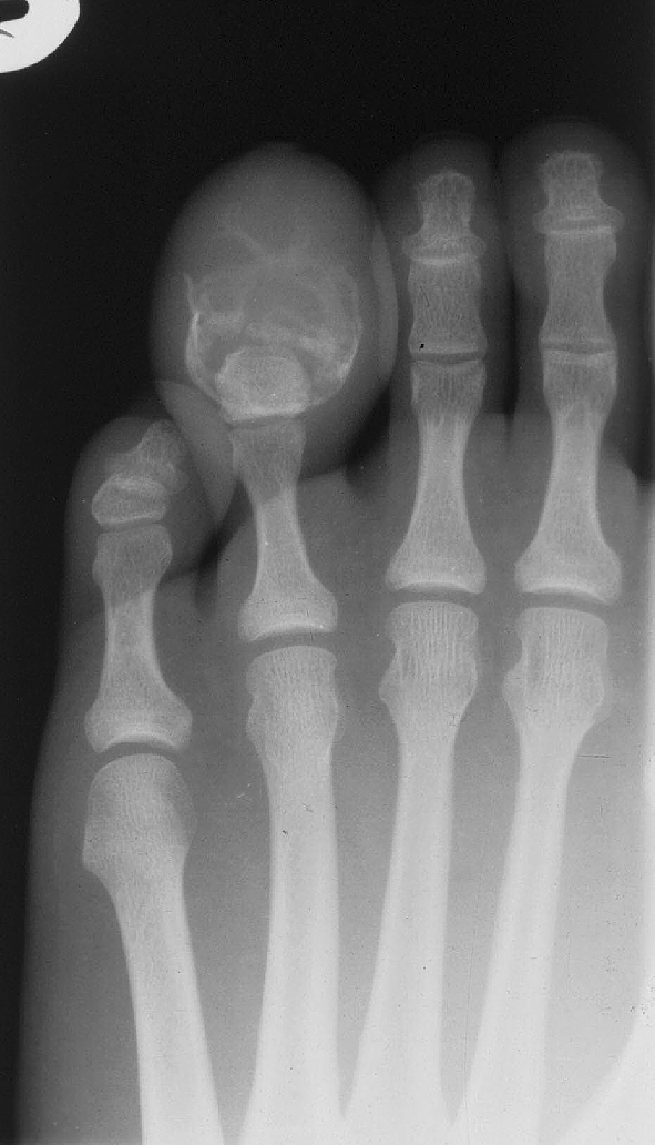
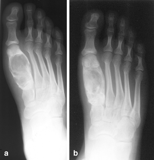
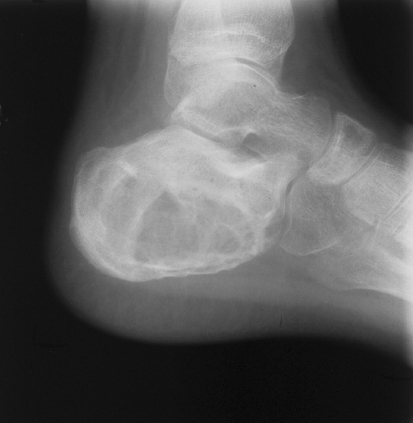
The patient's mean age was 19 years (range 11–33 years). Six patients were male and four were female. A right-sided preponderance was observed (7/10 cases). Five cases presented before and five after skeletal maturity. The mean age was 12.8 years for the skeletally immature patients, three of whom were male and two female. The same sex distribution was noted for the five skeletally mature patients, whose mean age was 25.2 years. The anatomical distribution of the lesions was five phalangeal (three proximal phalanx, one middle phalanx, one terminal phalanx), three metatarsal, one tarsal affecting os calcis and one case of distal tibial involvement (Figs. 1, 2, 3). The median duration of symptoms prior to seeking medical attention was 4.5 months. One patient ignored a painless lump for 10 years. Four cases presented with pain only and four with a painful lump. The remaining two patients had lumps only and no pain (Table 1). The typical radiographic finding was an expansile, lobulated, cystic lesion. Cortical erosion was documented in eight of the ten patients (Table 2).
Fig. 1.
a Preoperative radiograph revealing first metatarsal lesion; b radiograph 3 months after treatment with curettage and bone grafting
Fig. 2.
Radiograph of the os calcis chondromyxoid fibroma
Fig. 3.

Radiograph of the terminal phalanx chondromyxoid fibroma manifested as bulbous swelling with split toenail
Table 1.
Details of chondromyxoid fibroma of the foot and ankle
| Case no. | Age (years)/ sex/ side | Anatomical site | Duration of symptoms prior to presentation | Pain only | Lump only | Painful lump | Surgery | Bone grafting | Post-operative complication and its management | Total duration of follow-up |
|---|---|---|---|---|---|---|---|---|---|---|
| 1 | 11/M/R | Distal tibial metaphyseal | 6 weeks | − | + | − | Curettage | + | − | 15 months |
| 2 | 33/M/L | Third metatarsal diaphyseal | 4 weeks | + | − | − | Curettage | + | − | 18 years |
| 3 | 13/F/R | Body of os calcis | 5 months | − | − | + | Curettage | − | Infection, wound debridement and re-curettage | 5 years, 3 months |
| 4 | 20/M/L | Proximal phalanx of third toe | 6 months | + | − | − | Curettage | − | Recurrence, treated with re-curettage | 12 years |
| 5 | 22/M/L | Terminal phalanx of fourth toe | 10 years | − | + | − | Amputation at PIPJ level | − | − | 8 years, 6 months |
| 6 | 18/F/R | Proximal phalanx of big toe | 4 months | − | − | + | Curettage | + | − | 6 years |
| 7 | 15/M/R | First metatarsal diaphyseal | 2 months | + | − | − | Curettage | + | − | 2 years, 6 months |
| 8 | 14/M/R | Proximal phalanx of second toe | 2 months | − | − | + | Curettage | − | Recurrence, amputation through metatarsal neck | 5 years, 5 months |
| 9 | 33/F/R | Third metatarsal | 12 months | + | − | − | Curettage | + | − | 5 years |
| 10 | 11/F/R | Middle phalanx of second toe | 14 months | − | − | + | Curettage | − | − | 15 months |
NB: M-Male, F-Female, R-Right, L-Left, (+)-Yes, (−)-No
Table 2.
Radiological findings of chondromyxoid fibroma of the foot and ankle
| Radiological features | Case 1 | Case 2 | Case 3 | Case 4 | Case 5 | Case 6 | Case 7 | Case 8 | Case 9 | Case 10 |
|---|---|---|---|---|---|---|---|---|---|---|
| Size | 2x2cm | 3x1.5cm | 5x3cm | 2.5x1.5cm | 3x2cm | 2.5x1.5cm | 1x0.5cm | 2x1cm | 5x3cm | 0.5x0.25cm |
| Location | E | C | E | C | C | E | E | E | C | E |
| Bone expansion | S | S | S | S | I | I | S | S | S | I |
| Sclerotic rim | − | + | − | − | − | + | + | − | + | − |
| Trabecular pattern | − | + | + | + | + | − | − | − | + | − |
| Cortical thinning | + | + | + | + | + | + | + | + | + | + |
| Cortical erosion | − | + | + | + | + | + | + | + | − | + |
| Pathological fracture | − | − | − | − | − | − | + | − | − | − |
| Calcification within tumour | + | + | − | + | − | + | + | − | + | − |
| Intra-articular extension | − | − | − | + | − | + | − | − | − | + |
C Central, E eccentric, S smooth, I irregular, + present, − absent
Microscopy showed chondroid, myxoid, and fibrous tissues in variable amounts, the characteristic feature being a lobulated architecture with more cellular septa. Osteoclasts and chondrocytes were also found in sparsely cellular lobules of myxoid or chondroid matrix. More cellular zones of the tumour were found at the edges with some multinucleated giant cells. Histologically, chondroblastoma and chondrosarcoma can pose diagnostic problems in particular. A lesion initially diagnosed as chondroblastoma in one case was finally confirmed to be a CMF by two senior pathologists. None of the cases were misdiagnosed as chondrosarcoma. None of the sections revealed malignant change.
In four cases curettage alone was carried out, while five patients underwent curettage along with autogenous bone grafting. One patient with a distal phalangeal lesion had a primary toe amputation. Postoperatively, one patient with a calcaneal CMF developed infection, which was treated with wound debridement and re-curettage. Two patients experienced recurrences 9 and 16 months after their initial operation. Both were male patients with proximal phalangeal involvement, showed cortical erosion and were treated by curettage initially. One recurrence was treated by re-curettage, while the other toe was amputated through the metatarsal neck. Subsequently, neither patient developed further symptoms in the 12 years’ and 6.5 years’ follow-up respectively. The mean duration of follow-up was 6.1 years (range 15–192 months).
Discussion
In this series of ten cases with a mean follow-up of 6.1 years there was a 20% recurrence rate within 2 years of the initial operation. No patient was more than 40 years old. None developed malignant transformation.
CMF was first described by Jaffe and Lichtenstein in 1948 [11]. CMF presents in the second to third decade, has a male to female ratio of 2 to 1 and is found most often in the metaphysis around the knee in the proximal tibia, proximal fibula or distal femur [1, 4, 10, 25]. A cartilaginous origin, originally proposed on morphological grounds, was subsequently supported by ultrastructural studies and the demonstration of S-100 protein by immunohistochemical studies [27].
Involvement of the small bones of the feet has been reported as five times more common than involvement of the bones of the hands [25]. Various reports describe 15–31% of cases of CMF as originating in the foot and ankle: 21.05% by Wilson et al. [23], 21.9% by Wu et al. [25], 30.2% by Rahimi et al. [19], 31.2% by Schajowicz & Gallardo [20]. However no further details have been described with special regard to foot and ankle CMF.
The clinical presentation is usually chronic pain (85%), swelling (65%), restriction of motion and, more rarely, pathological fracture [6, 25]. In this regard, this series showed a close resemblance to the previously published literature in terms of the presence of pain (80%) and a lump (60%). The duration of pain averages approximately 22 months and the duration of swelling approximately 10 months [6, 25]. In our study, duration of presentation was strikingly different with pain averaging 5.7 months and swelling averaging 26.5 months.
The radiological findings of CMF in a long bone include a 3- to 10-cm, well-marginated, expansile, eccentric, lytic radiolucent medullary lesion with a sclerotic margin [16, 23, 26]. In contrast, the lesions in the small bones of the feet are typically osteolytic with scalloped bone erosions, bone expansion and coarse trabeculation [25]. The radiological findings of this study are comparable to those in the available literature. Although cortical erosion was found in 8 of 10 cases, pathological fracture occurred in only one patient. Further imaging was not performed in the current study, although CT/MRI are the preferred investigations at present. CT helps in defining cortical integrity and in confirming that there is no mineralisation of the matrix, in contrast to other cartilage tumours. MRI shows decreased signal on T1-weighted images and increased signal on T2-weighted images. MRI is helpful in preoperative planning and staging [1, 20, 25].
Histology remains the definitive way of differentiating these lesions. Commonly confusing histological differential diagnoses include chondroblastoma and chondrosarcoma [13, 15, 27]. In our series of CMF, chondroblastoma was initially diagnosed in one case. None of the cases were misdiagnosed as chondrosarcoma.
The treatment options for CMF are curettage alone, curettage with bone grafting, en bloc excision or amputation. When possible, curettage with bone grafting or en bloc excision is recommended to decrease the local recurrence rate [20, 25]. In this series, we treated four with curettage alone, five with curettage autogenous bone grafting and one by primary toe amputation. Radiation therapy was not used in this series owing to the inherent risk of radiation-induced sarcoma.
We found a 20% recurrence rate,comparable to the average of 15–20% in literature reports [6, 19, 20]. An 11% local recurrence rate after surgery was reported in the largest retrospective series of 278 patients [25]. Large or recurrent lesions may be locally aggressive. O’Connor et al. described four cases of CMF in the forefoot and reported that three of the four recurred, one at 19 years after surgery [17]. Malignant potential has been documented in the literature [2, 8], although we came across no case with malignant transformation. We believe that the cause for recurrence may have been multifactorial, due to incomplete removal, presence of cortical erosion by a locally aggressive lesion and high mitotic activity. We conclude that foot and phalangeal CMF initially treated with curettage only should be closely followed up. Toe amputation for recurrent CMF may be considered.
References
- 1.Beggs IG, Stoker DJ (1982) Chondromyxoid fibroma of bone. Clin Radiol 33:671–679 [DOI] [PubMed]
- 2.Bernd L, Ewerbeck V, Mau H, Cotta H (1994) Characteristics of chondromyxoid fibroma: are malignant courses possible? Presentation of personal cases and review of the literature. Unfallchirurg 97:332–335 [PubMed]
- 3.Crisafulli JA, Adams D, Sakhuja R (1990) Chondromyxoid fibroma of a metatarsal. J Foot Surg 29:164–168 [PubMed]
- 4.Dahlin DC (1956) Chondromyxoid fibroma of bone, with emphasis on its morphological relationship to benign chondroblastoma. Cancer 9:195–202 [DOI] [PubMed]
- 5.Feit EM, Dobbs BM (2000) Chondromyxoid fibroma of the fourth metatarsal. J Am Podiatr Med Assoc 90:211–216 [DOI] [PubMed]
- 6.Giudici M et al (1993) Cartilaginous bone tumors. Radiol Clin North Am 31:237–259 [PubMed]
- 7.Goldenhar AS, Neil J, Whittaker S (1994) Chondromyxoid fibroma of a metatarsal and cuneiform. J Am Podiatr Med Assoc 84:413–415 [DOI] [PubMed]
- 8.Grotepass FW, Wyma G, Nortje CJ, Farman AG (1988) Chondrosarcoma initially diagnosed as a chondromyxoid fibroma: malignant transformation? Dentomaxillofac Radiol 17:139–143 [DOI] [PubMed]
- 9.Gupta SC, Keswani NK, Mehrotra TN (1979) Chondromyxoid fibroma from the phalanx of toe. Indian J Pathol Microbiol 22:227–229 [PubMed]
- 10.Huvos A (1991) Bone tumors: diagnosis, treatment and prognosis. Saunders, Philadelphia
- 11.Jaffe HL, Lichtenstein L (1948) Chondromyxoid fibroma of bone: a distinctive benign tumor likely to be mistaken especially for chondrosarcoma. Arch Pathol 45:541 [PubMed]
- 12.Kim YS, Jeon SJ, Cha SH, Kim I (1998) Chondromyxoid fibroma of the distal phalanx of the great toe: a tumor with unusual histological findings. Pathol Int 48:739–743 [DOI] [PubMed]
- 13.Kreicbergs A, Lonnquist PA, Willems J (1985) Chondromyxoid fibroma: a review of the literature and a report on our own experience. Acta Pathol Microbiol Immunol Scand A 93:189–197 [PubMed]
- 14.Mitchell M, Sartoris DJ, Resnick D (1992) Case report 713. Chondromyxoid fibroma of the third metatarsal. Skeletal Radiol 21:252–255 [DOI] [PubMed]
- 15.Moser RP, Kransdorf MJ, Gilkey FW (1990) Chondromyxoid fibroma. In: Davidson AJ (ed) Cartilaginous tumors of the Skeleton: AFIP atlas of radiologic-pathologic correlation. Hanley & Belfus, pp 114–154
- 16.Murphy NB, Price CH (1971) The radiological aspects of chondromyxoid fibroma of bone. Clin Radiol 22:261–269 [DOI] [PubMed]
- 17.O’Connor PJ, Gibbon WW, Hardy G, Butt WP (1996) Chondromyxoid fibroma of the foot. Skeletal Radiol 25:143–148 [DOI] [PubMed]
- 18.Perdiue RL, Mason WH, Schroeder KE, McGee TP (1979) Chondromyxoid fibroma of the fourth metatarsal: case study and presentation. J Am Podiatry Assoc 69:385–388 [DOI] [PubMed]
- 19.Rahimi A, Beabout JW, Ivins JC, Dahlin DC (1972) Chondromyxoid fibroma: a clinicopathologic study of 76 cases. Cancer 30:726–736 [DOI] [PubMed]
- 20.Schajowicz F, Gallardo H (1971) Chondromyxoid fibroma (fibromyxoid chondroma) of bone: a clinicopathological study of thirty-two cases. J Bone Joint Surg Br 53:198–216 [PubMed]
- 21.Tang J, Gold RH, Mirra JM (1987) Case report 454: Chondromyxoid fibroma of the calcaneus. Skeletal Radiol 16:675–678 [DOI] [PubMed]
- 22.Van Horn JR, Lemmens JA (1986) Chondromyxoid fibroma of the foot. A report of a missed diagnosis. Acta Orthop Scand 57:375–377 [DOI] [PubMed]
- 23.Wilson AJ, Kyriakos M, Ackerman LV (1991) Chondromyxoid fibroma: radiographic appearance in 38 cases and in a review of the literature. Radiology 179:513–518 [DOI] [PubMed]
- 24.Wu KK (1995) Chondromyxoid fibroma of the foot bones. J Foot Ankle Surg 34:513–519 [DOI] [PubMed]
- 25.Wu CT, Inwards CY, O’Laughlin S, Rock MG, Beabout JW, Unni KK (1998) Chondromyxoid fibroma of bone: a clinicopathologic review of 278 cases. Hum Pathol 29:438–446 [DOI] [PubMed]
- 26.Yamaguchi T, Dorfman HD (1998) Radiographic and histologic patterns of calcification in chondromyxoid fibroma. Skeletal Radiol 27:559–564 [DOI] [PubMed]
- 27.Zillmer DA, Dorfman HD (1989) Chondromyxoid fibroma of bone: thirty-six cases with clinicopathologic correlation. Hum Pathol 20:952–964 [DOI] [PubMed]


