Abstract
In this paper, we wished to determine: (1) if a greater trochanteric epiphysiodesis (GTE) slows the growth of the greater trochanter; (2) if bone peg epiphysiodesis or screw epiphysiodesis is more effective at slowing greater trochanteric growth; and (3) if a GTE is most effective in young (<8 years) or older (>8) age. In this retrospective study, 35 children with unilateral Perthes disease underwent GTE. The height of the greater trochanter was measured radiographically at the time of surgery and at least two years after surgery. The untreated opposite side, which showed no radiographic evidence of Perthes disease, was used as a control. Epiphysiodesis was performed by using multiple drill holes and either a screw or a bone peg. Mean age at the time of surgery was 7 years (range, 3.2 to 12.2 years) and mean follow-up was 58 months (range, 24 to 104 months). We found that the growth of the greater trochanter that underwent epiphysiodesis was inhibited by 0.9 mm/year, as compared to the unaffected side (p = 0.007). Greater inhibition (1.8 mm/year) was noted in children who underwent a bone peg epiphysiodesis and also, surprisingly, in those over 8 years of age.
Résumé
Notre étude a pour but de déterminer : 1) Si une épiphysiodèse du grand trochanter (GTE) diminue la croissance de celui-ci ; 2) si l’épiphysiodèse de type osseuse (bouchon osseux trans cartilagineux ou vissage) est plus efficace que les autres méthodes dans le ralentissement de la croissance du grand trochanter, enfin 3) si l’épiphysiodèse du grand trochanter peut être pratiquée précocement (<8 ans) ou sur les sujets plus âgés. Cette étude est une étude rétrospective. 35 enfants présentant une maladie de LPC ont bénéficié d’une épiphysiodèse du grand trochanter. La hauteur du grand trochanter a été mesurée sur le plan radiographique, au moment de la chirurgie et le suivi s’est fait sur au moins deux ans post-opératoire. Le côté opposé à l’intervention ne montrait pas de signe de maladie de LPC et a été utilisé comme côté contrôle. L’épiphysiodèse a été réalisée par des perforations multiples ou par une vis ou par un bouchon osseux. L’âge moyen au moment de la chirurgie a été de 7 ans (3.2 à 12 ans), le suivi moyen a été de 58 mois (de 24 à 104 mois). Les résultats ont été les suivants : l’inhibition de la croissance du grand trochanter a été de 0.9 mm/an en comparaison avec le côté opposé (p = 0.007). Le résultat le plus important a été de 1.8 mm/an chez les enfants ayant bénéficié d’un blocage osseux et, de façon surprenante chez ceux dont l’âge était supérieur à 8 ans.
Introduction
Although the effects of greater trochanteric epiphysiodesis (GTE) have been studied for nearly a century, there is little consensus regarding the effectiveness, timing and technique of this procedure [1]. Previous studies have been small, have incorporated a variety of underlying aetiologies and have resulted in little objective documentation of the growth or inhibition of the greater trochanter [2, 4–8]. Nearly all previous studies have used the articulotrochanteric distance (ATD) to determine the growth of the greater trochanter [10]. The problem with using the ATD is that it is only an indirect measure of greater trochanteric growth and is determined, in part, by the growth of the capital femoral epiphysis, which is abnormal in Perthes disease [5, 7]. Additionally, there is no consensus as to what technique should be used or at what age a child should undergo the procedure [3, 5, 10, 13, 14].
The purpose of this study is threefold: (1) to determine if a GTE slows the growth of the greater trochanter; (2) to determine if bone peg epiphysiodesis or screw epiphysiodesis is more effective at slowing the growth of the greater trochanter; and (3) to determine if a GTE is most effective when the child is younger (less than 8 years of age) or older (greater than 8).
Methods
This is a retrospective study of children with unilateral Perthes disease who underwent a GTE. The contralateral hip was used for the control. Inclusion criteria were as follows: (1) diagnosis of unilateral Perthes disease and no known disease affecting the contralateral hip; (2) unilateral GTE by the senior author (DSW) between October 1980 and August 1990; (3) a minimum of two years radiographic follow-up.
Thirty-five patients met this criteria. The mean age at the time of surgery was 7 years (range, 3.2 to 12.2 years) and the mean follow-up was 58 months (range, 24 to 104 months). Fourteen (40%) of the patients were female and 21 (60%) were male. In 17 patients (49%), the left hip was affected and in 18 (51%) patients, the right hip was affected. A summary of the demographics is presented in Table 1.
Table 1.
Demographics of the study population
| No. of patients | Mean age (years) ±SD | Female:male ratio | Left:right hips | Mean follow-up (months) ±SD | |
|---|---|---|---|---|---|
| Overall | 35 | 7.0 ± 2.2 | 14:21 | 17:18 | 58 ± 24 |
| Age < 6 | 13 | 4.9 ± 0.9 | 6:7 | 5:8 | 59 ± 17 |
| 6–8 | 11 | 7.0 ± 0.5 | 5:6 | 5:6 | 57 ± 27 |
| >8 | 11 | 9.7 | 3:8 | 7:4 | 57 ± 28 |
| Bone peg | 5 | 6.9 ± 2.2 | 1:4 | 2:3 | 71 ± 16 |
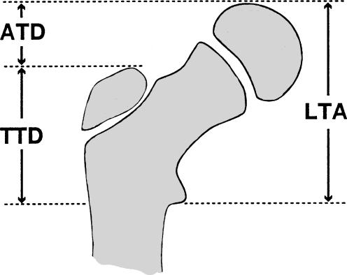
Radiographic measurements The growth of the greater trochanter was measured using the trochanter-to-trochanter distance (TTD) [5]. The TTD is the distance, measured radiographically, from the tip of the greater trochanter to the middle of the lesser trochanter along a line parallel with the anatomical axis of the femur (Fig. 1). The growth of the greater trochanter was determined by measuring the TTD at follow-up and subtracting the TTD measured at the time of surgery. This measurement is independent of the growth of the capital femoral epiphysis, as determined by Gage and Cary [5]. The growth of the greater trochanter that underwent an epiphysiodesis was compared to the growth of the opposite (unaffected) hip and the distance was considered as the growth inhibition. Also measured were the articulotrochanteric distance (ATD) [3], which is the distance between the articular surface of the femoral head and the tip of the greater trochanter, and the lesser trochanter-to-articular surface distance (LTA), which is used as a measurement of the growth of the capital femoral epiphysis [5] and is independent of the growth of the greater trochanter (Fig. 1).
Fig. 1.
Radiographic measurements of the articulotrochanteric distance (ATD), trochanter-to-trochanter distance (TTD) and lesser trochanter-to-articular surface distance (LTA)
Surgical technique In 30 patients, an epiphysiodesis was performed by drilling five to six holes across the greater trochanteric physis, followed by screw placement. Five epiphysiodeses were performed with a bone peg placed across the greater trochanteric epiphysis instead of the screw. The choice of surgical technique was based on the surgeon’s preference. In all cases, a varus osteotomy was also performed.
Comparisons by age The growth of the greater trochanter in the 13 patients who underwent GTE before the age of 6 years were compared to 11 patients who were older than 8 years of age at the time of surgery.
Clinical measurements Chart review and clinical examination were performed, which included epidemiological data, evaluation for Trendelenburg gait, operative procedure and complications.
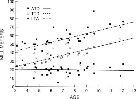
Control group All patients had unilateral Perthes disease, and the unaffected side was used as a paired control and was assumed to be normal. To determine the validity of this assumption, the ATD, TTD and LTA were measured and plotted for the unaffected hip (Fig. 2). We found that the ATD remained constant for different ages, at 20 mm, which is consistent with that reported by other authors [3, 5, 8]. Langenskiöld and Salenius [8] noted that the ATD varied with gender (the ATD for females was 16 mm and for males 23 mm). This compared to our measurements of 16.1 mm and 22.8 mm for females and males, respectively.
Fig. 2.
Scatter plot of the ATD, TTD and LTA measurements versus age for the unaffected hip
Statistical analysis Statistical comparisons were performed with StatMost 32 software. Significance was determined from paired t-tests between the affected and unaffected sides. For comparisons by surgical technique and age, non-parametric (Wilcoxon signed-rank) tests were performed.
Results
Radiographic measurements The greater trochanter that underwent epiphysiodesis grew 4.3 mm (SD ± 8.8) less than the contralateral hip over a 58-month period (p = 0.007). This corresponds to an inhibition of 0.9 mm/year. There was little change in ATD in the affected hip because there was a corresponding inhibition in the growth of the capital femoral epiphysis, presumably due to Perthes disease. In the hip undergoing epiphysiodesis, the greater trochanteric physis was found to be closed radiographically in 21 of 35 hips (60%). Closure on the unaffected hip was found in 11 of 35 hips (35%).
Surgical technique In five patients, an epiphysiodesis was performed using a bone peg instead of screws. In these patients, the growth of the greater trochanter was inhibited by 10.4 mm (SD ± 9.1) over a 71-month period (1.8 mm/year). In the 30 patients who underwent a screw epiphysiodesis, the greater trochanter was inhibited by 3.2 mm (SD ± 8.3) over a 56-month period (0.7 mm/year). Although this difference is large, it was not statistically significant due to the small number of bone peg epiphysiodeses performed.
Comparisons by age Epiphysiodesis performed in patients over the age of 8 years inhibited the growth of the greater trochanter by 1.7 mm/year (8 ± 13 mm over 57 months). Epiphysiodesis performed in patients under the age of 6 years inhibited the growth of the greater trochanter by 0.4 mm/year (1.3 ± 6 mm over 59 months), making this difference statistically significant (p = 0.047). Both groups had two patients who underwent a bone peg epiphysiodesis.
Discussion
The growth of the proximal femur has been previously described [1, 3, 9, 11]. Growth of the greater trochanter is divided equally between the appositional growth in the superior portion of the greater trochanter and growth in the metaphysis. Therefore, GTE, if complete, could, theoretically, arrest growth by 50% [3, 8]. Overall, the growth of the greater trochanter in our control hips (unaffected side) was 18.5 mm, with appositional growth of approximately 9.25 mm, or approximately 2 mm/year. Mean growth inhibition was 4.3 mm over a 57-month period (0.9 mm/year); therefore, 46% of appositional growth was arrested by epiphysiodesis. This is a mean and most likely reflects the fact that some procedures were more successful than others. When a bone peg epiphysiodesis was performed, 85% of the appositional growth was inhibited (1.8 mm/year).
Most other authors use the ATD to report their results of GTE. Unfortunately, this is only an indirect measure of the growth of the greater trochanter and also reflects the growth of the capital femoral epiphysis. Matan et al. [10] reported a mean ATD of 9 mm greater in patients with Perthes disease who underwent a GTE with bone peg, as compared to those who did not over a 4.8-year period. Therefore, the epiphysiodesis inhibited growth by about 1.9 mm/year.
Clinically, the benefit of GTE for Perthes disease is not clear. Several authors have discussed the clinical merits of GTE for the treatment of congenital dislocation of the hip [2–7, 10, 14]. Matan et al. [10] described statistically significant improvements in pain, range of motion, abductor strength and activity levels for patients with Perthes disease treated with intertrochanteric osteotomy and GTE compared to those treated with intertrochanteric osteotomy alone. Gage and Cary [5] state that the decrease in ATD is minimal after GTE, although the reduction of ATD is probably more significant in hips undergoing varus femoral osteotomy. Some authors [3, 5] feel that, for epiphysiodesis to have a beneficial clinical effect, it must be performed in younger children. Stevens and Coleman [13], in a review of 35 greater trochanteric epiphysiodeses for coxa breva, felt that effective stabilisation of the ATD was achieved if epiphysiodesis was performed before 8 years of age, but progressive loss of ATD would result if epiphysiodesis was performed after that. Although our study did not focus on the clinical results, we did find that a Trendelenburg gait was noted in only seven of the 27 patients tested and that the new ATD was less (9.1 mm vs. 14.4 mm) for those with a positive Trendelenburg gait.
Surprisingly, when we grouped our patients by age, we found that the epiphysiodesis was more effective in those over the age of 8 years than in those less than 6 years of age. The GTE in children from 6 to 8 years of age had less inhibition than those greater than 8 years of age and more than those less than 6 years of age. Matan et al. [10] demonstrated both clinical and radiographic benefits of GTE in a patient population with an average age of 8 years. The increased rate of inhibition in the older children may be due to the increased growth during this time period or due to the fact that the epiphysiodesis is technically easier at this age. As the younger children are followed to maturity, we would expect the overall growth inhibition to be greater in the younger children.
Most authors previously describe a technique for GTE as a modification of the Phemister technique [1, 12]. The technique of multiple drill holes and screw fixation is a modification of this technique. In our study, we had more success with bone peg epiphysiodesis.
Conclusion
From this study, the following conclusions can be drawn: (1) inhibition of the growth of the greater trochanter was obtained in children undergoing greater trochanteric epiphysiodesis (GTE); (2) the use of a bone peg epiphysiodesis tended to be more effective than the screw epiphysiodesis; and (3) epiphysiodesis after the age of 8 years may still have an effect on the growth of the greater trochanter.
References
- 1.Compere EL, Garrison M, Fahey JJ (1940) Deformities of the femur resulting from arrestment of growth of the capital and greater trochanteric epiphyses. J Bone Joint Surg Am 22:909–915
- 2.Cooperman DR, Stulberg SD (1986) Ambulatory containment treatment in Perthes’ disease. Clin Orthop Relat Res 203:289–299 [PubMed]
- 3.Edgren W (1965) Coxa plana. A clinical and radiographic investigation with particular reference to the importance of the metaphyseal changes for the final shape of the proximal part of the femur. Acta Orthop Scand Suppl 84:14–19 [PubMed]
- 4.Evans IK, Deluca PA, Gage JR (1988) A comparative study of ambulation-abduction bracing and varus derotation osteotomy in the treatment of severe Legg-Calve-Perthes disease in children over 6 years of age. J Pediatr Orthop 8(6):676–682 [DOI] [PubMed]
- 5.Gage JR, Cary JM (1980) The effects of trochanteric epiphyseodesis on growth of the proximal end of the femur following necrosis of the capital femoral epiphysis. J Bone Joint Surg Am 62(5):785–794 [PubMed]
- 6.Iwersen LJ, Kalen V, Eberle C (1989) Relative trochanteric overgrowth after ischemic necrosis in congenital dislocation of the hip. J Pediatr Orthop 9(4):381–385 [PubMed]
- 7.Kalamchi A, MacEwen GD (1980) Avascular necrosis following treatment of congenital dislocation of the hip. J Bone Joint Surg Am 62(6):876–878 [PubMed]
- 8.Langenskiöld A, Salenius P (1967) Epiphyseodesis of the greater trochanter. Acta Orthop Scand 38(2):199–219 [DOI] [PubMed]
- 9.Laurent LE (1959) Growth disturbances of the proximal end of the femur in the light of animal experiments. Acta Orthop Scand 28:255–261 [DOI] [PubMed]
- 10.Matan AJ, Stevens PM, Smith JT, Santora SD (1996) Combination trochanteric arrest and intertrochanteric osteotomy for Perthes’ disease. J Pediatr Orthop 16(1):10–14 [DOI] [PubMed]
- 11.Ogden JA (1981) Hip development and vascularity: relationship to chondro-osseous trauma in the growing child. Hip 9:139–187 [PubMed]
- 12.Phemister DB (1933) Operative arrestment of longitudinal growth of bones in the treatment of deformities. J Bone Joint Surg Am 15:1–15
- 13.Stevens PM, Coleman SS (1985) Coxa breva: its pathogenesis and a rationale for its management. J Pediatr Orthop 5(5):515–521 [PubMed]
- 14.Weiner SD, Weiner DS, Riley PM (1991) Pitfalls in treatment of Legg-Calve-Perthes disease using proximal femoral varus osteotomy. J Pediatr Orthop 11(1):20–24 [DOI] [PubMed]


