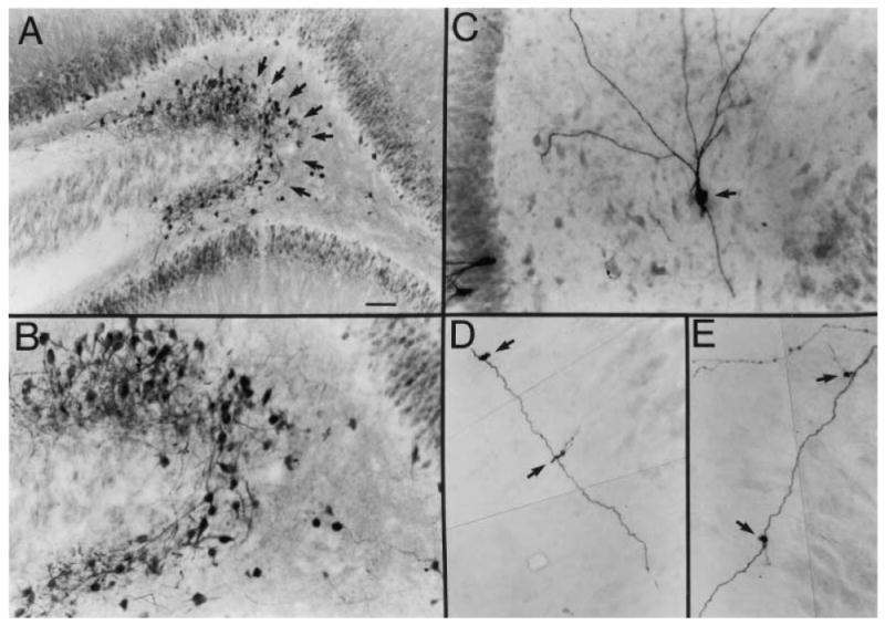
Fig. 2.

Newly born granule cells in the hilus after seizures. A, The dentate gyrus of a pilocarpine-treated rat after status epilepticus and spontaneous recurrent seizures, stained using an antibody to the calcium binding protein calbindin D28K. There is a novel population of cells at the hilar/CA3 border that is not normally present. Many of these cells were double labeled with bromodeoxyuridine (BrdU), a marker of cells undergoing cell division, following BrdU injection after pilocarpine-induced status epilepticus, which indicates that these cells were born after seizures (Scharfman and others 2000). GCL = granule cell layer; HIL = hilus; PCL = pyramidal cell layer. Calibration = 100 μm. B, A higher magnification of the region at the hilar/CA3 border in part A shows the calbindin-immunoreactive cells at the CA3/hilar border (arrows). Calibration (in A) = 50 μm. C, An intracellularly labeled hilar neuron (arrow), recorded in a hippocampal slice from a pilocarpine-treated rat that had spontaneous seizures. This neuron had the morphology and electrophysiology of a granule cell but was located in the hilus and oriented differently from normal granule cells, some of which were stained for comparison (asterisks). Calibration same as B. The end of the pyramidal cell layer is on the right. D, Mossy fiber “giant” boutons (arrow), which distinguish granule cell axons from axons of other cell types, were located along the axons of the hilar/CA3 granule cells such as those shown in part C. This piece of the mossy fiber had two giant boutons. Calibration (in A) = 25 μm.
From Scharfman and others (2000), with permission from the Society for Neuroscience.
