Abstract
We determined the effects of intravenous infusion of amino acids (AA) at serum insulin of 5, 30, 72, and 167 mU/l on anabolic signaling, expression of ubiquitin-proteasome components, and protein turnover in muscles of healthy young men. Tripling AA availability at 5 mU/l insulin doubled incorporation of [1-13C]leucine [i.e., muscle protein synthesis (MPS), P < 0.01] without affecting the rate of leg protein breakdown (LPB; appearance of d5-phenylalanine). While keeping AA availability constant, increasing insulin to 30 mU/l halved LPB (P < 0.05) without further inhibition at higher doses, whereas rates of MPS were identical to that at 5 mU/l insulin. The phosphorylation of PKB Ser473 and p70S6k Thr389 increased concomitantly with insulin, but whereas raising insulin to 30 mU/l increased the phosphorylation of mTOR Ser2448, 4E-BP1 Thr37/46, or GSK3β Ser9 and decreased that of eEF2 Thr56, higher insulin doses to 72 and 167 mU/l did not augment these latter responses. MAFbx and proteasome C2 subunit proteins declined as insulin increased, with MuRF-1 expression largely unchanged. Thus increasing AA and insulin availability causes changes in anabolic signaling and amounts of enzymes of the ubiquitin-proteasome pathway, which cannot be easily reconciled with observed effects on MPS or LPB.
Keywords: muscle protein synthesis, muscle protein breakdown
human muscle protein balance can be switched from negative to positive by the ingestion of a mixed meal (27). It is now well recognized that the components of mixed food are likely to have different effects on human muscle protein turnover. The stimulation of muscle protein synthesis (MPS) observed on consumption of mixed food appears to be mainly a result of the increased availability of amino acids; thus amino acids alone appear to be able to stimulate human MPS (3, 11, 30) in a dose-response manner (6, 8) and at fixed postabsorptive concentrations of plasma insulin (8). However, there is little consensus with regard to insulin; many workers have found that insulin alone at concentrations above those in the postabsorptive state, whether delivered systemically or by close arterial infusion, has no stimulatory effect upon human MPS (see Refs. 9, 14, 22, and 33), and its presence is not required for the stimulation resulting from increased availability of amino acids (6, 8). Others report a stimulation of MPS with close arterial infusion (4, 13, 26), and the difference has been suggested to be due to the preservation of increases in intramuscular amino acid availability (13, 26). Certainly, when amino acid delivery is increased together with insulin, there is no difficulty in observing a stimulation of MPS measured by incorporation of tracer amino acids into protein or by their disappearance from arterial blood (1, 16). However, insulin appears to inhibit muscle protein breakdown (MPB) independently of the presence of amino acids (14, 22), although amino acids may enhance the effect (10, 23).
Nevertheless, our current understanding of the molecular mechanisms by which human muscle protein turnover is modulated by amino acids and insulin is poor. There is evidence that the action of signaling molecules such as PKB, mammalian target of rapamycin (mTOR), p70 S6 kinase (p70S6k), and 4E-binding protein-1 (4E-BP1), as in rats (17, 19), is involved in the modulation of human MPS since these are also activated after increased amino acid provision (8, 12, 12, 20, 21). There is, however, very little data regarding the effects of insulin on anabolic signaling in human muscle. Raising insulin about 10-fold to a postprandial concentration (∼53 mU/l) increased p70S6k phosphorylation to about the same extent as doubling blood amino acid concentrations but without increasing activity of an index of eukaryotic initiation factor (eIF)-4E-BP1 (15). Unfortunately, these observations were made without any concomitant observations of MPS.
The current understanding of the molecular control of human muscle protein breakdown via amino acids and insulin availability is even more sparse. There is no mechanistic information from work in human muscle showing how insulin might reduce MPB, although there is evidence that insulin-stimulated PKB activity induces phosphorylation of the FOXO family of transcription factors (18, 32), which inhibits their transcriptional activity by localizing them to the cytoplasm. FOXO1 and -3 regulate the transcription of components of the ubiquitin-proteasome system, including the ubiquitin ligases muscle atrophy F-box (MAFbx) and muscle-specific RING finger-1 (MuRF-1), which are proposed to be central to the control of muscle proteolysis in animal muscle (5). Clearly, this could be a mechanism for reduction of MPB by insulin, although there is no direct evidence of this by concurrent measurements of proteolysis, and certainly none in human muscle. Thus there remain major gaps in our understanding of the molecular mechanisms underlying control of human muscle protein turnover. We wished to fill these gaps and chose to carry out our work not on rodents or in cultured muscle but in human beings in whom we could not only measure the effects of metabolic manipulation on protein turnover but also make measurements of muscle gene and protein expression and phosphorylation of signaling molecules. In particular, we wished to study the effects of insulin when they would be unaffected by variation in supply of amino acids, so we chose to make amino acids available at a high concentration, sufficiently high so that any effects of alteration of blood flow or delivery would be obviated.
Thus the purpose of the present work was to gain a more complete mechanistic, molecular understanding of how insulin promotes positive protein balance in human muscle in the presence of a sufficiency of amino acids. We have therefore measured indexes of muscle anabolic signaling (phosphorylation of proteins in the PKB-mTOR-p70S6k pathway), protein expression for the C2 subunit of the proteasome, and mRNA and protein expression for MAFbx and MuRF-1 while concurrently measuring MPS (as incorporation of stable isotope-labeled tracer leucine) and leg protein turnover [both leg protein synthesis (LPS; as rate of disappearance of phenylalanine) and leg protein breakdown (LPB; as rate of appearance of phenylalanine across the leg), both determined using stable isotope-labeled tracer phenylalanine]. Using this approach, we wished to determine 1) the effects of increasing blood amino acid availability while maintaining insulin at a fasting postabsorptive concentration and 2) the dose-response relationship between insulin availability and a) phosphorylation of signaling molecules, b) expression of components of the ubiquitin-proteasome system, and c) various indexes of muscle protein turnover. We hypothesized that there would be coherent changes in the extent of phosphorylation of signaling molecules and of the expression and amounts of proteolytic enzymes that would match the changes in size and direction of measured aspects of muscle protein turnover. The results of testing these hypotheses should provide valuable insights into the importance of these molecules for the regulation of MPS and MPB measured concurrently.
MATERIALS AND METHODS
Subjects
Eight healthy young men (age 20.4 ± 2.0 yr, body mass index 23.8 ± 2.6 kg·m−2) participated in the study, which was approved by the University of Nottingham Medical School Ethics Committee and carried out in accordance with the Declaration of Helsinki. Before taking part, all subjects underwent routine medical screening and completed a general health questionnaire. All gave their informed consent to take part in the study and were aware that they were free to withdraw from the experiment at any point.
Experimental Protocol
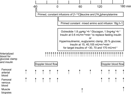
The subjects were asked to eat their normal diet and to refrain from strenuous exercise for 72 h before attending the laboratory on four occasions. The protocol (Fig. 1) was designed to allow the measurement of MPS (by incorporation of [1-13C]leucine into quadriceps protein), LPS as rate of disappearance of phenylalanine (i.e., LPB-leg balance), and LPB as rate of dilution of [2H5]phenylalanine (27) under graded steady-state serum insulin concentrations, but all with increased availability of blood amino acids resulting from infusion of amino acids at a fixed high rate of 18 g/h. Subjects were studied on the four occasions, with protocols that differed only in the nature of the procedures necessary to obtain target serum insulin concentrations of ∼5, 30, 70, and 170 mU/l, being equivalent to fasted, fed (30 and 70 mU/l), and physiologically high (170 mU/l) insulin concentrations.
Fig. 1.
Study protocol.
Upon arrival at the laboratory, in a postabsorptive state, not having eaten since 2000 the evening before, subjects were instructed to void their bladders, and their body mass was measured. Subjects then rested semisupine on a bed before femoral artery and vein catheters (ES-04150; Arrow Deutschland, Erding, Germany) were inserted for blood sampling. Venous catheters (Venflon, Viggo, Denmark) were also sited retrogradely in a dorsal hand vein for sampling of arterialized venous blood and antegradely at the antecubital fossa in both forearms for infusion of mixed amino acids as well as octreotide, glucose, and insulin to clamp insulin concentrations at various values. Patency of the lines was maintained using 0.9% NaCl infusions (Baxter Healthcare, Norfolk, UK).
On each occasion, primed constant infusions of stable isotope-labeled amino acids (Cambridge Isotope Laboratories, Andover, MA) were given throughout 270 min. Each experimental visit consisted of priming doses of [2H5]phenylalanine (0.3 mg/kg body mass, 98 atom%) with [1-13C]leucine (0.8 mg/kg body mass, 99 atom%) in 0.9% NaCl. Thereafter, constant infusions of 0.5 mg·kg−1·h−1 phenylalanine and 1.0 mg·kg−1·h−1 leucine in 0.9% NaCl were maintained throughout the experimental session. After 90 min, a primed constant infusion of mixed amino acids (Glamin; Fresenius-Kabi, Runcorn, UK) at 126 ml/h (to deliver 18 g of amino acids/h) was begun concomitantly with an insulin clamp (29), using Actrapid insulin (Novo Nordisk, Bagsvaerd, Denmark). The insulin infusion rates used were 0.6, 15, 40, or 105 mU·m body surface area−2·min−1; these were randomized between visits. Blood glucose concentration was measured at 5-min intervals in samples of arterialized venous blood, and a variable infusion of 20% glucose (Baxter Healthcare) was initiated to maintain a plasma glucose concentration of 4.5 mmol/l during the insulin infusion. An aliquot of the samples obtained every 10 min during the period of the insulin clamp was allowed to clot and used for serum insulin analysis. Endogenous insulin production was suppressed during the 0.6 mU·m−2·min−1 insulin infusion by concurrent infusion of octreotide (1.8 μg·kg body mass−1·h−1 Sandostatin; Novartis), and postabsorptive glucagon concentration was maintained by infusion of glucagon (15 ng−1·kg body mass−1·h−1 GlucaGen, HypoKit; Novo Nordisk). Femoral arterial blood flow was measured in the leg contralateral to that in which the cannulae were inserted at frequent intervals throughout the 270-min protocol using Doppler ultrasound (5 mHz convex linear array transducer, Diasonics Prisma; Diasonics International, Les Ulis, France). Arterial and femoral venous blood samples (2 ml) were taken for blood tracer measurements at baseline, −60, −50, −40, −30, −5, +30, +60, +90, +120, +150, +165, and +180 min (Fig. 1). Quadriceps muscle biopsy samples were taken from the leg in which arterial and venous cannulae were placed, using a 5-mm Bergstrom needle after appropriate local anesthesia with 1% lignocaine on two occasions, at 60 and again at 180 min after amino acid infusion began.
Sample Processing and Analysis
Sampled blood was placed in preweighed tubes containing 4 ml of 1 M HClO4; the tubes containing the blood were weighed again and centrifuged (3,000 rpm, 10 min) to precipitate the blood debris. The supernatant was neutralized with KOH/KHCO3 and stored until analysis. Blood for insulin analysis was allowed to clot, and after centrifugation (3,000 rpm, 10 min) the serum was stored frozen at −80°C.
All muscle samples were, on collection, immediately freed from blood and visible connective and fat tissue, rapidly frozen in liquid nitrogen, and stored at −80°C before analyses.
Blood Glucose and Serum Insulin
Glucose was measured using a glucose analyzer (YSI 2300 STATplus; Yellow Springs Instruments, Yellow Springs, OH). Insulin was measured with a radioimmunoassay kit (Coat-a-Count Insulin; Diagnostics Products).
Gene Expression
RNA extraction.
Total RNA was extracted from frozen muscle biopsy samples obtained after 180 min of amino acid and insulin infusion, as described by constantin et al. (7). Approximately 30 mg of frozen wet muscle tissue was homogenized for 30 s using a polytron homogenizer in 800 μl of Trizol reagent (Invitrogen, Paisley, UK) containing 200 μg of glycogen. An aliquot of each extracted RNA sample was then quantified spectrophotometrically to determine purity and concentration by absorbance at 260 nm and the 260:280 nm ratio.
Reverse transcription.
Reverse transcription was carried out with 0.5 μg total RNA, using Moloney murine leukemia virus reverse transcriptase (Promega, Southampton, UK) and random hexamer primers, in a total volume of 30 μl containing RNAse inhibitor (Promega) at room temperature for 10 min. This was followed by 1-h incubation at 42°C, after which 25 μl of cDNA was diluted fourfold with RNAse-free H2O to give a 100-μl cDNA pool.
Real-time quantitative PCR.
Real-time PCR was performed using PCR universal mix (Applied Biosystems) in a MicroAmp 96-well reaction plate. Each well contained a final total reaction volume of 25 μl. The probes for detection of mRNA species of interest (Table 1) were purchased from Applied Biosystems (Taqman probe). All samples and nontemplate control reactions were performed in triplicate. All reactions were performed in the ABI Prism 7000 (Applied Biosystems). The PCR products were run on a 3% agarose gel and stained with ethidium bromide to check for band size and product purity. Finally, the standard curve method was utilized to measure relative mRNA changes. Ribosomal 18S RNA was used as a housekeeping gene. All values were expressed relative to the 18S expression.
Table 1.
Absolute mRNA expression concentrations at the end of each insulin clamp for selected genes implicated in human skeletal muscle protein turnover
| Gene |
Serum Insulin Achieved, mU/l |
|||
|---|---|---|---|---|
| 5 | 30 | 72 | 167 | |
| PKB | 3.96±0.69 | 5.63±1.28 | 3.68±0.39 | 5.45±0.16 |
| mTOR | 3.76±0.74 | 3.38±0.47 | 3.28±0.71 | 4.46±0.94 |
| p70S6k | 4.11±0.86 | 4.04±0.78 | 3.88±0.62 | 5.61±1.13 |
| GSK-3β | 0.72±0.30 | 0.43±0.16 | 0.85±0.38 | 0.59±0.24 |
| MAFbx | 3.72±1.38 | 3.13±1.29 | 2.70±1.17 | 4.22±1.74 |
| MuRF-1 | 1.00±0.58 | 0.76±0.44 | 0.92±0.49 | 0.77±0.39 |
| Calpain 2 | 7.25±3.15 | 7.31±2.88 | 5.44±2.53 | 8.91±3.82 |
| Calpain 3 | 0.70±0.27 | 0.76±0.24 | 0.79±0.33 | 0.74±0.26 |
| Calpastatin | 0.74±0.38 | 0.70±0.35 | 0.66±0.32 | 0.71±0.35 |
Data expressed as means ± SE. mTOR, mammalian target of rapamycin; p70S6k, p70 S6 kinase; MAFbx, muscle atrophy F-box; MuRF-1, muscle-specific RING finger-1. No significant difference was observed between any treatment groups.
Western Analysis of Signaling Proteins, the C2 Proteasome Subunit, and Muscle-Specific E3 Ligases
Approximately 50 mg of each muscle biopsy sample obtained after 180 min of amino acid and insulin infusion was homogenized on ice in a phosphatase inhibitor buffer (unless otherwise stated, all reagents were purchased from Sigma-Aldrich, Poole, UK; 50 mM Tris·HCl, 1% Triton X-100, 1 mM EDTA, 1 mM EGTA, 50 mM sodium fluoride, 10 mM β-glycerophosphate, 0.1% 2-mercaptoethanol, 100 nM okadaic acid) at pH 7.5 for 15 s. The samples were then rotated for 1 h at 4°C and centrifuged at 13,000 g for 10 min at 4°C. The supernatant was removed, and protein concentrations were quantified with a Bradford assay.
For phosphorylated forms of PKB (PKB Ser473), mTOR (mTOR Ser2448), p70S6k (p70S6k Thr389), eIF-4E-BP1 (eIF-4E-BP1 Thr37/46), eukaryotic elongation factor 2 (eEF2 Thr56), and glycogen synthase kinase 3-β (GSK-3β Ser9), 20 μg of protein was electrophoresed on a 4 to 12% NuPAGE Novex Bis-Tris gel (Invitrogen, Paisley, UK) for 2.5 h at 40 mA. For total protein assay of MAFbx (or atrogin 1) and MuRF-1, 5 μg of protein was electrophoresed on a 4 to 12% NuPAGE Novex Bis-Tris gel, and for the C2 proteasome subunit, 50 μg protein was separated on a 12% Tris·HCl criterion gel (Bio-Rad, Hemel Hempstead, UK). Proteins were then transferred by electrophoresis overnight onto nitrocellulose membrane (Hybond-C Extra; Amersham, Buckinghamshire, UK) or 0.2 μm PVDF for the C2 subunit, after which the membrane was placed in a blocking buffer [5% BSA-TBS-T (Tris-buffered saline and 0.1% Tween-20)] for 1 h before incubation overnight at 4°C with primary antibody in 5% BSA-TBS-T. Antibodies for PKB Ser473, mTOR Ser2448, p70S6k Thr389, 4E-BP1 Thr37/46, eEF2 Thr56, and GSK-3β Ser9 were obtained from Cell Signaling Technology (Danvers, MA), for the C2 proteasome from Abcam (Cambridge, UK), and for α-actin from Sigma-Aldrich (Poole, UK). MuRF-1 and MAFbx polyclonal anti-rabbit antibodies were produced in house by Pfizer (Groton, CT) and were provided as a gift. The following day, membranes were washed (5 × 5 min) in TBS-T before incubation for 1 h (1:2,000 5% BSA-TBS-T, with the exception of actin at 1:5,000) in secondary anti-rabbit antibody (DAKO).
The blots were then washed (5 × 5 min) and incubated for 2 min in Western Lightning Chemiluminescence Reagent Plus (PerkinElmer Life Sciences, Boston, MA) and developed using chemiluminescence film (Hyperfilm-enhanced chemiluminescence; Amersham). Chemiluminescent films were scanned and quantified using the Syngene densitometry software (Syngene, Cambridge, UK). Results are expressed as the ratio between phosphorylated protein level and α-actin, with the exception of MAFbx, MuRF-1, and the C2 proteasome, all expressed as the ratio of total protein level and α-actin (loading control).
MPS and Leg Protein Turnover
Muscle protein.
Muscle (25 mg) was ground in liquid nitrogen, and mixed muscle protein was precipitated using 1 M perchloric acid and the pellet washed twice with 70% ethanol.
Muscle protein was hydrolyzed in 0.05 M HCl-Dowex 50W-X8-200 (Sigma, Poole, UK), and the liberated amino acids were purified on cation exchange columns (Dowex 50W-X8-200) (31, 32). The amino acids were then converted to their n-acetyl-N-propyl ester derivative and the leucine labeling determined by gas chromatography-combustion-isotope ratio mass spectrometry (Delta-plus XL; Thermofinnigan, Hemel Hempstead, UK) (33, 34).
Plasma amino acid labeling.
To determine labeling (atom %excess) of plasma and tissue free leucine, plasma α-ketoisocaproate (KIC) and phenylalanine proteins were precipitated, and the supernatant, containing free amino acids, was collected to prepare the tert-butyldimethylsilyl (t-BDMS; leucine) and OPDA-t-BDMS (KIC) derivative for analysis by gas chromatography-mass spectrometry (MD800; Fisons, Ipswich, UK) by using electron ionization and selective ion monitoring (35, 36). Blood leucine and phenylalanine concentrations were determined using gas chromatography-mass spectrometry and appropriate standard curves.
Calculations.
The rate of MPS between the biopsies was calculated using the standard equation fractional protein synthesis (%/h) = (ΔEm/Ep × 1/t) × 100, where ΔEm is the change in labeling of muscle protein leucine between two biopsy samples, Ep is the mean enrichment over time of the precursor for protein synthesis (taken as venous α-KIC 13C labeling), and t is the time between biopsies. Venous α-KIC was chosen to represent the immediate precursor for protein synthesis, i.e., leucyl-tRNA (37, 38).
The net amino acid balance was calculated as the difference in arterial and venous concentrations multiplied by the blood flow. LPB was calculated from the arterio-venous dilution of [2H2]phenylalanine tracer amino acid using the following equation: {[(EA/EV) − 1] × Cv × BF}, where Ea and Ev (1) are the values of phenylalanine labeling at steady state in arterial and femoral venous plasma, respectively, where Ca is the mean concentration in the arterial blood, and BF is the blood flow in ml/leg. The values for LPS were calculated as the disappearance into the leg of phenylalanine, i.e., {[(Ea/Ev) − 1] × Ca } − (Ca − Cv) × BF, where Cv is the concentration of phenylalanine in femoral venous blood. Each set of samples taken with accompanying blood flows was used to calculate the values of leg protein metabolism over the hour of basal (postabsorptive) and varied insulin availability. The basal values from all studies were averaged so that these represent the mean of 8 × 4 × 4 individual estimates of basal muscle protein metabolism. The values presented from measurements at various insulin concentrations were the mean of 8 × 4 estimates.
Statistical Analysis
All data are reported as means ± SE. Comparisons between intervention groups were performed using one-way ANOVA. When a significant F ratio was achieved, differences were then located using the least significent difference post hoc test. Where appropriate, Student's paired t-test was used. Significance of difference was defined as P ≤ 0.05 on all occasions.
RESULTS
Serum Insulin
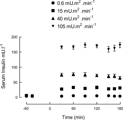
Serum insulin in the postabsorptive state before the amino acid infusion or the insulin clamps were instituted was 5.0 ± 0.47 mU/l (Fig. 2). The experimental procedures performed to achieve steady-state serum insulin concentrations of ∼5, 30, 70, and 170 mU/l were successful. Actual mean steady-state values achieved over the final 150 min of insulin clamp were 5, 30, 72, and 167 mU/l.
Fig. 2.
Arterialized venous serum insulin concentrations before and during 3 h of continuous insulin infusion aimed at achieving steady-state insulin concentrations of ∼5, 30, 70, and 170 mU/l. Values are means ± SE (some error bars lie within the symbols).
Blood Amino Acid Concentrations and Leg Blood Flow
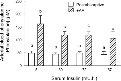
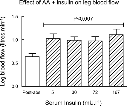
Blood amino acid concentrations rose rapidly from postabsorptive values to a new steady-state value of about three times the basal values (Fig. 3). The changes in phenylalanine were indicative of the changes in all essential amino acids, as reported previously (8). Postabsorptive leg blood flow was 0.64 ± 0.07 l/min and increased when amino acids were given, and when insulin was clamped at the postabsorptive serum insulin concentration (5 mU/l), to 1.00 ± 0.10 l/min (P < 0.01) (Fig. 4). There were no further increases of leg blood flow when insulin was raised to 30, 72, or 167 mU/l.
Fig. 3.
Blood phenylalanine concentrations (means ± SE) in postabsorptive state and 3 h after infusion of mixed amino acids (AA; Glamin) at 18 g/h. Different letters denote significance of difference, at least P < 0.01.
Fig. 4.
Leg blood flow (means ± SE) in the postabsorptive (post-abs) state and after insulin infusion to serum concentrations shown. The values during insulin infusion are collectively different from the post-abs value, i.e., no added AA (ANOVA, P < 0.007), and the values at 5 mU/l are significantly different from the post-abs value ( P < 0.01).
Muscle mRNA Expression
No significant difference was observed between any treatment groups for mRNA expression for PKB, mTOR, p70S6k, GSK-3β, MAFbx, MuRF-1, calpain-2, calpain-3/p94, or calpastatin in biopsies taken at the end of the clamps (Table 1).
Effect of Amino Acid and Insulin Availability on Anabolic Signaling
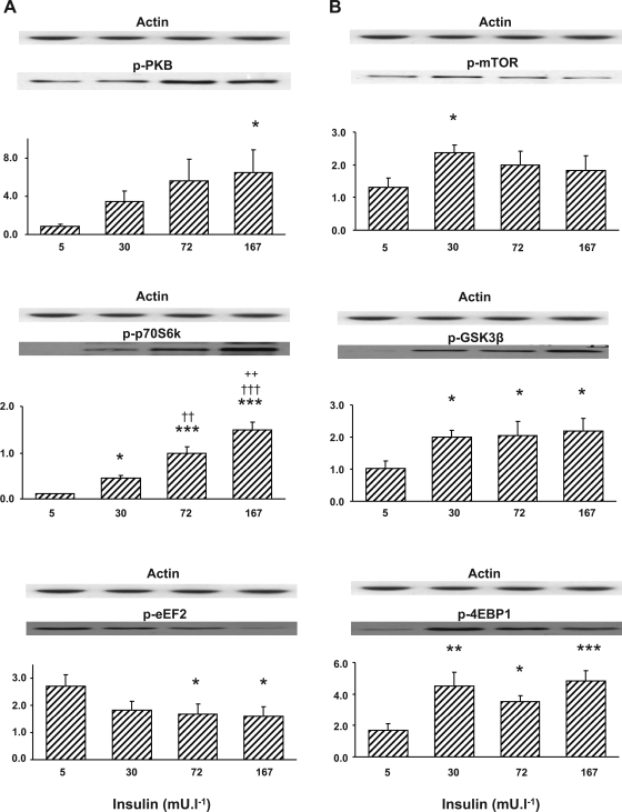
Increasing serum insulin concentration during the mixed amino acid infusion produced insulin dose-related increases in the phosphorylation status of PKB Ser473 and p70S6k Thr389 and dose-related decreases in eEF2 Thr56 phosphorylation status relative to the values at 5 mU/l (Fig. 5A). The phosphorylation of mTOR Ser2448, GSK-3β Ser9, and 4E-BP1 Thr37/46 all showed an increase at an insulin concentration of 30 mU/l (Fig. 5B); no further changes in the phosphorylation status of these signaling molecules were observed with increasing insulin availability.
Fig. 5.
A: PKB Ser473, p70 S6 kinase (p70S6k) Thr389, and eukaryotic elongation factor-2 (eEF2) Thr56 phosphorylation after a 3-h insulin clamp at 5, 30, 72, and 167 mU/l. Significantly different from 5 mU/l: *P < 0.05; ***P < 0.001. Significantly different from 30 mU/l−1: ††P < 0.01; †††P < 0.001. Significantly different from 72 mU/l: ++P < 0.01. Values are means ± SE (arbitrary units) corrected for actin. Representative Western blots are shown above the bars. B: mammalian target of rapamycin (mTOR) Ser2448, GSK-3β Ser9, and 4E-binding protein-1 (4E-BP1) Thr37/46 phosphorylation after a 3-h insulin clamp at 5, 30, 72, and 167 mU/l. Significantly different from 5 mU/l: *P < 0.05; **P < 0.01; ***P < 0.001. Values are means ± SE (arbitrary units) corrected for actin. Representative Western blots are shown above the bars.
Effect of Increased Amino Acid and Insulin Availability on Protein Expression, the Proteasome C2 subunit, and MAFbx and MuRF-1
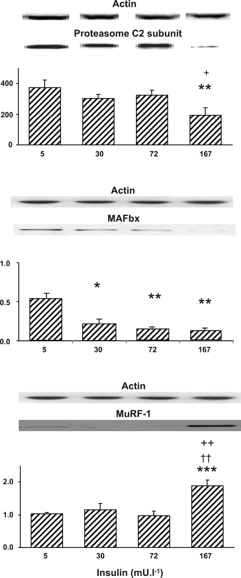
Increasing serum insulin concentrations to 30, 72, and 167 mU/l produced graded dose-related reductions in MAFbx protein expression to 40 (P < 0.05), 25 (P = 0.01), and 18% (P < 0.01), respectively, of values measured at 5 mU/l (Fig. 6). However, MuRF-1 total protein was the same at 5, 30, and 72 mU/l, and, paradoxically, it rose significantly at 167 mU/l. There was a trend for expression of the proteasome C2 subunit protein to fall at insulin concentrations of 30–72 mU/l, and this fall became significant at an insulin concentration of 167 mU/l, when its expression declined by ∼50% compared with ∼5 mU/l (P < 0.01).
Fig. 6.
Proteasome C2 subunit, muscle atrophy F-box (MAFbx), and muscle-specific RING finger-1 (MuRF-1) total protein concentrations after a 3-h insulin clamp at 5, 30, 72, and 167 mU/l. Significantly different from 5 mU/l: *P < 0.05; **P < 0.01; ***P < 0.001. Significantly different from 30 mU/l: ††P < 0.01. Significantly different from 72 mU/l: +P < 0.05; ++P < 0.01. Values are means ± SE (arbitrary units) corrected for actin. Representative Western blots are shown above the bars.
Leg Protein Turnover and MPS
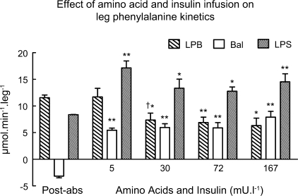
In the postabsorptive state, net leg protein balance was negative, with LPB exceeding LPS. Infusion of amino acids at a clamped serum insulin concentration of 5 mU/l doubled LPS (P < 0.01), with no effect on LPB, and so leg protein balance became significantly positive (P < 0.01; Fig. 7). The effect shown by phenylalanine kinetics for LPB was confirmed by leucine kinetics (data not shown). However, a relatively modest increase in insulin concentration (to 30 mU/l) halved LPB (determined with both amino acid tracers; leucine data not shown) by ∼50% (P < 0.05). Manipulation of insulin concentrations to steady values higher than those normally seen in the postprandial state (i.e., 72 and 167 mU/l) maintained the net positive leg protein balance through increased LPS and reduced LPB, without any differences in the measured variables between 5 and 167 mU/l for LPS or between 30 and 167 mU/l for LPB.
Fig. 7.
Leg protein turnover as leg phenylalanine kinetics. LPB, leg protein breakdown as phenylalanine appearance; LPS, leg protein synthesis as phenylalanine disappearance; Bal, leg phenylalanine balance. Significantly different from postabsorptive: *P < 0.05; **P < 0.01. †P < 0.05, significantly different from value at 5 mU/l. The values of LPS, LPB, and Bal were not significantly different from each other at 30, 72, and 167 mU/ml of insulin. Values are means ± SE.
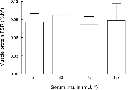
Rates of mixed MPS were effectively identical at all values of serum insulin between 5 and 167 mU/l at a mean value 0.088 ± 0.07%/h (Fig. 8). This is approximately double the value likely to have existed before the amino acid infusion of 0.044%/h, which has been observed routinely in postabsorptive men (e.g., see Refs. 3, 25, and 31). Although we did not have a measurement of MPS in the basal postabsorptive state, the values achieved after amino acids were increased in conjunction with various insulin concentrations were similar to those we and others have reported by increasing amino acid availability in previous work (3, 31).
Fig. 8.
Muscle protein synthesis as incorporation of [1-13C]leucine. Values are not different from one another but about twice the expected value in post-abs human muscle, i.e., 0.044%/h (see text). FSR, fractional synthesis rate.
DISCUSSION
The main purpose of the present work was to gain a more complete mechanistic molecular understanding of how insulin promotes positive protein balance in human muscle by varying its availability during constant availability of amino acids and simultaneously measuring muscle protein turnover, gene expression, phosphorylation of key anabolic signaling molecules, and amounts of proteolytic enzymes. We hypothesized that there would be coherent changes in the extent of phosphorylation of signaling molecules and of the expression and amounts of proteolytic enzymes that would match the changes in size and direction of measured aspects of muscle protein turnover.
Some of the present results were predictable from what had previously been reported [e.g., the effects of amino acids on MPS (1–3, 8), insulin plus amino acids and the effects of insulin on LPB (14, 22)]. However, it was surprising that adding insulin at higher than systemic postabsorptive concentrations had no further effects on MPS or LPS. Some workers have reported increases of MPS and LPS with close arterial insulin contraction without making additional amino acids available (4, 13). We cannot account for these differences except to raise the possibility that in our studies the stimulatory effects of amino acids stimulated protein synthesis to a maximal extent and that further addition of insulin had no additional effect. That this is true is supported by the results of studies in which the effects of insulin plus hyperaminoacidemia vs. hyperaminoacidemia were studied in the forearms of healthy young men (11); raising insulin 10-fold had no significantly greater effect on phenylalanine rate of disappearance compared with doubling amino acid delivery. However, presentation of supraphysiological insulin concentrations in the presence of a doubling of postprandial amino acid concentrations did increase phenylalanine disappearance twofold above the basal rate (16).
The results we obtained for gene expression, insulin signaling, and expression of mRNA and proteins associated with the ubiquitin-proteasome proteolytic pathway are, taken together, highly surprising. These included the dose-related increases in the extent of phosphorylation of PKB and p70S6k without any change in muscle protein synthesis and the dose-related decreases in expression of MAFbx and C2 proteasome subunit protein without any change in muscle protein breakdown. So far as we are aware, no workers have made measurements of human muscle protein turnover and attempted to combine this with information about relevant anabolic and catabolic signaling events determined simultaneously in the same muscle. Many workers have assumed that the results from animal studies are directly extrapolatable to human muscle. This is manifestly not the case.
In response to increased availability of amino acids and insulin, there appear to be changes in the phosphorylation of anabolic signaling molecules and amounts of enzymes associated with the ubiquitin-proteasome pathway that cannot be easily reconciled with alterations in MPS, LPS, and LPB and that change our view of the nature of regulation of muscle protein balance by amino acids and insulin. Specifically, the insulin dose-related increases in the phosphorylation of PKB and p70S6k and dose-dependent decreases in the phosphorylation of eEF2 are not reflected in any way by dose-related changes in MPS or leg turnover. Although this pattern was not seen for the changes in phosphorylation of mTOR, 4E-BP1, and GSK-3β (instead there were “square wave” increases, i.e., a similar increase at insulin values >5 mU/l), there were in fact square wave changes in leg protein turnover that to some extent matched the changes seen in the signaling phosphorylation. Thus the current view that increases in MPS follow from proportionate alterations in the activity of signaling molecules (indicated by alteration of phosphorylation status after manipulation of insulin and amino acid availability) may be an oversimplification; it is certainly not supported by the present results. However, we recognize that the interpretation of the present findings is made without knowledge of the relationship between the degree of phosphorylation and activity of the signaling kinases, the extent of phosphorylation required to elicit a maximum response, or the temporal relationship between protein phosphorylation and resulting anabolic effects. However, in the meantime, workers in this field may not be justified in using changes in phosphorylation status of members of signaling pathways to imply changes in human MPS, and even with direct measurement of the latter, care will be required to distinguish functional from coincidental changes.
In attempting to match up our observed changes in LPB with alterations in protein expression of the ubiquitin ligases and the C2 proteasome unit, we discovered anomalies as severe as those discussed above. The expression of the proteins appear to show insulin dose-related falls, whereas LPB was decreased by insulin at 30 mU/l and thereafter unchanged at higher insulin availability. In all cases, the rate of LPB was lower than in the postabsorptive condition, except when insulin was maintained at the postabsorptive concentration, which is consonant with the 40% reduction in MAFbx expression; however, further decreases in the expression of MAFbx protein were not associated with any physiological effect on protein breakdown. The lack of any effect on MuRF-1 protein expression and the paradoxical rise at the highest insulin availability are also puzzling, since they do not match the observed behavior of LPB. No clue to the proteolytic activity of the ubiquitin ligases comes from the results for their mRNA expression, which was identical at all insulin concentrations. This lack of an effect of insulin on mRNA expression is surprising given that gene array analysis of the effects of insulin at high doses has been reported to stimulate, within 2–3 h, expression of genes that participate in protein synthesis and breakdown (28, 34). However, this discrepancy is probably attributable to the insulin infusion rate in those studies being markedly above those that we used; certainly, at physiological insulin availability, there is no stimulation or inhibition of gene expression for the proteolytic enzymes we studied. As for the anabolic processes, again it appears that there is a disjunction between the measured behavior of protein turnover and observed molecular changes in enzyme expression; these should provide caution to those who use expression of enzymes as indexes of proteolysis without actually measuring it.
This leaves us with the following question: Is there any physiological significance to the observed changes with elevated insulin in signaling molecule phosphorylation and enzyme protein expressions for protein turnover? One possible link appears to be between the altered phosphorylation of the majority of the signaling molecules at 30 mU/l insulin and above and the decreased expression of MAFbx and possibly the C2 proteasome subunit. No previous workers have observed a fall in the expression of these proteins concomitantly with a fall in bulk proteolysis such as what we observed in human muscle, and this novel observation is in itself important in helping us understand the control of muscle protein balance. It may be that increased insulin availability causes a decrease in proteolysis via a decrease in the activity of the ubiquitin-proteasome pathway through alterations in the phosphorylation status of members of what we normally think of as anabolic signaling pathways. PKB-induced phosphorylation of FOXO transcription factors control ubiquitin ligase expression, resulting in inhibition of the ubiquitin-proteasome protein degradation system in myotubes and mouse muscle (29). There is also evidence from work on myotubes and in mice that FOXO coordinates activities of both the ubiquitin-proteasome pathways and lysosomal proteolysis (24, 35). However, if these functions occur in human muscle, then why the graded responses of phosphorylation of PKB or MAFbx do not result in graded reductions in LPB is puzzling. From our results, the counterargument that the atrogins have a limited role physiologically, at the very least in the noninflammatory state, is strengthened by the lack of any change in mRNA of the ubiquitin ligases and the distinctly different behaviors of MAFbx and MuRF-1 protein, which in many circumstances show similar expression changes (7). In an inflammatory state, the situation may be different. Furthermore, the peroxisome proliferator-activated receptor-δ agonist-mediated switch in rat muscle fuel utilization toward increased fat oxidation also results in decreased PKB protein expression concomitantly with upregulation of MAFbx and MuRF-1 mRNA and protein expression without any obvious change in muscle protein/DNA ratio or proteasome activity (7). Thus, under some physiological conditions, expressions of muscle anabolic signaling proteins and ubiquitin ligases may be modulated by alterations in fuel metabolism without being linked to muscle protein turnover. Such modulation by insulin may underlie the molecular changes we observed in the present study (i.e., the expressions of muscle anabolic signaling proteins and ubiquitin ligases in human muscle may simply reflect changes in insulin availability, rather than have an intrinsic physiological role, particularly at insulin concentrations >30 mU/l). A further possibility is that, although we measured bulk proteolysis, the ubiquitin ligase enzymes may be concerned only in the marking for proteolysis of a minor, possibly slower, tuning over pool of protein. However, it is hard to imagine what this pool could be, because myofibrillar proteins constitute two-thirds of all muscle protein, and although their turnover is slower than that of sarcoplasmic protein, it is still sizeable at about 60% of the mixed protein rate.
In conclusion, we have shown that increasing availability of amino acids and of insulin over the range of 5–167 mU/l doubled LPS and halved LPB without any dose-response relationship with insulin. Changes in signaling activity (indicated by phosphorylation of members of the PKB-mTOR-P70S6k-eIF4F pathway) were not consistent with the observed changes in protein turnover and may simply be reflecting insulin availability. Decreases in the amounts of MAFbx protein and the C2 proteasome unit in response to increased insulin availability were observed for the first time, which provided some mechanistic explanation for the fall in proteolysis, but further alterations in protein expression at higher insulin availability appear to be decoupled from any effect on proteolysis. This is the first demonstration that tight coupling does not exist between muscle insulin availability, the activation status of signaling proteins regulating translation initiation, amounts of proteolytic enzymes, and directly measured rates of protein turnover in human muscle. These findings should stimulate careful examinations of the relationships between the acute temporal changes and dose responses of muscle protein turnover to amino acids and insulin and provide caution to those who use indirect markers of protein turnover as indicative surrogates.
GRANTS
This study was supported principally by research funding provided by Iovate, Mississauga, ON, Canada, and also by the United Kingdom (UK) Medical Research Council (G 99000163), the UK Biotechnology and Biological Sciences Research Council (BB/X510697/1 and BB/C516779/1), and The Wellcome Trust (056885/Z/99) and EXEGENESIS program of the European Community.
Acknowledgments
We thank Pfizer for the supply of MAFbx and MuRF-1 antibodies.
The costs of publication of this article were defrayed in part by the payment of page charges. The article must therefore be hereby marked “advertisement” in accordance with 18 U.S.C. Section 1734 solely to indicate this fact.
REFERENCES
- 1.Bennet WM, Connacher AA, Scrimgeour CM, Jung RT, Rennie MJ. Euglycemic hyperinsulinemia augments amino acid uptake by human leg tissues during hyperaminoacidemia. Am J Physiol Endocrinol Metab 259: E185–E194, 1990. [DOI] [PubMed] [Google Scholar]
- 2.Bennet WM, Connacher AA, Scrimgeour CM, Rennie MJ. The effect of amino acid infusion on leg protein turnover assessed by l-[15N]phenylalanine and l-[1-13C]leucine exchange. Eur J Clin Invest 20: 41–50, 1990. [DOI] [PubMed] [Google Scholar]
- 3.Bennet WM, Connacher AA, Scrimgeour CM, Smith K, Rennie MJ. Increase in anterior tibialis muscle protein synthesis in healthy man during mixed amino acid infusion: studies of incorporation of [1-13C]leucine. Clin Sci (Lond) 76: 447–454, 1989. [DOI] [PubMed] [Google Scholar]
- 4.Biolo G, Declan Fleming RY, Wolfe RR. Physiologic hyperinsulinemia stimulates protein synthesis and enhances transport of selected amino acids in human skeletal muscle. J Clin Invest 95: 811–819, 1995. [DOI] [PMC free article] [PubMed] [Google Scholar]
- 5.Bodine SC, Latres E, Baumhueter S, Lai VK, Nunez L, Clarke BA, Poueymirou WT, Panaro FJ, Na E, Dharmarajan K, Pan ZQ, Valenzuela DM, DeChiara TM, Stitt TN, Yancopoulos GD, Glass DJ. Identification of ubiquitin ligases required for skeletal muscle atrophy. Science 294: 1704–1708, 2001. [DOI] [PubMed] [Google Scholar]
- 6.Bohe J, Low A, Wolfe RR, Rennie MJ. Human muscle protein synthesis is modulated by extracellular, not intramuscular amino acid availability: a dose-response study. J Physiol 552: 315–324, 2003. [DOI] [PMC free article] [PubMed] [Google Scholar]
- 7.Constantin D, Constantin-Teodosiu D, Layfield R, Tsintzas K, Bennett AJ, Greenhaff PL. PPARδ agonism induces a change in fuel metabolism and activation of an atrophy programme, but does not impair mitochondrial function in rat skeletal muscle. J Physiol 583: 381–390, 2007. [DOI] [PMC free article] [PubMed] [Google Scholar]
- 8.Cuthbertson D, Smith K, Babraj J, Leese G, Waddell T, Atherton P, Wackerhage H, Taylor PM, Rennie MJ. Anabolic signaling deficits underlie amino acid resistance of wasting, aging muscle. FASEB J 19: 422–424, 2005. [DOI] [PubMed] [Google Scholar]
- 9.Denne SC, Liechty EA, Liu YM, Brechtel G, Baron AD. Proteolysis in skeletal muscle and whole body in response to euglycemic hyperinsulinemia in normal adults. Am J Physiol Endocrinol Metab 261: E809–E814, 1991. [DOI] [PubMed] [Google Scholar]
- 10.Flakoll PJ, Kulaylat M, Frexes-Steed M, Hourani H, Brown LL, Hill JO, Abumrad NN. Amino acids augment insulin's suppression of whole body proteolysis. Am J Physiol Endocrinol Metab 257: E839–E847, 1990. [DOI] [PubMed] [Google Scholar]
- 11.Fryburg DA, Jahn LA, Hill SA, Oliveras DM, Barrett EJ. Insulin and insulin-like growth factor-I enhance human skeletal muscle protein anabolism during hyperaminoacidemia by different mechanisms. J Clin Invest 96: 1722–1729, 1995. [DOI] [PMC free article] [PubMed] [Google Scholar]
- 12.Fujita S, Dreyer HC, Drummond MJ, Glynn EL, Cadenas JG, Yoshizawa F, Volpi E, Rasmussen BB. Nutrient signalling in the regulation of human muscle protein synthesis. J Physiol 582: 813–823, 2007. [DOI] [PMC free article] [PubMed] [Google Scholar]
- 13.Fujita S, Rasmussen BB, Cadenas JG, Grady JJ, Volpi E. Effect of insulin on human skeletal muscle protein synthesis is modulated by insulin-induced changes in muscle blood flow and amino acid availability. Am J Physiol Endocrinol Metab 291: E745–E754, 2006. [DOI] [PMC free article] [PubMed] [Google Scholar]
- 14.Gelfand RA, Barrett EJ. Effect of physiologic hyperinsulinemia on skeletal muscle protein synthesis and breakdown in man. J Clin Invest 80: 1–6, 1987. [DOI] [PMC free article] [PubMed] [Google Scholar]
- 15.Hillier T, Long W, Jahn L, Wei L, Barrett EJ. Physiological hyperinsulinemia stimulates p70(S6k) phosphorylation in human skeletal muscle. J Clin Endocrinol Metab 85: 4900–4904, 2000. [DOI] [PubMed] [Google Scholar]
- 16.Hillier TA, Fryburg DA, Jahn LA, Barrett EJ. Extreme hyperinsulinemia unmasks insulin's effect to stimulate protein synthesis in the human forearm. Am J Physiol Endocrinol Metab 274: E1067–E1074, 1998. [DOI] [PubMed] [Google Scholar]
- 17.Jefferson LS, Kimball SR. Translational control of protein synthesis: implications for understanding changes in skeletal muscle mass. Int J Sport Nutr Exerc Metab 11 Suppl: S143–S149, 2001. [DOI] [PubMed] [Google Scholar]
- 18.Kim DH, Kim JY, Yu BP, Chung HY. The activation of NF-kappaB through Akt-induced FOXO1 phosphorylation during aging and its modulation by calorie restriction. Biogerontology 9: 33–47, 2008. [DOI] [PubMed] [Google Scholar]
- 19.Kimball SR, Jefferson LS. Control of protein synthesis by amino acid availability. Curr Opin Clin Nutr Metab Care 5: 63–67, 2002. [DOI] [PubMed] [Google Scholar]
- 20.Krebs M, Brunmair B, Brehm A, Artwohl M, Szendroedi J, Nowotny P, Roth E, Furnsinn C, Promintzer M, Anderwald C, Bischof M, Roden M. The mammalian target of rapamycin pathway regulates nutrient-sensitive glucose uptake in man. Diabetes 56: 1600–1607, 2007. [DOI] [PubMed] [Google Scholar]
- 21.Liu Z, Jahn LA, Wei L, Long W, Barrett EJ. Amino acids stimulate translation initiation and protein synthesis through an Akt-independent pathway in human skeletal muscle. J Clin Endocrinol Metab 87: 5553–5558, 2002. [DOI] [PubMed] [Google Scholar]
- 22.Louard RJ, Fryburg DA, Gelfand RA, Barrett EJ. Insulin sensitivity of protein and glucose metabolism in human forearm skeletal muscle. J Clin Invest 90: 2348–2354, 1992. [DOI] [PMC free article] [PubMed] [Google Scholar]
- 23.Louard RJ, Barrett EJ, Gelfand RA. Overnight branched-chain amino acid infusion causes sustained suppression of muscle proteolysis. Metabolism 44: 424–429, 1995. [DOI] [PubMed] [Google Scholar]
- 24.Mammucari C, Milan G, Romanello V, Masiero E, Rudolf R, Del PP, Burden SJ, Di LR, Sandri C, Zhao J, Goldberg AL, Schiaffino S, Sandri M. FoxO3 controls autophagy in skeletal muscle in vivo. Cell Metab 6: 458–471, 2007. [DOI] [PubMed] [Google Scholar]
- 25.Petersen AMW, Magkos F, Atherton P, Selby A, Smith K, Rennie MJ, Pedersen BK, Mittendorfer B. Smoking impairs muscle protein synthesis and increases the expression of myostatin and MAFbx in muscle. Am J Physiol Endocrinol Metab 293: E843–E848, 2007. [DOI] [PubMed] [Google Scholar]
- 26.Rasmussen BB, Fujita S, Wolfe RR, Mittendorfer B, Roy M, Rowe VL, Volpi E. Insulin resistance of muscle protein metabolism in aging. FASEB J 20: 768–769, 2006. [DOI] [PMC free article] [PubMed] [Google Scholar]
- 27.Rennie MJ, Edwards RH, Halliday D, Matthews DE, Wolman SL, Millward DJ. Muscle protein synthesis measured by stable isotope techniques in man: the effects of feeding and fasting. Clin Sci (Lond) 63: 519–523, 1982. [DOI] [PubMed] [Google Scholar]
- 28.Rome S, Clement K, Rabasa-Lhoret R, Loizon E, Poitou C, Barsh GS, Riou JP, Laville M, Vidal H. Microarray profiling of human skeletal muscle reveals that insulin regulates approximately 800 genes during a hyperinsulinemic clamp. J Biol Chem 278: 18063–18068, 2003. [DOI] [PubMed] [Google Scholar]
- 29.Sandri M, Sandri C, Gilbert A, Skurk C, Calabria E, Picard A, Walsh K, Schiaffino S, Lecker SH, Goldberg AL. Foxo transcription factors induce the atrophy-related ubiquitin ligase atrogin-1 and cause skeletal muscle atrophy. Cell 117: 399–412, 2004. [DOI] [PMC free article] [PubMed] [Google Scholar]
- 30.Smith K, Barua JM, Watt PW, Scrimgeour CM, Rennie MJ. Flooding with l-[1-13C]leucine stimulates human muscle protein incorporation of continuously infused l-[1-13C]valine. Am J Physiol Endocrinol Metab 262: E372–E376, 1992. [DOI] [PubMed] [Google Scholar]
- 31.Smith K, Rennie MJ. Protein turnover and amino acid metabolism in human skeletal muscle. In: Ballière's Clinical Endocrinology and Metabolism: Muscle Metabolism, edited by Harris JB and Turnbull DM. London, UK: Ballière Tindall, 1990, p. 461–498. [DOI] [PubMed]
- 32.Tang ED, Nunez G, Barr FG, Guan KL. Negative regulation of the forkhead transcription factor FKHR by Akt. J Biol Chem 274: 16741–16746, 1999. [DOI] [PubMed] [Google Scholar]
- 33.Tessari PI, Biolo G, Vincenti E, Sabadin L. Effects of acute systemic hyperinsulinemia on forearm muscle proteolysis in healthy men. J Clin Invest 88: 27–33, 1991. [DOI] [PMC free article] [PubMed] [Google Scholar]
- 34.Wu X, Wang J, Cui X, Maianu L, Rhees B, Rosinski J, So WV, Willi SM, Osier MV, Hill HS, Page GP, Allison DB, Martin M, Garvey WT. The effect of insulin on expression of genes and biochemical pathways in human skeletal muscle. Endocrine 31: 5–17, 2007. [DOI] [PubMed] [Google Scholar]
- 35.Zhao J, Brault JJ, Schild A, Cao P, Sandri M, Schiaffino S, Lecker SH, Goldberg AL. FoxO3 coordinately activates protein degradation by the autophagic/lysosomal and proteasomal pathways in atrophying muscle cells. Cell Metab 6: 472–483, 2007. [DOI] [PubMed] [Google Scholar]


