Abstract
The ability of an individual to sense pain is fundamental for its capacity to adapt to its environment and to avoid damage. The sensation of pain can be enhanced by acute or chronic inflammation. In the present study, we have investigated whether inflammatory pain, as measured by hypernociceptive responses, was modified in the absence of the microbiota. To this end, we evaluated mechanical nociceptive responses induced by a range of inflammatory stimuli in germ-free and conventional mice. Our experiments show that inflammatory hypernociception induced by carrageenan, lipopolysaccharide, TNF-α, IL-1β, and the chemokine CXCL1 was reduced in germ-free mice. In contrast, hypernociception induced by prostaglandins and dopamine was similar in germ-free or conventional mice. Reduction of hypernociception induced by carrageenan was associated with reduced tissue inflammation and could be reversed by reposition of the microbiota or systemic administration of lipopolysaccharide. Significantly, decreased hypernociception in germ-free mice was accompanied by enhanced IL-10 expression upon stimulation and could be reversed by treatment with an anti-IL-10 antibody. Therefore, these results show that contact with commensal microbiota is necessary for mice to develop inflammatory hypernociception. These findings implicate an important role of the interaction between the commensal microbiota and the host in favoring adaptation to environmental stresses, including those that cause pain.
Keywords: cytokines, germ-free mice, hyperalgesia, nociception
The ability of an individual to sense pain is fundamental for its capacity to adapt to its environment and to avoid damage. The sensation of pain can be enhanced by acute or chronic inflammation, an effect due to the sensitization of primary sensory nociceptive neurons (1–4). Sensitization of nociceptors leads to a state of hyperalgesia (increased sensation to painful stimuli) or allodynia (pain from stimuli that are not normally painful), better described as hypernociception (decrease of behavioral nociceptive threshold) in animal models (5–7). Hypernociception is induced by the direct action of the final mediators—prostaglandins and sympathetic amines—on peripheral nociceptors (8, 9). Secondary signaling pathways (cAMP, PKA, and PKC) are then triggered, lowering the nociceptor threshold and increasing neuronal membrane excitability (10–12). In inflamed tissue, direct-acting hypernociceptive mediators are ultimately stimulated by a cascade of cytokines (TNF-α, IL-1β and CXC chemokines) produced by resident and incoming cells (6, 7, 13–16). In addition to this cascade of mediators that induce inflammatory pain, there is a parallel release of mediators that control the process, including the cytokine IL-10 (17). IL-10 down-modulates hypernociception via inhibition of the production of inflammatory cytokines and prevention of the expression of COX-2.
In mice, reperfusion of the ischemic superior mesenteric artery is followed by severe tissue pathology and systemic inflammation (18, 19). In contrast, germ-free mice that have no detectable bacteria (and, indeed, no other known pathogen) in their gut developed little local or systemic injury after intestinal ischemia and reperfusion (20). The low-inflammatory responsiveness of germ-free mice in response to systemic lipopolysaccharide (LPS) administration or reperfusion-induced injury was largely due to the innate capacity of these mice to produce large quantities of IL-10 (20). Reposition of the microbiota was accompanied by loss of the ability to produce IL-10 and regained ability to inflame in response to diverse stimulation (20). More importantly, blockade of IL-10 production in germ-free mice was accompanied by normal inflammatory responsiveness to intestinal reperfusion or LPS administration. Thus, the latter results suggested that the lack of microbiota was accompanied by a state of active IL-10-mediated inflammatory hyporesponsiveness.
In the present study, we have investigated whether inflammatory pain, as measured by hypernociceptive responses, was modified in germ-free mice. Our experiments show that inflammatory hypernociception induced by diverse stimuli, including carrageenan, LPS, TNF-α, IL-1β, and the chemokine, CXCL1, is reduced in germ-free mice. In contrast, hypernociception induced by prostaglandins and dopamine was similar in germ-free or conventional mice. Reduction of hypernociception induced by carrageenan was associated with reduced tissue inflammation and could be reversed by reposition of the microbiota or systemic administration of LPS. Significantly, the decreased inflammatory hypernociception in germ-free mice could be prevented by treatment with an anti-IL-10 antibody. Therefore, these results show that contact with commensal microbiota is necessary for mice to develop inflammatory hypernociception. These findings implicate an important role of the interaction between the commensal microbiota and the host in favoring adaptation to environmental stresses, including those that cause pain.
Results and Discussion
Diminished Inflammatory Hypernociception in Germ-Free Mice.
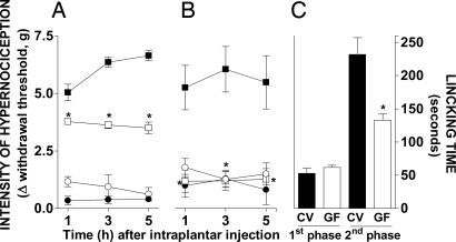
In conventional Swiss/NIH mice, injection of 100 μg per paw of carrageenan in the foot pad induced hypernociception that peaked at 3 h (Fig. 1A) and was resolved by 7 h (data not shown). In germ-free mice, there was a significant reduction of hypernociception induced by injection of carrageenan at all time points observed (Fig. 1A). Similarly, the hypernociceptive response induced by injection of LPS (100 ng per paw) into conventional mice was virtually absent in germ-free animals (Fig. 1B). The s.c. administration of formalin is a well known nociceptive test characterized by two distinct phases of nociceptive responses: an initial neurogenic phase that lasts for 5 min, followed by an inflammatory phase from 15 min (21). Whereas the initial neurogenic response to injection of formalin was preserved in germ-free mice, there was a significant reduction of the secondary response (Fig. 1C). Altogether these results clearly demonstrate that inflammatory hypernociception induced by a range of stimuli is reduced in germ-free mice when compared with their conventional counterparts.
Fig. 1.
Mechanical hypernociception and edema formation induced by injection of carrageenan, LPS, or formalin in germ-free (GF, open symbols) or conventional (CV, filled symbols) mice. (A) Time–response curve of hypernociception induced by i.pl. injection of 30 μl of carrageenan (squares, 100 μg per paw) or 30 μl of saline (circles, vehicle). The hypernociceptive effects were determined at 1, 3, and 5 h after injection. (B) Time-response curve of hypernociception induced by i.pl. injection of 30 μl of LPS (squares, 100 ng per paw) or 30 μl saline (circles, vehicle). (C) Nociceptive responses induced by i.pl. injection of formalin (2%; 30 μl per paw) or 30 μl of saline (vehicle). The nociceptive responses were determined from 0 to 5 min and from 15 to 30 min after formalin injection. Results are expressed by the mean ± SEM of at least six animals per group. *, P < 0.05 when comparing GF and CV mice.
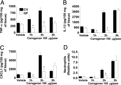
The administration of carrageenan in the footpad of mice induces a local inflammatory response characterized by edema formation, neutrophil influx, and the release of a cascade of cytokines that precedes inflammatory hypernociception (6). Indeed, previous studies from our group have shown that the injection of carrageenan induced the initial release of TNF-α and CXCL1 and subsequent production of IL-1β. Whereas TNF-α and IL-1β are important to drive prostaglandin production, CXCL1 is relevant for the production of sympathomimetic amines (6). Together prostaglandins and sympathomimetic amines act as final mediators of inflammatory hypernociception (6). More recently, we also have shown that the neutrophil influx induced by carrageenan, TNF-α, IL-1β, and CXCL1 was important in the cascade of events leading to the local production of prostaglandins and sympathomimetic amines and the induction of hypernociception (F.Q.C. and M.M.T., unpublished data). Therefore, we measured the edema, local level of cytokines, and neutrophil influx induced by intraplantar injection of carrageenan in conventional and germ-free mice. Edema formation in response to carrageenan injection was lower by 52% in germ-free than in conventional mice (data not shown). Similarly, there was lower local production of TNF-α and the neutrophil-active chemokine, CXCL1, in germ-free mice (Fig. 2). There also was a delayed local production of IL-1β and a delayed recruitment of neutrophils, albeit the levels of IL-1β and the number of neutrophils were similar at 5 h after carrageenan injection (Fig. 2). Histological evaluation of paws injected with carrageenan demonstrated edema formation and leukocyte influx consisting mainly of neutrophils in conventional mice. In contrast, neutrophil influx and edema formation were suppressed in germ-free mice at 3 h after carrageenan injection (data not shown). Altogether, it is clear that the generation of mediators of inflammation and the induction of neutrophil influx are diminished and delayed in germ-free mice injected with carrageenan when compared with their conventional counterparts.
Fig. 2.
Production of TNF-α, IL-1β, and CXCL1 and recruitment of neutrophils after injection of carrageenan in GF (open bars) or CV (filled bars) mice. (A–C) Concentrations of TNF-α (A), IL-1β (B), and CXCL1 (C) in paws injected with 100 μg of Cg or saline (vehicle). (D) Recruitment of neutrophils in soft paw tissue injected with 100 μg of Cg or saline (vehicle). Mice were culled 1, 3, and 5 h after injection, and soft paw tissue was used to measure cytokines by ELISA and neutrophil influx by myeloperoxidase. Results are expressed as the mean ± SEM of five animals per group. *, P < 0.05 when comparing GF and CV mice.
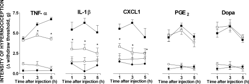
Because the production of TNF-α, IL-1β, and CXCL1 was deficient, we next examined whether injection of the latter mediators would induce hypernociception in germ-free mice. As seen in Fig. 3, hypernociception was significantly reduced in germ-free mice injected with the mediators. Thus, not only is production of TNF-α, IL-1β, and CXCL1 reduced after carrageenan injection, but the function of the released mediators is reduced. The observation that TNF-α, IL-1β, and CXCL1 also need to induce other mediators and neutrophil influx to induce hypernociception (6, 7) is consistent with the previous finding. In contrast, injection of final mediators of hypernociception, prostaglandin E2 (PGE2) and dopamine, induced similar hypernociceptive responses in both conventional and germ-free mice (Fig. 3). These findings demonstrate that neuronal pathways necessary for the induction of hypernociception are intact in germ-free mice. Moreover, the normal initial response of germ-free mice to formalin injection suggests that perception of nociceptive stimuli is preserved in these animals. Moreover, it is suggested that the diminished hypernociceptive response to complex inflammatory stimuli is secondary to the diminished or delayed local production of cytokines and recruitment of leukocytes necessary to trigger hypernociception. The latter possibility is consistent with findings in germ-free mice submitted to ischemia and reperfusion injury and injected with LPS in which there is greatly diminished neutrophil influx and production of TNF-α and CXCL1 (20).
Fig. 3.
Mechanical hypernociception induced by TNF-α, IL-1β, CXCL1, PGE2, and dopamine in GF (open symbols) and CV (filled symbols) mice. Time–response curves of hypernociception induced by i.pl. injection of the mediators (squares) TNF-α (100 pg per paw), IL-1β (100 pg per paw), CXCL1 (10 ng per paw), prostaglandin E2 (PGE2; 100 ng per paw), dopamine (Dopa; 10 μg per paw), or vehicle (circles, 30 μl saline). Results are expressed as the mean ± SEM of six animals per group. Asterisks denote statistically significant differences compared with the control group (P < 0.05).
Reposition of Microbiota or LPS Administration Reverses the Diminished Hypernociceptive Responses of Germ-Free Mice.
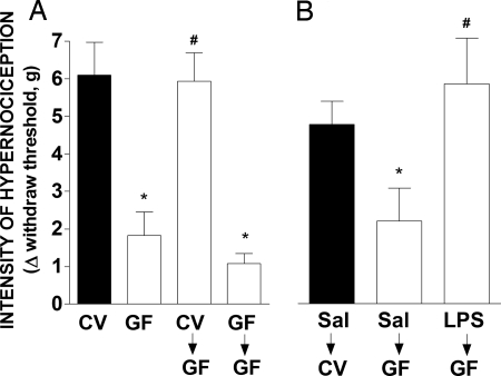
Reposition of the microbiota by the administration of feces from conventional to germ-free mice is accompanied by a reversal of inflammatory hyporesponsiveness (20). This conventionalization process takes 21 days to complete and is followed by a virtual normalization of inflammatory responses of conventionalized mice. The administration of feces from conventional to germ-free mice was accompanied by normalization of hypernociceptive responses to carrageenan injection (Fig. 4A). Administration of feces from germ-free mice had no effect on nociceptive responses (Fig. 4A).
Fig. 4.
Reversion of antihypernociceptive phenotype of GF (open bars) mice compared with CV (filled bars) ones. (A) Two weeks after feces administration (10 mg/kg; by oral gavage) from conventional or germ-free mice, mechanical hypernociception induced by i.pl. injection carrageenan (100 μg per paw) was evaluated in GF mice and compared with CV and GF vehicle-treated. (B) Then, 48 h after systemic LPS administration (1 mg/kg; s.c.), mechanical hypernociception induced by i.pl. injection of carrageenan (100 μg per paw) was evaluated in GF mice and compared with CV and GF vehicle-treated. The hypernociceptive responses were taken 3 h after carrageenan injection. The results are expressed by the mean ± SEM of at least five animals per group. Asterisks denote statistically significant differences compared with the control group (P < 0.05).
Bacteria and other gut-living microorganisms are recognized by the immune system via pattern-recognition receptors (PRRs), including the Toll-like receptors (TLRs) (22). Indeed, activation of PRRs by pathogen-associated molecular patterns is essential for adequate inflammatory responses to pathogens and adequate mounting of an adaptive immune response (22). LPS derived from Gram-negative bacteria induces inflammation, costimulation, and immune priming via activation of TLR4 (22–24). To evaluate whether activation of a single PRR was sufficient to reverse the diminished nociceptive responses of germ-free mice, animals were given a single dose of LPS 48 h before the injection of carrageenan. Previous experiments in the laboratory identified this dose and interval to be optimal for administration of this TLR4 ligand (data not shown). There was no difference in the expression of TLR4 in spleen leukocytes (CD11b+, CD11c+, B220+, NK1.1+, and GR1+ leukocytes) from germ-free or conventional mice (data not shown). Basal expression of TLR4, as assessed by quantitative PCR, also was similar in paw tissue from germ-free or conventional mice (data not shown). Administration of LPS to conventional mice caused 100% lethality by 24 h, whereas all germ-free mice were still alive 1 week after LPS administration (data not shown). As seen in Fig. 4B, injection of LPS 48 h before carrageenan greatly enhanced the nociceptive response of germ-free mice. Indeed, responses in LPS-injected germ-free mice were virtually identical to those of conventional mice. Altogether the results in Fig. 4 demonstrate that the phenotype of germ-free mice can be reversed by the reposition of microbiota or administration of TLR ligands, suggesting that continuous activation of TLRs by the commensal microbiota is sufficient and, perhaps, necessary for expression of an adequate hypernoniceptive response to inflammatory stimulation.
Innate IL-10 Production Accounts for the Diminished Hypernociception of Germ-Free Mice.
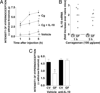
Previous studies from our group have shown that the cytokine IL-10 limited inflammatory hyperalgesia induced by a range of inflammatory mediators, including the cytokines TNF-α and IL-1β (17). As seen in Fig. 5A, injection of IL-10 significantly diminished the inflammatory hypernociception induced by carrageenan in conventional mice. Germ-free mice when stimulated with LPS or undergoing reperfusion injury produce large amounts of IL-10, which accounts for most of the inflammatory hyporesponsiveness of these mice (20). In this context, injection of carrageenan into the footpad of germ-free but not conventional mice was accompanied by an increase in IL-10 gene expression, as demonstrated by quantitative PCR (Fig. 5B). More importantly, germ-free mice given an anti-IL-10 monoclonal antibody (50 μg per mouse, s.c.) had hypernociceptive responses to carrageenan that were similar to those of conventional animals (Fig. 5C). Thus, the innate higher production of IL-10 by germ-free mice accounts for the lower hypernociceptive responses to injection of carrageenan observed in germ-free mice.
Fig. 5.
IL-10 mediates the diminished hypernociceptive response of GF mice. (A) CV mice were injected with recombinant murine IL-10 (100 ng per paw, s.c.) before the injection of carrageenan (100 μg per paw). (B) Expression of IL-10 as measured by quantitative PCR in paw skin of CV and GF mice after i.pl. injection of carrageenan (100 μg per paw). (C) GF or CV mice were treated with a monoclonal anti-IL-10 antibody (50 μg per mouse, s.c.) or control antibody (50 μg per mouse) and hypernociception after i.pl. injection of carrageenan (100 μg per paw) evaluated 40 min later. Data are mean ± SEM of measurements of at least five mice per group (except in B) and are from one of two representative experiments performed.
Conclusion and Implications
Altogether these results clearly demonstrate that germ-free mice have reduced perception of pain after different inflammatory stimulation. Reduced hypernociception depends on IL-10 expression and is secondary to reduced production and action of mediators of the inflammatory process and reduced influx of inflammatory cells. Reposition of microbiota or activation of TLR4 reverses the hyporesponsive phenotype of germ-free mice.
Adaptation to environmental adversities is fundamental for the survival of an organism. Organisms may associate with each other to deal with stressful conditions. When one considers the relationship between a host and its microbiota, associations may be either beneficial or detrimental to the host (25). In the present study, we showed that, in the absence of interaction of mice with their microbiota, as observed in germ-free mice, perception of inflammatory pain was decreased. Therefore, these results show that contact with commensal microbiota is necessary for mice to develop inflammatory hypernociception possibly in a TLR-dependent manner. These findings implicate an important role of the interaction between the commensal microbiota and the host in favoring adaptation to environmental stresses that inflict tissue damage and inflammation and result in enhanced perception of pain.
Materials and Methods
Animals.
Germ-free Swiss/NIH mice were derived from a germ-free nucleus (Taconic Farms) and maintained in flexible plastic isolators (Standard Safety Equipment) by using classical gnotobiology techniques (26). Conventional Swiss/NIH mice were derived from germ-free matrices and considered conventional only after two generations in the conventional facility. All animals were 8–10 weeks old. Experimental procedures in germ-free mice were conducted under aseptic conditions to avoid infection of animals. Animal care and handling procedures were in accordance with the guidelines of the International Association for Study of Pain and had prior approval from the local animal ethics committee.
Experimental Procedures.
Mechanical hypernociception (see Nociceptive Mechanical Test) was measured in mice before and after the intraplantar (i.pl.) injection of one of the following substances: carrageenan (Cg; 100 μg per paw), TNF-α (100 pg per paw), IL-1β (100 pg per paw), KC (10 ng per paw), PGE2 (100 ng per paw), dopamine (Dopa; 10 μg per paw), and LPS (100 ng per paw). Edema formation was measured before and after i.pl. injection of Cg (300 μg per paw). For detection of cytokines and chemokine levels, soft paw tissue was collected after Cg injection, processed according to the protocol described below, and assayed according to the procedures supplied by the manufacturer. To measure neutrophil accumulation on paw skin, residual processed tissue used to quantify cytokine was used to evaluate neutrophil accumulation by assaying myeloperoxidase activity (18).
A series of experiments were conducted to evaluate whether the loss of hypernociception by germ-free mice could be reversed by conventionalization or LPS administration. To conventionalize germ-free mice, feces from conventional animals were given to germ-free animals (20). Fourteen days later, hypernociceptive responses induced by i.pl. injection of carrageenan were evaluated; 1 mg/kg LPS was given s.c. 48 h before the mechanical hypernociceptive test (20).
To evaluate the role of IL-10 in mediating the loss of hypernociception, a monoclonal antibody against IL-10 was given systemically (50 μg per mouse, s.c.) 40 min before mechanical hypernociception was evaluated. To evaluate the activity of IL-10 in a hypernociceptive test in conventional mice, IL-10 was injected (100 ng per paw) 40 min before carrageenan (100 μg per paw) injection, and the response was analyzed up to 5 h after stimulation.
Nociceptive mechanical test.
The term “hypernociception” was used to define the decrease of nociceptive withdrawal threshold (6). Mechanical hypernociception was tested in mice as reported previously (27). In a quiet room, mice were placed in 12 × 10 × 17-cm acrylic cages with wire grid floors 15–30 min before the start of testing. The test consisted of evoking a hindpaw flexion reflex with a hand-held force transducer (electronic anesthesiometer, Insight mod. EFF-301) adapted with a 0.5-mm2 polypropylene tip. The investigator was trained to apply the tip perpendicularly to the central area of the hindpaw with a gradual increase in pressure. The endpoint was characterized by the removal of the paw, followed by clear flinching movements. After the paw withdrawal, the intensity of the pressure was automatically recorded. The value for the response was obtained by averaging three measurements. Animals were tested before and after treatments. Results are expressed as Δ withdrawal threshold (in g) calculated by subtracting zero-time mean measurements from the time interval mean measurements.
Formalin-induced nociception.
This test was based on the method of Dubuisson and Dennis (28) adapted for mice by Hunskaar et al. (21). Briefly, formalin solution (2% in PBS, 30 μl per paw) was injected into the hind paw via its plantar surface (i.pl. injection). The time (s) spent licking or biting the affected paw was rated during two time intervals: 0–5 min (first phase or neurogenic pain) and 15–30 min (second phase or inflammatory pain).
Quantification of cytokines, chemokines, and neutrophil accumulation.
The concentration of TNF-α, IL-1β, and KC in samples was measured in the soft skin tissue of animals by using commercially available antibodies and according to the procedures supplied by the manufacturer (R&D Systems). At various times after carrageenan injection (1, 3, and 5 h), animals were killed and soft paw tissue was collected. These samples were weighted and homogenized in 1 ml of PBS (0.4 M NaCl and 10 mM NaPO4) containing antiproteases (0.1 mM phenylmethylsulfonyl fluoride, 0.1 mM benzethonium chloride, 10 mM EDTA, and 20 KI/ml aprotinin A) and 0.05% Tween 20. Samples were then centrifuged for 10 min at 3,000 × g, and the supernatant was immediately used for ELISAs at a 1:3 dilution in PBS. The pellet was used to evaluate neutrophil accumulation by assaying myeloperoxidase activity as previously described (18). Briefly, pellets were snap-frozen in liquid nitrogen. Upon thawing and processing, the tissue was assayed for myeloperoxidase activity by measuring the change in OD at 450 nm by using tetramethylbenzidine. Results were expressed as the neutrophil index that denotes MPO activity of a known number of casein-elicited murine peritoneal neutrophils processed in the same way. That neutrophils were the main infiltrating leukocytes in carrageenan-injected paws was confirmed by evaluation of the H&E-stained sections.
Conventionalization of germ-free mice.
The process of colonizing germ-free mice with microbiota from conventional mice is a process referred to as conventionalization; this process was performed as described previously (26). Briefly, fecal samples removed from the large intestine of conventional mice were homogenized in saline (10%) and administered by oral gavage to germ-free mice. Fourteen days later, mechanical hypernociception was measured after carrageenan injection into the hind paw. To assess whether there was adequate conventionalization of germ-free mice, fecal samples were assayed by using a thioglycolate test (26).
Real-time PCR.
Paws were removed 1, 3, and 5 h after carrageenan injection into germ-free and conventional mice for IL-10 mRNA expression. Total RNA was isolated from paw skin by using an Illustra RNAspin Mini RNA Isolation Kit (GE Healthcare). The RNA obtained was resuspended in diethyl pyrocarbonate-treated water and stocked at −70°C until use. Real-time RT-PCR was performed on an ABI PRISM 7900 sequence-detection system (Applied Biosystems) by using SYBR Green PCR Master Mix (Applied Biosystems) after a reverse transcription reaction of 1 μg of RNA by using M-MLV reverse transcriptase (Promega). The relative level of gene expression was determined by the comparative threshold cycle method as described by the manufacturer, whereby data for each sample were normalized to hypoxanthine phosphoribosyltransferase and expressed as a fold change compared with sham-operated controls. The following primer pairs were used: hypoxanthine phosphoribosyltransferase, 5′-GTTGGTTACAGGCCAGACTTTGTTG-3′ (forward) and 5′-GAGGGTAGGCTGGCCTATAGGCT-3′ (reverse); and il-10, 5′-GCTCTTACTGACTGGCATGAG-3′ (forward) and 5′-CGCAGCTCTAGGAGCATGTG-3′ (reverse).
Statistical Analysis.
Results are shown as means ± SEM. The differences between the experimental groups were compared by one-way ANOVA. In the case of statistical significance, individual comparisons were subsequently made with a Student–Newman–Keuls post hoc test. The level of significance was set at P < 0.05.
ACKNOWLEDGMENTS.
This work was supported by the Fundação de Amparo a Pesquisas do Estado de Minas Gerais (FAPEMIG, Brazil), Fundação de Amparo a Pesquisas do Estado de São Paulo (FAPESP, Brazil), Conselho Nacional de Desenvolvimento Científico e Tecnologico (CNPq, Brazil), and the Guggenheim Foundation (to M.M.T.).
Footnotes
The authors declare no conflict of interest.
References
- 1.Martin HA, Basbaum AI, Kwiat GC, Goetzl EJ, Levine JD. Leukotriene and prostaglandin sensitization of cutaneous high-threshold C- and A-delta mechanonociceptors in the hairy skin of rat hindlimbs. Neuroscience. 1987;22:651–659. doi: 10.1016/0306-4522(87)90360-5. [DOI] [PubMed] [Google Scholar]
- 2.Schaible HG, Schmidt RF. Time course of mechanosensitivity changes in articular afferents during a developing experimental arthritis. J Neurophysiol. 1988;60:2180–2195. doi: 10.1152/jn.1988.60.6.2180. [DOI] [PubMed] [Google Scholar]
- 3.Davis KD, Meyer RA, Campbell JN. Chemosensitivity and sensitization of nociceptive afferents that innervate the hairy skin of monkey. J Neurophysiol. 1993;69:1071–1081. doi: 10.1152/jn.1993.69.4.1071. [DOI] [PubMed] [Google Scholar]
- 4.Rueff A, Dray A. Sensitization of peripheral afferent fibres in the in vitro neonatal rat spinal cord-tail by bradykinin and prostaglandins. Neuroscience. 1993;54:527–535. doi: 10.1016/0306-4522(93)90272-h. [DOI] [PubMed] [Google Scholar]
- 5.Vivancos GG, et al. An electronic pressure-meter nociception paw test for rats. Braz J Med Biol Res. 2004;37:391–399. doi: 10.1590/s0100-879x2004000300017. [DOI] [PubMed] [Google Scholar]
- 6.Cunha TM, et al. A cascade of cytokines mediates mechanical inflammatory hypernociception in mice. Proc Natl Acad Sci USA. 2005;102:1755–1760. doi: 10.1073/pnas.0409225102. [DOI] [PMC free article] [PubMed] [Google Scholar]
- 7.Verri WA, Jr, et al. Hypernociceptive role of cytokines and chemokines: Targets for analgesic drug development? Pharmacol Ther. 2006;112:116–138. doi: 10.1016/j.pharmthera.2006.04.001. [DOI] [PubMed] [Google Scholar]
- 8.Ferreira SH, Nakamura M, Abreu Castro MS. The hyperalgesic effects of prostacyclin and prostaglandin E2. Prostaglandins. 1978;16:31–37. doi: 10.1016/0090-6980(78)90199-5. [DOI] [PubMed] [Google Scholar]
- 9.Khasar SG, McCarter G, Levine JD. Epinephrine produces a beta-adrenergic receptor-mediated mechanical hyperalgesia and in vitro sensitization of rat nociceptors. J Neurophysiol. 1999;81:1104–1112. doi: 10.1152/jn.1999.81.3.1104. [DOI] [PubMed] [Google Scholar]
- 10.Cunha FQ, Teixeira MM, Ferreira SH. Pharmacological modulation of secondary mediator systems–cyclic AMP, cyclic GMP–on inflammatory hyperalgesia. Br J Pharmacol. 1999;127:671–678. doi: 10.1038/sj.bjp.0702601. [DOI] [PMC free article] [PubMed] [Google Scholar]
- 11.Aley KO, Levine JD. Role of protein kinase A in the maintenance of inflammatory pain. J Neurosci. 1999;19:2181–2186. doi: 10.1523/JNEUROSCI.19-06-02181.1999. [DOI] [PMC free article] [PubMed] [Google Scholar]
- 12.Parada CA, Reichling DB, Levine JD. Chronic hyperalgesic priming in the rat involves a novel interaction between cAMP and PKCepsilon second messenger pathways. Pain. 2005;113:185–190. doi: 10.1016/j.pain.2004.10.021. [DOI] [PubMed] [Google Scholar]
- 13.Ferreira SH, Lorenzetti BB, Bristow AF, Poole S. Interleukin-1 beta as a potent hyperalgesic agent antagonized by a tripeptide analogue. Nature. 1988;334:698–700. doi: 10.1038/334698a0. [DOI] [PubMed] [Google Scholar]
- 14.Cunha FQ, Lorenzetti BB, Poole S, Ferreira SH. Interleukin-8 as a mediator of sympathetic pain. Br J Pharmacol. 1991;104:765–767. doi: 10.1111/j.1476-5381.1991.tb12502.x. [DOI] [PMC free article] [PubMed] [Google Scholar]
- 15.Cunha FQ, Poole S, Lorenzetti BB, Ferreira SH. The pivotal role of tumour necrosis factor alpha in the development of inflammatory hyperalgesia. Br J Pharmacol. 1992;107:660–664. doi: 10.1111/j.1476-5381.1992.tb14503.x. [DOI] [PMC free article] [PubMed] [Google Scholar]
- 16.Lorenzetti BB, et al. Cytokine-induced neutrophil chemoattractant 1 (CINC-1) mediates the sympathetic component of inflammatory mechanical hypersensitivitiy in rats. Eur Cytokine Netw. 2002;13:456–461. [PubMed] [Google Scholar]
- 17.Poole S, Cunha FQ, Selkirk S, Lorenzetti BB, Ferreira SH. Cytokine-mediated inflammatory hyperalgesia limited by interleukin-10. Br J Pharmacol. 1995;115:684–688. doi: 10.1111/j.1476-5381.1995.tb14987.x. [DOI] [PMC free article] [PubMed] [Google Scholar]
- 18.Souza DG, et al. Increased mortality and inflammation in tumor necrosis factor-stimulated gene-14 transgenic mice after ischemia and reperfusion injury. Am J Pathol. 2002;160:1755–1765. doi: 10.1016/s0002-9440(10)61122-4. [DOI] [PMC free article] [PubMed] [Google Scholar]
- 19.Souza DG, et al. Role of PAF receptors during intestinal ischemia and reperfusion injury. A comparative study between PAF receptor-deficient mice and PAF receptor antagonist treatment. Br J Pharmacol. 2003;139:733–740. doi: 10.1038/sj.bjp.0705296. [DOI] [PMC free article] [PubMed] [Google Scholar]
- 20.Souza DG, et al. The essential role of the intestinal microbiota in facilitating acute inflammatory responses. J Immunol. 2004;173:4137–4146. doi: 10.4049/jimmunol.173.6.4137. [DOI] [PubMed] [Google Scholar]
- 21.Hunskaar S, Fasmer OB, Hole K. Formalin test in mice, a useful technique for evaluating mild analgesics. J Neurosci Methods. 1985;14:69–76. doi: 10.1016/0165-0270(85)90116-5. [DOI] [PubMed] [Google Scholar]
- 22.Medzhitov R. Recognition of microorganisms and activation of the immune response. Nature. 2007;449:819–826. doi: 10.1038/nature06246. [DOI] [PubMed] [Google Scholar]
- 23.Poltorak A, et al. Defective LPS signaling in C3H/HeJ and C57BL/10ScCr mice: mutations in Tlr4 gene. Science. 1998;282:2085–2088. doi: 10.1126/science.282.5396.2085. [DOI] [PubMed] [Google Scholar]
- 24.Hoshino K, et al. Cutting edge: Toll-like receptor 4 (TLR4)-deficient mice are hyporesponsive to LPS: Evidence for TLR4 as the Lps gene product. J Immunol. 1999;162:3749–3752. [PubMed] [Google Scholar]
- 25.Casadevall A, Pirofski LA. Microbial virulence results from the interaction between host and microorganism. Trends Microbiol. 2003;11:157–158. doi: 10.1016/s0966-842x(03)00008-8. [DOI] [PubMed] [Google Scholar]
- 26.Pleasants JR. Germfree animals and their significance. Endeavour. 1973;32:112–116. [PubMed] [Google Scholar]
- 27.Cunha TM, et al. An electronic pressure-meter nociception paw test for mice. Braz J Med Biol Res. 2004;37:401–407. doi: 10.1590/s0100-879x2004000300018. [DOI] [PubMed] [Google Scholar]
- 28.Dubuisson D, Dennis SG. The formalin test: A quantitative study of the analgesic effects of morphine, meperidine, and brain stem stimulation in rats and cats. Pain. 1977;4:161–174. doi: 10.1016/0304-3959(77)90130-0. [DOI] [PubMed] [Google Scholar]


