Abstract
Glucose ingestion after an overnight fast triggers an insulin-dependent, homeostatic program that is altered in diabetes. The full spectrum of biochemical changes associated with this transition is currently unknown. We have developed a mass spectrometry-based strategy to simultaneously measure 191 metabolites following glucose ingestion. In two groups of healthy individuals (n=22 and 25), 18 plasma metabolites changed reproducibly, including bile acids, urea cycle intermediates, and purine degradation products, none of which were previously linked to glucose homeostasis. The metabolite dynamics also revealed insulin's known actions along four key axes—proteolysis, lipolysis, ketogenesis, and glycolysis—reflecting a switch from catabolism to anabolism. In pre-diabetics (n=25), we observed a blunted response in all four axes that correlated with insulin resistance. Multivariate analysis revealed that declines in glycerol and leucine/isoleucine (markers of lipolysis and proteolysis, respectively) jointly provide the strongest predictor of insulin sensitivity. This observation indicates that some humans are selectively resistant to insulin's suppression of proteolysis, whereas others, to insulin's suppression of lipolysis. Our findings lay the groundwork for using metabolic profiling to define an individual's 'insulin response profile', which could have value in predicting diabetes, its complications, and in guiding therapy.
Keywords: glucose homeostasis, insulin sensitivity, metabolic profiling
Introduction
Glucose homeostasis is a complex physiologic process involving the orchestration of multiple organ systems. During an overnight fast, for instance, glucose levels are maintained through both glycogenolysis and gluconeogenesis, and the liver begins to generate ketone bodies (e.g. acetoacetate, β-hydroxybutyrate) from fatty acids released by adipose tissue (Cahill, 2006). Ingestion of glucose after overnight fasting then triggers the rapid release of insulin from the pancreas, which promotes glucose uptake in peripheral tissues and switches the body from catabolism to anabolism. For example, proteolysis in skeletal muscle and associated release of amino acids (which support gluconeogenesis) are replaced by amino-acid uptake and protein synthesis (Felig, 1975; Fukagawa et al, 1985). Also, triacylglycerol lysis in adipose tissue and hepatic synthesis of ketone bodies are inhibited and replaced by fatty acid uptake and re-esterification. Hence the transition from fasting to feeding is accompanied by many changes in metabolite concentrations as the body makes adjustments to achieve glucose homeostasis.
To obtain a systematic view of the physiologic response to glucose ingestion in health and disease, we wished to simultaneously monitor the concentrations of a large and diverse set of metabolites. Such an integrated analysis can aid in the classification of metabolic states, reveal new pathways, and potentially improve the sensitivity for detection of abnormalities. Traditionally, single metabolites or classes of small molecules have been detected using dedicated analytical assays. With these methods, relationships among diverse metabolites and pathways have been missed, and a comprehensive picture of a complex physiologic program has not been possible. Such relationships could, in principle, be important in understanding disease pathogenesis and in aiding in the diagnosis of disease.
Recent advances in analytical chemistry and computation now enable the simultaneous measurement of a large number of metabolites. Two technologies are commonly used for metabolic profiling: nuclear magnetic resonance (NMR) spectroscopy and mass spectrometry (MS) (Dunn et al, 2005). Although NMR spectroscopy has a number of advantages, primarily its non-destructive nature, its ability to provide information on chemical structure and its better suitability for absolute quantification, it tends to have low sensitivity. MS technology, on the other hand, affords sensitive and specific analysis of metabolites in complex biological samples, particularly when implemented as tandem MS and coupled with high-performance liquid chromatography (LC), a combination termed LC-MS/MS. Metabolic profiling with LC-MS/MS technology has already been successfully used for identifying human plasma markers of myocardial ischemia (Sabatine et al, 2005) as well as for characterizing the metabolic response to starvation in model organisms (Brauer et al, 2006).
We have developed an LC-MS/MS metabolic profiling system capable of quantifying metabolites in plasma (see Materials and methods), and we report here its application to studying the human response to an oral glucose load. Our system can measure 191 endogenous human metabolites spanning diverse chemical classes (Supplementary information). This collection of metabolites includes those previously studied in the context of glucose homeostasis, as well as many metabolites not previously linked to this area. We first applied this technology to healthy individuals to characterize the normal human response to an oral glucose challenge, and then asked how this response is affected by reduced insulin sensitivity in individuals with impaired glucose tolerance.
Results
Eighteen plasma metabolites change significantly and reproducibly during an oral glucose challenge
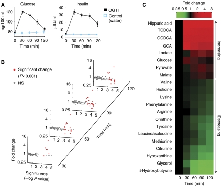
To systematically characterize the normal biochemical response to glucose ingestion in humans, we obtained plasma samples for metabolic profiling from an ongoing study, Metabolic Abnormalities in College Students (MACS, see Materials and methods). In this study, young adults undergo a series of metabolic evaluations, including a questionnaire for metabolic syndrome risk factors, indirect calorimetry, measurement of body composition, and a fasting blood lipid profile. As part of the metabolic assessment, MACS subjects also undergo a 2-h oral glucose tolerance test (OGTT) with multiple blood draws. To control for the fasting condition and for the effects of fluid ingestion, a subset of MACS subjects selected at random were given an identical volume of water instead of the glucose solution. Venous blood was drawn during fasting and then every 30 min following glucose or water ingestion for the 2-h duration of the test. We obtained samples from 22 subjects ingesting glucose and 7 control subjects ingesting water (Table I). Serum concentrations of glucose and insulin were measured throughout the test (Figure 1A). All subjects had normal fasting glucose levels, and all glucose-ingesting subjects showed normal glucose tolerance, as currently defined by the American Diabetes Association (2007).
Table 1.
Demographic and clinical characteristics of human subjects
| Clinical study | MACS |
FOS |
||
|---|---|---|---|---|
| Group | Glucosea (n=22) | Waterb (n=7) | FOS-NGT (n=25) | FOS-IGT (n=25) |
| Age (years) | 23±3 (18–30) | 24±4 (20–30) | 45±3 (40–49) | 46±3 (40–50) |
| Gender | 9 women, 13 men | 3 women, 4 men | 13 women, 12 men | 13 women, 12 men |
| Ancestry | Wh: 9; As: 6; Un: 7 | Wh: 3; Aa: 1; As: 1; Un: 2 | Wh: 25 | Wh: 25 |
| BMI | 22.4±2.1 (18.3–26.9) | 22.1±2.7 (17.8–26.0) | 24.6±3.4 (19.0–31.5) | 26.8±4.8 (18.8–41.2) |
| Fasting glucose (mg/100 ml) | 78±5 (71–90) | 77±7 (70–86) | 89±6 (76–100) | 100±9 (87–115) |
| 120 min glucose (mg/100 ml) | 86±16 (66–119) | 80±9 (71–92) | 88±21 (43–122) | 153±12 (140–180) |
| Fasting insulin (μIU/ml) | 4.6±2.9 (2.8–14.2) | 3.6±0.7 (2.8–4.8) | 4.2±2.7 (1.0–10.7) | 10.3±8.1 (1.0–25.7) |
| 120 min insulin (μIU/ml) | 18.1±16.5 (3.0–75.9) | 3.6±1.0 (2.9–5.5) | 29.7±20.5 (1.0–93.3) | 102.8±51.8 (35.0–202.3) |
| IGT/NGTc | 0/22 | NA | 0/25 | 25/0 |
Abbreviations: MACS, Metabolic Abnormalities in College Students, conducted at MIT Clinical Research Center; FOS, Framingham Offspring Study; BMI, body mass index. Ancestry abbreviations: Aa, African American; As, Asian; Un, unknown; Wh, White.
Quantitative variables are expressed as mean±s.d. (range).
Subjects ingesting glucose (OGTT).
Subjects ingesting water (control).
Number of subjects in each glucose tolerance category. IGT, impaired glucose tolerance (American Diabetes Association, 2007); NGT, normal glucose tolerance.
Figure 1.
Temporal response to an oral glucose challenge in individuals with normal glucose tolerance (MACS). (A) Kinetics of blood glucose and insulin in response to glucose ingestion (mean±s.e.m.). (B) Magnitude and significance of metabolite change over time. Dots represent the 97 metabolites detected in plasma. Change is with respect to the fasting metabolite levels. Significant (P<0.001) changes are colored red. (C) Significant metabolite changes. In total, 21 metabolites changed significantly (P<0.001) when compared to their fasting levels and showed a significantly (P<0.05) distinct response compared to control (water ingestion). Color intensity reflects the median fold change compared to the fasting levels. Metabolites are ordered according to the magnitude of change.
We performed LC-MS/MS metabolic profiling of the OGTT time course in MACS subjects. Out of the 191 metabolites monitored by our platform, 97 were detected in at least 80% of subjects in all time points (Figure 1B). The levels of 21 metabolites changed significantly (P<0.001) from the fasting levels and were also significantly (P<0.05) different when compared to the response to water (Figure 1C). These 21 significantly altered metabolites span pathways previously studied in the context of glucose homeostasis, as well as some never linked to this program.
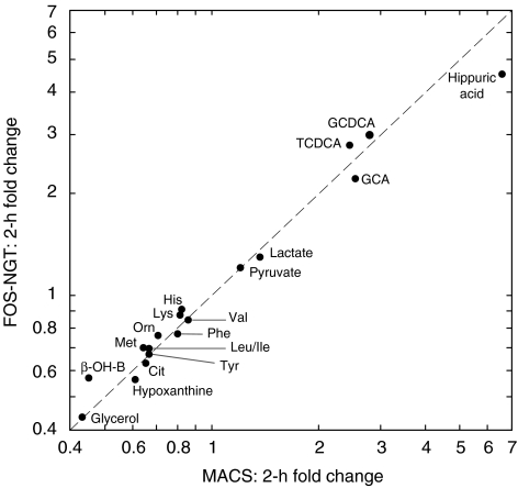
To validate these findings, we profiled fasting and 2-h OGTT samples from an independent cohort. The 2-h time point has clinical significance, and can aid in the diagnosis of impaired glucose tolerance and diabetes (American Diabetes Association, 2007). FOS-NGT (Table I) is a group of individuals with normal glucose tolerance, derived from the Framingham Offspring Study (Kannel et al, 1979). This group is similar to MACS in size, gender composition, and glucose tolerance, but these individuals are approximately 20 years older and differ in their ancestry (Table I). Of the 21 metabolites displaying significant change in MACS at any time point during OGTT (Figure 1C), the levels of 20 (glucose excluded) remained significantly (P<0.05) altered at the 2-h time point. In total, 18 of these 20 metabolite changes replicated significantly (P<0.05) and in the same direction in FOS-NGT (Figure 2). The remaining two metabolites, malate and arginine, fell below our significance threshold. We have thus identified 18 plasma metabolites exhibiting highly reproducible and likely robust responses to glucose ingestion in healthy individuals.
Figure 2.
Validation of metabolite changes at the 2-h time point. In total, 18 out of the 20 metabolites that changed significantly in MACS (Figure 1C) replicated (P<0.05) in FOS-NGT. Dots correspond to the median fold change of metabolites at the 2-h time point. Abbreviations: TCDCA, taurochenodeoxycholic acid; GCDCA, glycochenodeoxycholic acid; GCA, glycocholic acid, Orn: ornithine, Cit: citrulline, β-OH-B: β-hydroxybutyrate.
Metabolic profiling reveals novel biochemical changes during the OGTT
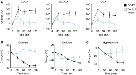
The systematic profiling approach has enabled us to identify a number of plasma metabolites, not previously associated with glucose homeostasis, that change reproducibly in response to an oral glucose challenge. Perhaps most striking were the observed changes in bile acids. The levels of three bile acids—glycocholic acid, glycochenodeoxycholic acid and taurochenodeoxycholic acid—more than doubled during the first 30 min after glucose ingestion, and remained elevated for the entire 2 h (Figure 3A). Water ingestion produced a smaller increase in bile acids, which was not sustained beyond the 30 min time point. All three compounds are primary bile acids conjugated to glycine or taurine.
Figure 3.
Metabolic responses not previously linked to glucose homeostasis. Kinetic patterns in the MACS group are shown (mean±s.e.m.). (A) Bile acids. Abbreviations: TCDCA, taurochenodeoxycholic acid; GCDCA, glycochenodeoxycholic acid; GCA, glycocholic acid. (B) Citrulline and ornithine, urea cycle intermediates. (C) Hypoxanthine, a product of purine nucleotide degradation.
The levels of citrulline and ornithine, two non-proteinogenic amino acids that participate in hepatic urea synthesis, decreased by 35 and 29%, respectively during the 2-h test (Figure 3B). Gluconeogenesis from amino acids (primarily alanine), which supports 25–40% of the non-glycogen-derived hepatic glucose output after an overnight fast (Felig, 1975), is coupled to urea synthesis (Devlin, 2002). The decreases in citrulline and ornithine may thus be associated with the reduction in gluconeogenesis and urea synthesis following glucose ingestion.
Hypoxanthine, a purine base generated from degradation of adenine and guanine nucleotides, decreased in MACS by 39% within 2 h of glucose ingestion (Figure 3C), and this pattern was replicated in FOS-NGT. Xanthine, a purine base generated from hypoxanthine by oxidation, also decreased in both groups (MACS: −9%, P<0.05; FOS-NGT: −41%, P<10−4). The decreases in hypoxanthine and xanthine may be explained by a combination of attenuated release and accelerated uptake. Hypoxanthine taken up by tissues can support nucleotide biosynthesis through the purine salvage pathway (Mateos et al, 1987; Yamaoka et al, 1997) and may also be indicative of a switch from catabolism to anabolism of nucleic acids, analogous to the simultaneous transitions in fat and protein metabolism.
Interestingly, hippuric acid increased by over 1000% during the first 30 min and decreased gradually thereafter. Most likely this response is not related to glucose, but rather reflects the presence of the preservative benzoic acid, a precursor of hippuric acid (Kubota and Ishizaki, 1991), found in the glucose beverage used for OGTT (see Materials and methods).
Changes in plasma metabolites span four arms of insulin action
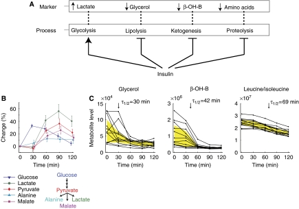
Much of the biochemical response to glucose ingestion, which we studied in an unbiased way, can be attributed to the broad actions of insulin (Figure 4A). Specifically, we have detected an increase in lactate and decreases in glycerol, β-hydroxybutyrate and amino acids. These metabolite changes correspond to the stimulation of glucose metabolism and to the suppression of lipolysis, ketogenesis, and proteolysis (as well as stimulation of amino-acid uptake), all of which are known to be elicited by insulin (Fukagawa et al, 1985; Nurjhan et al, 1986).
Figure 4.
Metabolites reflecting four axes of insulin action. (A) Four axes of insulin action and their associated metabolic markers. (B) The kinetics of glucose and pyruvate derivatives (MACS, mean±s.e.m.). (C) The kinetics of insulin's suppression of catabolism. Each line corresponds to 1 of 12 individual MACS subjects. τ1/2: time to half-maximal decrease (median of all subjects). The inter-quartile range of metabolite levels is shaded in yellow. Abbreviations: β-OH-B, β-hydroxybutyrate.
Our method captured the temporal relationship between glucose and intermediates of glycolysis (Figure 4B). Specifically, the increase in pyruvate, lactate, and alanine occurred between 30 and 60 min, lagging ∼30 min behind the glucose rise, consistent with previous reports (Kelley et al, 1988). Interestingly, the kinetics of malate, an intermediate in the Krebs cycle, closely resembled the kinetics of lactate and pyruvate, suggesting that a fraction of the pyruvate formed through glycolysis was carboxylated to generate malate through oxaloacetate. To our knowledge, elevation of plasma malate levels due to glucose ingestion has not been previously reported.
To gain insight into the kinetics of insulin action, we compared temporal patterns for metabolites indicative of the suppression of fat and protein catabolism. The levels of glycerol, β-hydroxybutyrate, and multiple amino acids all declined after glucose ingestion, but the kinetic pattern of glycerol and β-hydroxybutyrate was remarkably different from amino acids (Figure 4C). Over the 2 h, the decrease in glycerol and β-hydroxybutyrate levels was 57 and 55%, respectively, whereas the drop in amino acids was moderate (between 14 and 36%). The branched-chain amino acids leucine/isoleucine (indistinguishable by our method), for example, decreased by 33%. Interestingly, the time to reach half-maximal decrease in the amino acids (50–72 min) was greater than in β-hydroxybutyrate (42 min) and glycerol (30 min). Moreover, the inter-individual variance in metabolite levels shrunk dramatically over the 2 h in glycerol and β-hydroxybutyrate (84 and 95% reduction of inter-quartile range, respectively), whereas in amino acids the maximal reduction was 53%. These findings suggest that the suppression of lipolysis and ketogenesis is more sensitive to the action of insulin compared to suppression of protein catabolism.
Metabolite excursions reflect multiple dimensions of insulin sensitivity
Having identified 18 metabolites that exhibit robust 2-h excursions, we were interested in determining whether the metabolites might be useful in understanding insulin sensitivity. Insulin sensitivity is traditionally defined as the ability of insulin to promote the uptake of glucose into peripheral tissues such as skeletal muscle and fat or to suppress gluconeogenesis in the liver. A decline of insulin sensitivity is one of the earliest signs of type 2 diabetes mellitus (T2DM). This decline is often manifest as elevated levels of fasting insulin, which is strongly correlated with measurements of insulin sensitivity (Hanson et al, 2000). Considering that several metabolic processes taking place following glucose ingestion are mediated by insulin, we hypothesized that insulin sensitivity could be reflected not only by changes in glucose but also by the OGTT response of multiple other metabolites. Because our initial studies were focused on normal, healthy individuals spanning a narrow range of fasting insulin levels, we performed a third analysis on a group of individuals with impaired glucose tolerance from the Framingham Offspring Study, FOS-IGT, who spanned a broader range of fasting insulin concentrations (Table I).
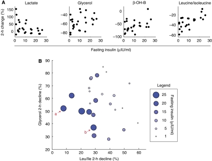
First, to systematically evaluate the relationship between individual metabolite excursions and fasting insulin, we performed linear regression of the fasting insulin concentration on each of the 18 2-h excursions. Out of the 18, 6 showed a statistically significant (P<0.05) correlation with fasting insulin, and included the excursions in lactate, β-hydroxybutyrate, amino acids (leucine/isoleucine, valine, and methionine), and a bile acid (GCDCA) (Table II). Taken together with the glycerol excursion, which scored (P=0.07) slightly below the significance threshold, the response of four distinct insulin action markers correlated with fasting insulin (Figure 5A). Individuals with high fasting insulin exhibited a blunted excursion in all seven metabolites: they had a smaller change both in increasing metabolites (lactate and GCDCA) and in decreasing metabolites (the other five). Notably, the glucose excursion was not correlated with fasting insulin (P=0.20). These findings suggest that resistance to the action of insulin on the metabolism of glucose, fat, and protein is reflected by the metabolite response to OGTT.
Table 2.
Regression models relating fasting insulin to 2-h metabolite change in individuals with impaired glucose tolerance (FOS-IGT)
| Predictor(s) | R2adj | P-value | Prediction errora |
|---|---|---|---|
| Δb Leucine/isoleucine | 0.36 | 9E−4 | 6.65 |
| Δ Valine | 0.17 | 3E−2 | 7.74 |
| Δ Lactate | 0.16 | 3E−2 | 7.60 |
| Δ Glycochenodeoxycholic acid | 0.14 | 4E−2 | 7.86 |
| Δ Methionine | 0.14 | 4E−2 | 7.68 |
| Δ β-Hydroxybutyrate | 0.14 | 4E−2 | 7.90 |
| Δ Leucine/isoleucine+Δ glycerolc | 0.54 | 7E−5 | 5.66 |
| PLSd | 0.46 | 1E−4 | 6.89 |
| BMI | 0.33 | 1E−3 | 6.74 |
The prediction error is expressed as the root mean square error of prediction (RMSEP), in micro-international units per milliliter insulin.
Δ denotes log of the 2-h fold change of metabolite levels.
A bivariate model consisting of the 2-h changes in leucine/isoleucine and in glycerol.
Partial least squares based on changes in the 18 validated metabolites.
Figure 5.
Correlation between fasting insulin and 2-h metabolite changes in individuals with impaired glucose tolerance (FOS-IGT). (A) 2-h changes in markers of insulin action are correlated with fasting insulin concentration. Each dot corresponds to an individual. (B) A bivariate model explaining fasting insulin using the 2-h decline of Leu/Ile and glycerol. Each circle represents an individual, and the circle size is proportionate to fasting insulin levels. aA representative individual exhibiting a blunted decline in Leu/Ile (resistant to proteolysis suppression). bA representative individual exhibiting a blunted decline in glycerol (resistant to lipolysis suppression).
Next, we sought to determine whether a combination of two or more of the 18 metabolite excursions might be more predictive of insulin sensitivity than are individual excursions. We used forward stepwise linear regression to discover an optimal linear model relating 2-h metabolite changes to fasting insulin levels. The top regression model identified consisted of the excursions in Leu/Ile and glycerol (R2adj=0.54, P=0.0001). In this bivariate model, the independent contribution from each metabolite excursion was significant (Leu/Ile: P=8 × 10⁻⁵, glycerol: P=4 × 10⁻³), and the two predictors were not correlated with each other (P=0.6). The Leu/Ile–glycerol model predicted fasting insulin levels better than any individual metabolite excursion (Table II). BMI, which is known to be a strong predictor of fasting insulin, was less predictive than the bivariate model. Notably, the explanatory power of the Leu/Ile and glycerol excursions was significant even after controlling for BMI (P=2 × 10⁻³ and 3 × 10⁻³, respectively). A graphical representation of the Leu/Ile–glycerol model (Figure 5B) demonstrates that some individuals with high fasting insulin exhibit a blunted decline in glycerol, whereas others exhibit a blunted decline in Leu/Ile.
To identify linear combinations of all 18 metabolite excursions that might be predictive of fasting insulin, we used partial least squares (PLS). PLS is a multivariate method that finds orthogonal linear combinations (called components) of the original predictor variables (here, metabolite excursions), which are most correlated with a response variable (here, fasting insulin). To determine how many components should be included in the model while keeping it general, the model is tested on new data (a method called cross-validation). In the current PLS model, the smallest prediction error was achieved with the first component alone (Table II), and a similar error was obtained with the first two components. Interestingly, the Leu/Ile excursion had the largest coefficient in the first component, whereas the glycerol excursion contributed primarily to the second. The prediction error of the bivariate model consisting of the excursions in Leu/Ile and glycerol was lower than the PLS model error (Table II). These findings indicate that the excursions in Leu/Ile and glycerol are sufficient to capture the correlation of 2-h metabolite changes with fasting insulin.
In summary, we have used a univariate approach as well as two multivariate approaches to investigate the correlates of insulin sensitivity. Remarkably, all three methods spotlighted the change in Leu/Ile and change in glycerol as being predictive of fasting insulin. Changes in these two metabolites are blunted in insulin-resistant individuals during the OGTT.
Discussion
In the current study, we have applied metabolic profiling to investigate the kinetics of human plasma biochemicals in response to an oral glucose challenge. To our knowledge, this is the first study to apply a profiling technology to characterize this physiologic program. The systematic approach we have taken has enabled us to recapitulate virtually all known polar metabolite changes associated with the OGTT, while spotlighting some pathways never linked to this program. Importantly, simultaneous measurement of multiple metabolites made it possible to explore connections among metabolic pathways, providing novel insights into normal physiology and disease.
One of the most surprising findings of this investigation is the dramatic increase in bile acid levels following glucose ingestion. In response to eating, the gallbladder contracts and releases bile into the intestines. Bile acids are then absorbed and travel through the portal vein back to the liver, where the enterohepatic cycle completes. Although the uptake of bile acids to the liver is fairly efficient, a constant fraction (10–30%) reaches the systemic circulation (Angelin et al, 1982). In a previous investigation, human subjects ingesting a standard liquid meal containing fat and protein exhibited an increase of 320–330% in individual serum bile acids (De Barros et al, 1982). In the current study, we found that ingesting glucose alone elicits a bile acid response of similar magnitude (Figure 3A), which is sustained for 2 h. Our finding is supported by previous work showing that glucose ingestion can increase the plasma concentration of cholecystokinin, a hormone signaling the gallbladder to contract (Liddle et al, 1985). Interestingly, a recent investigation has shown that bile acids are a signal for fat and muscle cells to increase their energy expenditure through activation of thyroid hormone (Watanabe et al, 2006). Given these findings, and our observation of bile acid release following glucose ingestion, we tested the hypothesis that bile acids influence peripheral glucose uptake. We could not detect an effect of bile acids on basal or insulin-stimulated glucose uptake, however, in cultured adipocytes (data not shown). At present, the physiologic role of sustained bile acid release following glucose ingestion is unknown.
Although insulin resistance is traditionally defined as the reduced ability of insulin to promote glucose uptake or as the impaired suppression of gluconeogenesis, resistance can emerge in other insulin-dependent processes. In prior studies, for example, inadequate suppression of lipolysis was observed in women with a history of gestational diabetes (Chan et al, 1992), and an elevated proteolysis rate was seen in individuals with HIV-associated insulin resistance (Reeds et al, 2006). In obesity, manifestations of insulin resistance include elevated rates of lipolysis (Robertson et al, 1991) and proteolysis (Jensen and Haymond, 1991; Luzi et al, 1996). We have used metabolic profiling to monitor insulin action across multiple axes (Figure 4A), and with markers of each axis in hand, were able to detect kinetic differences between them. In healthy individuals, insulin's suppression of lipolysis and ketogenesis is rapid compared to its suppression of proteolysis (Figure 4C), consistent with the different concentrations of insulin required to inhibit each of these processes (Fukagawa et al, 1985; Nurjhan et al, 1986). Such differences in the activation thresholds of metabolic pathways could be contributing to the diversity in the clinical presentation of insulin resistance. Mouse models of insulin-resistant T2DM provide another example for inter-pathway differences in insulin sensitivity: in the livers of these mice, gluconeogenesis is resistant to suppression by insulin, whereas lipogenesis is responsive, resulting in hyperglycemia and hypertriglyceridemia (Brown and Goldstein, 2008). Collectively, these observations demonstrate that environmental or genetic perturbations could lead to selectivity in insulin sensitivity and contribute to pathogenesis.
Our study demonstrates that an individual's ‘insulin response profile', namely, the vector of sensitivities to insulin action along multiple physiologic axes, can be revealed by metabolite excursions in response to a glucose challenge. Specifically, we have shown that excursions in Leu/Ile and glycerol, reflecting the sensitivity of proteolysis and lipolysis to the action of insulin, are jointly predictive of fasting insulin, with each of the two excursions offering complementary and significant explanatory power (Figure 5B). These findings demonstrate that in two individuals, different axes could be responsible for the same elevation of fasting insulin, and simultaneous measurement of metabolites can distinguish between the two conditions. Monitoring the response to glucose ingestion across multiple axes in larger, prospective clinical studies of pre-diabetics could establish links between insulin sensitivity profiles and disease progression, thus helping to predict future diabetes and its complications as well as to guide therapeutic interventions.
Materials and methods
Human subjects
MACS
Subjects were young adults in the age range of 18–30 years who volunteered for the study during the academic year 2006–2007. Control subjects (ingesting water instead of glucose solution) were assigned at random, balancing gender. Metabolic profiling analysis was limited to those subjects with normal fasting glucose concentrations (below 100 mg/100 ml) and normal glucose tolerance (2-h glucose concentration below 140 mg/100 ml).
FOS
Subjects were selected at random (balancing gender) from among all participants in the fifth FOS examination cycle (1991–1995) aged 40–49 years who had no diabetes mellitus, hypertension or prior cardiovascular disease. Additional selection criteria for FOS-NGT were normal fasting glucose concentrations and normal glucose tolerance. Additional selection criterion for FOS-IGT was impaired glucose tolerance (2-h glucose concentration between 140 and 199 mg/100 ml).
Table I lists demographic and metabolic characteristics of cohorts from the MACS and FOS studies.
OGTT
MACS
Subjects were admitted to the Massachusetts Institute of Technology Clinical Research Center (CRC) after a 10 h overnight fast. An intravenous catheter was inserted into an antecubital vein or a wrist vein and fasting samples were drawn. Next, each subject ingested a glucose solution (Trutol, 75 g in 296 ml; NERL Diagnostics, East Providence, RI; contains citric acid and sodium benzoate) or an identical volume of bottled water (Poland Spring Water, Wilkes Barre, PA) over a 5-min period. Additional blood samples were drawn from the inserted catheter 30, 60, 90 and 120 min after ingestion. Subjects remained at rest throughout the test. The study protocol was approved by the MIT Committee on the Use of Humans as Experimental Subjects and the CRC Scientific Advisory Committee.
FOS
Subjects underwent an OGTT as part of the FOS (Arnlov et al, 2005). After 12 h overnight fast, subjects ingested 75 g glucose in solution. Blood samples were drawn fasting and 120 min after glucose ingestion. The study protocol was approved by the Institutional Review Board at Boston Medical Center.
Metabolic profiling
Blood processing
Blood was drawn into EDTA-coated tubes. In MACS, blood was centrifuged for 10 min at 6°C and 2000 g. In FOS, blood was centrifuged for 30 min at 4°C and 1950 g. Plasma samples were stored at −80°C.
Sample preparation and analysis
Plasma samples were thawed gradually, and 165 μl from each sample was mixed with 250 μl of ethanol solution (80% ethanol, 19.9% H2O, and 0.1% formic acid) to precipitate out proteins. After 2 h at 4°C, the samples were centrifuged at 15 000 g for 15 min, and 300 μl of the supernatant was extracted and evaporated under nitrogen. Samples were reconstituted in 60 μl HPLC-grade water, and separated on three different HPLC columns. The columns were connected in parallel to a triple quadrupole mass spectrometer (4000 Q Trap; Applied Biosystems) operated in selected reaction-monitoring mode. Each metabolite was identified by a combination of chromatographic retention time, precursor ion mass, and product ion mass. Metabolite quantification was performed by integrating the peak areas of product ions using MultiQuant software (Applied Biosystems). Additional details on the analytical methodology are provided in the Supplementary information.
Glucose and insulin
Plasma glucose concentration was measured with a hexokinase assay (MACS: Quest Diagnostics, Cambridge, MA; FOS: Abbott Laboratories, IL). Insulin international units were determined using a radioimmunoassay (Diagnostic Product Corporation, Los Angeles, CA). In MACS, sodium fluoride–potassium oxalate blood tubes were used for glucose analysis, and blood tubes with no additive were used for insulin analysis.
Statistical analysis
Univariate analysis
The significance of a change from the fasting metabolite level was calculated using the paired Wilcoxon signed-rank test. The significance of a difference between glucose and water ingestion was calculated using the unpaired Wilcoxon rank sum test. Where all ∼100 detected metabolites were tested, a significance threshold of α=0.001 was used to account for multiple hypotheses testing. A threshold of α=0.05 was used elsewhere.
Multivariate analysis and linear regression
Two multivariate approaches were used to identify combinations of 2-h metabolite changes predictive of fasting insulin: forward stepwise linear regression and PLS regression. In stepwise linear regression, the significance threshold for addition of a variable to the model was α=0.05. In PLS regression, the number of components to include in the model was determined using leave-one-out cross-validation. The prediction error of a model was expressed as the root mean square error of prediction. Missing values (less than 2% of the data) were replaced with the mean of present values.
Statistical analysis was performed in R (The R Project for Statistical Computing) and in Matlab (The MathWorks Inc.).
Supplementary Material
Supplementary Data 1
Supplementary Data 2
Acknowledgments
We thank Joseph Avruch, Ronald Kahn, Barbara Kahn, Sudha Biddinger, and Mark Herman for helpful comments on the paper; Toshimori Kitami for glucose uptake measurements; and Alice M McKenney for assistance in preparing figures. OS was supported by a training grant for Bioinformatics and Integrative Genomics from the National Human Genome Research Institute. TJW was supported by the National Institutes of Health (NIH) (R01-HL-086875 and R01-HL-083197). GDL was supported by the Heart Failure Society of America and the Harvard/MIT Clinical Investigator Training Program. REG was supported by the NIH (U01HL083141), the Donald W Reynolds Foundation, and Fondation Leducq. VKM was supported by a Burroughs Wellcome Career Award in the Biomedical Sciences and a Howard Hughes Medical Institute Early Career Physician Scientist Award. This study was supported by a generous grant from the Broad Institute Scientific Planning and Allocation of Resources Committee, a General Clinical Research Center grant awarded by the NIH to the Massachusetts Institute of Technology General Clinical Research Center (MO1-RR01066), and a NIH/National Heart Lung and Blood Institute contract supporting the Framingham Heart Study (N01-HC-25195).
Conflict of interest The authors declare there is no financial conflict.
References
- American Diabetes Association (2007) Diagnosis and classification of diabetes mellitus. Diabetes Care 30 (Supplement 1): S42–S47 [DOI] [PubMed] [Google Scholar]
- Angelin B, Bjorkhem I, Einarsson K, Ewerth S (1982) Hepatic uptake of bile acids in man. Fasting and postprandial concentrations of individual bile acids in portal venous and systemic blood serum. J Clin Invest 70: 724–731 [DOI] [PMC free article] [PubMed] [Google Scholar]
- Arnlov J, Pencina MJ, Nam BH, Meigs JB, Fox CS, Levy D, D'Agostino RB, Vasan RS (2005) Relations of insulin sensitivity to longitudinal blood pressure tracking: variations with baseline age, body mass index, and blood pressure. Circulation 112: 1719–1727 [DOI] [PubMed] [Google Scholar]
- Brauer MJ, Yuan J, Bennett BD, Lu W, Kimball E, Botstein D, Rabinowitz JD (2006) Conservation of the metabolomic response to starvation across two divergent microbes. Proc Natl Acad Sci USA 103: 19302–19307 [DOI] [PMC free article] [PubMed] [Google Scholar]
- Brown MS, Goldstein JL (2008) Selective versus total insulin resistance: a pathogenic paradox. Cell Metab 7: 95–96 [DOI] [PubMed] [Google Scholar]
- Cahill GF Jr (2006) Fuel metabolism in starvation. Annu Rev Nutr 26: 1–22 [DOI] [PubMed] [Google Scholar]
- Chan SP, Gelding SV, McManus RJ, Nicholls JS, Anyaoku V, Niththyananthan R, Johnston DG, Dornhorst A (1992) Abnormalities of intermediary metabolism following a gestational diabetic pregnancy. Clin Endocrinol (Oxf) 36: 417–420 [DOI] [PubMed] [Google Scholar]
- De Barros SG, Balistreri WF, Soloway RD, Weiss SG, Miller PC, Soper K (1982) Response of total and individual serum bile acids to endogenous and exogenous bile acid input to the enterohepatic circulation. Gastroenterology 82: 647–652 [PubMed] [Google Scholar]
- Devlin TM (2002) Textbook of Biochemistry with Clinical Correlations, 5th edn. New York: Wiley-Liss [Google Scholar]
- Dunn WB, Bailey NJ, Johnson HE (2005) Measuring the metabolome: current analytical technologies. Analyst 130: 606–625 [DOI] [PubMed] [Google Scholar]
- Felig P (1975) Amino acid metabolism in man. Annu Rev Biochem 44: 933–955 [DOI] [PubMed] [Google Scholar]
- Fukagawa NK, Minaker KL, Rowe JW, Goodman MN, Matthews DE, Bier DM, Young VR (1985) Insulin-mediated reduction of whole body protein breakdown. Dose–response effects on leucine metabolism in postabsorptive men. J Clin Invest 76: 2306–2311 [DOI] [PMC free article] [PubMed] [Google Scholar]
- Hanson RL, Pratley RE, Bogardus C, Narayan KM, Roumain JM, Imperatore G, Fagot-Campagna A, Pettitt DJ, Bennett PH, Knowler WC (2000) Evaluation of simple indices of insulin sensitivity and insulin secretion for use in epidemiologic studies. Am J Epidemiol 151: 190–198 [DOI] [PubMed] [Google Scholar]
- Jensen MD, Haymond MW (1991) Protein metabolism in obesity: effects of body fat distribution and hyperinsulinemia on leucine turnover. Am J Clin Nutr 53: 172–176 [DOI] [PubMed] [Google Scholar]
- Kannel WB, Feinleib M, McNamara PM, Garrison RJ, Castelli WP (1979) An investigation of coronary heart disease in families. The Framingham offspring study. Am J Epidemiol 110: 281–290 [DOI] [PubMed] [Google Scholar]
- Kelley D, Mitrakou A, Marsh H, Schwenk F, Benn J, Sonnenberg G, Arcangeli M, Aoki T, Sorensen J, Berger M, Sonksen P, Gerich J (1988) Skeletal muscle glycolysis, oxidation, and storage of an oral glucose load. J Clin Invest 81: 1563–1571 [DOI] [PMC free article] [PubMed] [Google Scholar]
- Kubota K, Ishizaki T (1991) Dose-dependent pharmacokinetics of benzoic acid following oral administration of sodium benzoate to humans. Eur J Clin Pharmacol 41: 363–368 [DOI] [PubMed] [Google Scholar]
- Liddle RA, Goldfine ID, Rosen MS, Taplitz RA, Williams JA (1985) Cholecystokinin bioactivity in human plasma. Molecular forms, responses to feeding, and relationship to gallbladder contraction. J Clin Invest 75: 1144–1152 [DOI] [PMC free article] [PubMed] [Google Scholar]
- Luzi L, Castellino P, DeFronzo RA (1996) Insulin and hyperaminoacidemia regulate by a different mechanism leucine turnover and oxidation in obesity. Am J Physiol 270 (2 Part 1): E273–E281 [DOI] [PubMed] [Google Scholar]
- Mateos FA, Puig JG, Jimenez ML, Fox IH (1987) Hereditary xanthinuria. Evidence for enhanced hypoxanthine salvage. J Clin Invest 79: 847–852 [DOI] [PMC free article] [PubMed] [Google Scholar]
- Nurjhan N, Campbell PJ, Kennedy FP, Miles JM, Gerich JE (1986) Insulin dose–response characteristics for suppression of glycerol release and conversion to glucose in humans. Diabetes 35: 1326–1331 [DOI] [PubMed] [Google Scholar]
- Reeds DN, Cade WT, Patterson BW, Powderly WG, Klein S, Yarasheski KE (2006) Whole-body proteolysis rate is elevated in HIV-associated insulin resistance. Diabetes 55: 2849–2855 [DOI] [PMC free article] [PubMed] [Google Scholar]
- Robertson DA, Singh BM, Nattrass M (1991) Effect of obesity on circulating intermediary metabolite concentrations in the absence of impaired glucose tolerance. Int J Obes 15: 635–645 [PubMed] [Google Scholar]
- Sabatine MS, Liu E, Morrow DA, Heller E, McCarroll R, Wiegand R, Berriz GF, Roth FP, Gerszten RE (2005) Metabolomic identification of novel biomarkers of myocardial ischemia. Circulation 112: 3868–3875 [DOI] [PubMed] [Google Scholar]
- Watanabe M, Houten SM, Mataki C, Christoffolete MA, Kim BW, Sato H, Messaddeq N, Harney JW, Ezaki O, Kodama T, Schoonjans K, Bianco AC, Auwerx J (2006) Bile acids induce energy expenditure by promoting intracellular thyroid hormone activation. Nature 439: 484–489 [DOI] [PubMed] [Google Scholar]
- Yamaoka T, Kondo M, Honda S, Iwahana H, Moritani M, Ii S, Yoshimoto K, Itakura M (1997) Amidophosphoribosyltransferase limits the rate of cell growth-linked de novo purine biosynthesis in the presence of constant capacity of salvage purine biosynthesis. J Biol Chem 272: 17719–17725 [DOI] [PubMed] [Google Scholar]
Associated Data
This section collects any data citations, data availability statements, or supplementary materials included in this article.
Supplementary Materials
Supplementary Data 1
Supplementary Data 2


