Abstract
Cases of cardiac hydatid cyst disease are uncommon, occurring in approximately 0.5% to 2% of patients with hydatid disease. Most cardiac hydatid cysts are located in the left ventricle and interventricular septum. Cardiac involvement may have serious consequences. Both the disease and its surgical treatment carry a high complication rate, including rupture leading to cardiac tamponade, anaphylaxis and also death. In the present report, a 10-year-old girl with cardiac tamponade secondary to a pericardial hydatid cyst is described.
Keywords: Cardiac tamponade, Hydatid cyst, Pericarditis
Abstract
Les cas de kystes hydatiques cardiaques sont rares; ils ne s’observent en effet chez guère plus de 0,5 % à 2 % des patients atteints d’hydatidose. La plupart des kystes hydatiques cardiaques sont situés sur le ventricule gauche et le septum interventriculaire. L’atteinte cardiaque peut avoir de graves conséquences. La maladie et son traitement chirurgical comportent un fort taux de complications, y compris la rupture, entraînant la tamponnade cardiaque, l’anaphylaxie et la mort. On décrit ici le cas d’une fillette de 10 ans victime d’une tamponnade secondaire à un kyste hydatique.
Hydatid disease is a parasitic infestation caused by Echinococcus granulosus larvae (1). It is endemic in subtropical and tropical regions, such as the Mediterranean, the Middle East, South America, Africa and Australia. People are infected with the intermediate stage of the parasite by ingesting water or food contaminated with eggs or by direct contact with infected dogs. Once the parasite passes through the intestinal wall to reach the portal venous system or lymphatic system, the liver acts as the first line of defense and is therefore the most frequently involved organ (1,2). In people, hydatid disease involves the liver in approximately 75%, the lung in 15% and other anatomical locations in 10% of cases (2).
Cardiac hydatid cysts represent only 0.5% to 2% of cases of systemic echinococcal infection (3,4). The most common location is the left ventricle, followed by the interventricular septum and right ventricle. Pericardial localization without myocardial involvement is also extremely uncommon. Patients with a cardiac hydatid cyst may remain asymptomatic for many years or have minor nonspecific complaints, but it is associated with an increased risk of lethal complications, including rupture leading to cardiac tamponade, anaphylaxis and also death, if undiagnosed and untreated (3–7).
In the present report, a patient with cardiac tamponade due to a pericardial hydatid cyst without myocardial involvement is presented.
CASE PRESENTATION
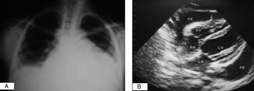
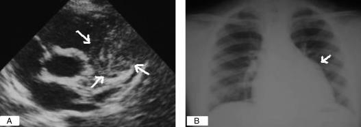
A 10-year-old girl was admitted to the hospital with fever, dry cough, chest pain and severe exertional dyspnea. Her complaints had started seven days earlier and her exertional dyspnea increased gradually. On physical examination, the patient had a temperature of 39°C, heart rate of 112 beats/min and blood pressure of 80/60 mmHg. She had respiratory distress, with orthopnea, mild peripheral face and leg edema, jugular venous distention, pulsus paradoxus, a 10 cm palpable liver and distant heart sounds, but no distinct murmur or friction rub. Chest x-ray revealed gross cardiomegaly (Figure 1A). Electrocardiography showed low QRS voltage and a normal PR interval. Transthoracic echocardiography demonstrated a massive pericardial effusion and right atrial collapse (Figure 1B). Given these clinical and echocardiographic findings, the patient was diagnosed to have cardiac tamponade; a pericardiocentesis was immediately performed and approximately 0.82 L of yellow serofibrinous fluid was withdrawn. Pericardial fluid cytology and cultures for bacteria were negative. A tuberculin skin test, and throat and blood cultures were all negative. Echocardiography repeated one day after pericardiocentesis revealed decreased pericardial fluid with no pathology of the mitral or aortic valve. Because the main complaints of the patient presenting with cardiac tamponade were fever, chest pain and dyspnea, our initial diagnosis was pericarditis. In spite of no supporting evidence for recent group A streptococcal infection, acute rheumatic fever was thought to be the probable cause and corticosteroid therapy was initiated. Clinical improvement was rapid, and repeat echocardiography performed three weeks after steroid therapy showed minimal pericardial effusion but revealed the presence of a solid mass inside the pericardial cavity (Figure 2A). The mass was located on the right ventricle outflow tract. It had a regular border measuring 3.8 cm by 4.5 cm and contained mixed echogenic circles characteristic of a hydatid cyst. A chest x-ray showed a bulge along the left cardiac border (Figure 2B). A computed tomography scan of the chest also showed the solid mass inside the pericardium. There were no cysts detected in the liver, lungs or brain. Laboratory tests did not show eosinophilia, and the hydatic serology was negative. The patient subsequently underwent surgery. Anterior thoracotomy and pericardiotomy were performed, and approximately 0.3 L of yellow serofibrinous hydatic fluid was drained. The pericardial hydatic lesion was extracted, with the histology confirming a hydatid cyst. Postoperatively, the patient recovered without complications and was discharged with oral albendazol prophylaxis treatment. No recurrence was observed on echocardiography at the six-month follow-up visit.
Figure 1).
A Chest x-ray of a patient with a large pericardial effusion showing gross cardiomegaly. B Two-dimensional echocardiography showing a large pericardial effusion and collapse on the free wall of the right atrium. LA Left aorta; LV Left ventricle; RA Right aorta; RV Right ventricle; PE Pericardial effusion
Figure 2).
Hydatid cyst mimicking a solid mass. A Echocardiographic image of the parasternal short axis showing a round lesion (arrows) with mixed echogenic circles and serpentine structures within the matrix, representing collapsed membranes on the right ventricle outlet tract and treaded main pulmonary artery inside the pericardial cavity. B Chest x-ray showing a bulge (arrow) along the left cardiac border where the mass was located
DISCUSSION
Cardiac hydatid cysts are uncommon in cases of hydatid disease. The most frequent location of the cyst is the myocardial region, particularly the interventricular septum and left ventricular free wall. Pericardial involvement in cardiac hydatid cysts is extremely uncommon (5–7). In the present patient, the hydatid cyst was located inside the pericardial cavity without myocardial involvement.
Clinical presentation of cardiac hydatid cysts depends on the location, size and number of cysts and presence of complications (2–8). Most patients remain asymptomatic for many years or have minor nonspecific complaints, such as fever, chest pain and weakness. However, anaphylactic shock may develop due to cyst rupture into the bloodstream. Other complications include systemic or pulmonary hydatid embolism, valve obstruction, mitral regurgitation secondary to papillary muscle involvement, atrioventricular conduction defects and arrhythmias. Symptoms of a pericardial hydatid cyst are generally due to the pressure exerted on the myocardium by an enlarging cyst or due to rupture of the cyst. Rupture of hydatid cysts into the pericardial cavity may lead to pericarditis with effusion, cardiac tamponade and formation of secondary cysts (7,8). In the present case, the disease manifested with pericarditis with effusion and cardiac tamponade. The diagnosis of a pericardial hydatid cyst was established by echocardiography.
Diagnosis is based on cardiac imaging techniques, such as echocardiography, computed tomography scans, magnetic resonance imaging and serological tests (2). Echocardiography is the diagnostic tool of choice because it is noninvasive, easily performed, and has a high sensitivity in the determination of intracardiac hydatid cysts and in planning surgical intervention (4,7,8).
On ultrasonography, the appearance of a round, thin-walled, multiloculated mass is characteristic of the echinococcal cyst (2). A hydatid cyst should always be considered when there is an intracardiac cystic image. But, the echolucent and multiseptate nature of hydatid cysts may sometimes be absent, and they may appear as a tumour-like mass (2,9). The recommended treatment is excision of the cyst because of the possibility of severe complications, including cyst rupture and sudden death, even in asymptomatic patients (6,7,10).
CONCLUSION
Cardiac hydatid cysts should always be considered in the differential diagnosis of pericarditis or pericardial effusion, especially in regions where hydatid disease is endemic.
REFERENCES
- 1.Blanton R. Echinococcosis. In: Behrman RE, Kliegman RM, Jenson HB, editors. Nelson Textbook of Pediatrics. 17. Philadelphia: WB Saunders Company; 2004. pp. 1173–4. [Google Scholar]
- 2.Pedrosa I, Saiz A, Arrazola J, Ferreiros J, Pedrosa CS. Hydatid disease: Radiologic and pathologic features and complications. Radiographics. 2000;20:795–817. doi: 10.1148/radiographics.20.3.g00ma06795. [DOI] [PubMed] [Google Scholar]
- 3.Perez-Gomez F, Duran H, Tamames S, Perrote JL, Blanes A. Cardiac echinococcosis: Clinical picture and complications. Br Heart J. 1973;35:1326–31. doi: 10.1136/hrt.35.12.1326. [DOI] [PMC free article] [PubMed] [Google Scholar]
- 4.Kudaiberdiev T, Djoshibaev S, Yankovskaya L, Djumanazarov A. Multiple hydatid cysts of epicardium and pericardium. Int J Cardiol. 2001;81:265–7. doi: 10.1016/s0167-5273(01)00554-x. [DOI] [PubMed] [Google Scholar]
- 5.Noah MS, el Din Hawas N, Joharjy I, Abdel-Hafez M. Primary cardiac echinococcosis: Report of two cases with review of the literature. Ann Trop Med Parasitol. 1988;82:67–73. doi: 10.1080/00034983.1988.11812211. [DOI] [PubMed] [Google Scholar]
- 6.Thameur H, Abdelmoula S, Chenik S, et al. Cardiopericardial hydatid cysts. World J Surg. 2001;25:58–67. doi: 10.1007/s002680020008. [DOI] [PubMed] [Google Scholar]
- 7.Birincioglu CL, Bardakci H, Kucuker SA, et al. A clinical dilemma: Cardiac and pericardiac echinococcosis. Ann Thorac Surg. 1999;68:1290–4. doi: 10.1016/s0003-4975(99)00692-x. [DOI] [PubMed] [Google Scholar]
- 8.De Martini M, Nador F, Binda A, Arpesani A, Odero A, Lotto A. Myocardial hydatid cyst ruptured into the pericardium: Cross-sectional echocardiographic study and surgical treatment. Eur Heart J. 1988;9:819–24. doi: 10.1093/eurheartj/9.7.819. [DOI] [PubMed] [Google Scholar]
- 9.Birincioglu CL, Tarcan O, Nisanoglu V, Bardakci H, Tasdemir O. Is it cardiac tumor or echinococcosis? Tex Heart Inst J. 2001;28:230–1. [PMC free article] [PubMed] [Google Scholar]
- 10.Onursal E, Elmaci TT, Tireli E, Dindar A, Atilgan D, Ozcan M. Surgical treatment of cardiac echinococcosis: Report of eight cases. Surg Today. 2001;31:325–30. doi: 10.1007/s005950170153. [DOI] [PubMed] [Google Scholar]


