Abstract
A cytomegalovirus (CMV) was isolated from its natural host, Peromyscus maniculatus, and was designated Peromyscus CMV (PCMV). A recombinant PCMV was constructed that contained Sin Nombre virus glycoprotein G1 (SNV-G1) fused in frame to the enhanced green fluorescent protein (EGFP) gene inserted into a site homologous to the human CMV UL33 (P33) gene. The recombinant CMV was used for expression and immunization of deer mice against SNV-G1. The results of the study indicate that P. maniculatus could be infected with as few as 10 virus particles of recombinant virus. Challenge of P. maniculatus with either recombinant or wild-type PCMV produced no overt pathology in infected animals. P. maniculatus immunized with recombinant virus developed an antibody response to SNV and EGFP. When rechallenged with recombinant virus, animals exhibited an anamnestic response against SNV. Interestingly, a preexisting immune response against PCMV did not prevent reinfection with recombinant PCMV.
Human cytomegalovirus (HCMV) has been used to express marker genes such as β-galactosidase or green fluorescent protein (GFP) in order to localize CMV in cells and animal tissues or to produce mutant viruses (4, 17, 31, 32). Our laboratory is interested in developing an animal model for hantavirus infections in order to explore the possible use of CMV as a vaccine vector. Advantages to the use of CMV as a vector include the following: (i) CMV normally does not cause severe disease in an immunocompetent host; (ii) CMV exists as a latent or persistent infection for the life of the host, resulting in continuous antigenic stimulation (1); (iii) healthy animals can be infected with more than one strain of CMV, indicating that the presence of preexisting immunity against the virus does not preclude immunization with a second strain expressing the same or alternative antigens (19); (iv) in cell culture, the virus has a relatively slow replication cycle, allowing expression of a protein for several days before the death of permissive cells; and (v) CMV can abortively infect many cell types, which can result in the expression of immediate-early and some early genes without resulting in cell death. This overcomes the problems with a vector such as vaccinia virus, which is toxic to many cell types.
We isolated a novel CMV from its natural host, the deer mouse (Peromyscus maniculatus), and designated this virus PCMV. It was confirmed to be a herpesvirus by electron microscopy and a CMV based on homology with other CMVs. The deer mouse is a reservoir for a severe human disease, hantavirus pulmonary syndrome, caused by several unique new world hantaviruses. Sin Nombre virus (SNV) is the predominant cause of hantavirus pulmonary syndrome in humans in North America. SNV causes an epizootic disease resulting in a chronic infection, with some transient mild pathology in its primary host, the deer mouse P. maniculatis (16, 20, 27). However, SNV infection in humans results in a severe life-threatening disease (8, 21). In deer mice the maternal antibodies of seropositive, lactating females provide protection from infection to the offspring (5). Similarly, maternal antibody may protect against hantavirus infection in humans (23). These data suggested that the humoral immune response is an important factor in determining the course of infection and in the outcome of the disease in both rodent hosts and humans. We were interested in using this as an animal model to examine the humoral immune response against hantavirus proteins. Our intent was to use this unique PCMV vector to express hantavirus proteins in deer mice to determine whether an immune response could be initiated by using a CMV vector in the natural rodent host. This approach has a number of advantages over directly challenging deer mice with SNV, including the following: (i) animal studies with hantaviruses associated with pulmonary syndrome require a biosafety level 4 facility (BSL-4), whereas expression of SNV proteins in a CMV vector only requires BSL-2 or BSL-3 conditions, and (ii) it would allow us to determine which of the virus-coded proteins is important in developing a protective immune response.
We used the PCMV P33 gene, a homolog of HCMV UL33 and murine CMV (MCMV) M33, as an insertion site. This site has been identified by several investigations as a region of the genome not essential for CMV replication in tissue culture, and insertions in this region of MCMV do not block in vivo replication. The P33 gene was interrupted by homologous recombination with a fusion gene consisting of SNV glycoprotein G1 (SNV-G1) and enhanced GFP (EGFP), thereby creating a recombinant PCMV(ΔP33:G1EGFP). SNV-G1 was selected since it has been shown to play an important role in generating neutralizing antibodies in mice (10, 28). Although expression of Old World hantavirus glycoproteins has been reported (9, 29), for New World viruses such as SNV the expression of full-length glycoproteins in mammalian cells has typically been difficult. Recombinant PCMV(ΔP33:G1EGFP) was used to infect deer mice in order to induce an immune response against SNV in the presence and in the absence of a preexisting antibody response against wild-type virus.
MATERIALS AND METHODS
Cells and virus culture.
PCMV was grown on P. maniculatis embryonic cells (PMEC). Virus stocks were prepared by infecting roller bottles with PMEC in Iscove modified Dulbecco medium (IMDM) with 2% fetal calf serum (FCS), at a multiplicity of infection (MOI) between 0.05 and 0.1 at 37°C. When 100% of cells demonstrated cytopathology but were still attached, the cells were scraped off, and the suspension was clarified by pelleting it in a Sorvall GLC centrifuge at 1,500 × g for 5 min. Cell pellets were resuspended in 10 ml of IMDM with 2% FCS and subjected to three freeze-thaw cycles. The cell lysate was clarified by pelleting in a Sorvall GLC centrifuge at 1,500 × g for 5 min. The titer of the virus was determined by inoculating PMEC on 24-well plates with a serial dilution of virus stock in IMDM-2% FCS at 100 μl/well for 1 h at 37°C. The 24-well plates were cultured with an overlay containing IMDM with 0.5% agarose, 2% FCS, and 50 μg of kanamycin/ml for 1 week, after which the plates were fixed with 4% formaldehyde in phosphate-buffered saline (PBS) for 1 h, the overlay was removed, and the cells stained with 0.6% methylene blue. After a wash with water, the plates were dried and the plaques were counted.
Preparation of PCMV genomic clones.
PCMV virus stock was prepared as described above, after which, the PCMV was pelleted at 15,000 rpm in an SW27 rotor (Beckman) for 1 h at room temperature. PCMV genomic DNA was extracted with the PureGene DNA extraction kit (Gentra Systems) according to the protocol of the manufacturer.
Virus genomic DNA was digested with NotI for 2 h, and the reaction was stopped by heating at 65°C for 20 min. The restricted virus DNA was ligated into pET29c (Novagen) that was linearized with NotI and treated with shrimp alkaline phosphatase (USB). Electrocompetent DH10B Escherichia coli (Life Technologies) was transformed with the ligation mix at 330 μF and 400 V (with the voltage booster set at 4 kΩ). Clones were selected with NotI inserts of ≥10 kb. The viral origin of the NotI inserts was verified by Southern blotting (not shown).
PCR, cloning, and sequencing of the P33 gene and DNA polymerase fragment.
Virus genomic DNA was used as a template for PCR. The UL33 homolog was subloned by PCR with forward primer 5′-ATGATA-C/G-G/A/C-TGGTTCTAC-3′ and reverse primer 5′-GCGTACAG-C/G-A-C/T-GGGATT-3′ (Life Technologies). The PCR mix contained 50 ng of template DNA, each primer at 100 ng, 2.5 mM MgCl2, 0.2 mM deoxynucleoside triphosphates, and 5 U of Tag in 1× reaction mix (Promega). Cycles consisted of a denaturation at 94°C for 1 min, followed by annealing at 50°C for 1 min and an extension at 72°C for 1 min for 35 cycles. The same PCR was repeated with eight viral NotI clones as a template. The viral NotI genomic clones were cut with BamHI overnight and separated on a 1.0% agarose gel. The gel was transferred to Zetaprobe GT nylon membrane (Bio-Rad) according to manufacturer's recommendations. In order to detect the P33-gene, we used a 31-mer, 5′-TGGGACGTCACCTGCGAATCTATCAACAACA-3′, which was derived from the sequence obtained from one end of the PCR product. The oligonucleotide was end labeled by using [γ-32P]ATP (NEN Life Sciences) and T4 polynucleotide kinase (Life Technologies). The hybridization was performed at 65°C in hybridization buffer containing 10.36% (wt/vol) polyethylene glycol 8000, 7.25% sodium dodecyl sulfate (SDS), 0.233 M sodium chloride, 0.155 M sodium phosphate (pH 7.4), 1.55 mM EDTA, and 100 μg of salmon sperm DNA. The membrane was washed in 0.2× SSC (1× SSC is 0.15 M NaCl plus 0.015 M sodium citrate)-0.1% SDS at 42°C. After washing, the membrane was dried and exposed to X-ray film. Based on the result from the Southern blot, NotI clone #121 was cleaved with BamHI (New England Biolabs). The cleaved DNA fragments were separated on a 1.0% agarose gel, and the 3-kb band was excised from the gel and purified by using the Qiaex-II kit (Qiagen). The BamHI fragment was cloned into the BamHI linearized pPUR (Clontech), creating pPUR3Kb. The P33 gene was sequenced, with gene-specific primers, based on an initial sequence from the P33 PCR, by using a rhodamine sequencing kit (PE Biosystems) and the Prism 310 automated sequencer (PE Biosystems). An additional sequence spanning the entire P33 gene was obtained by primer walking in both directions. The DNA polymerase gene fragment was PCR amplified with forward primer 5′-C/G-CG-C/T-GGCGTG-A/T-T-A/G-TA-C/T-GA-C/T-GG-3′ and reverse primer 5′-GATCTG-C/T-TG-T/G-CC-C/G-TCGAAGATGAC-3′. The PCRs were done by using Platinum Taq (Life Technologies). The cycles consisted of denaturation at 94°C for 30 s, followed by annealing at 60°C for 30 s and an extension at 68°C for 1 min for 35 cycles. Sequence comparisons were done by using the BLASTN, BLASTX, and BLASTP programs (National Center for Biotechnology Information). All sequence analysis, primer design restriction analysis, and protein coding evaluation were done by using the DNA-Star computer package (Lasergene).
RT-PCR of P33 gene.
Total RNA from PCMV-infected PMEC was extracted by using Trizol (Life Technologies) according to the protocol supplied by the manufacturer. The RNA was reverse transcribed with the Thermoscript reverse transcription-PCR (RT-PCR) system (Life Technologies) by using the random hexamers supplied with the kit. The resulting cDNA was treated with DNase I (Promega). The 5′ end of the gene was subsequently amplified by PCR with the forward primer 5′-CATACACAATAAAAGCCTCCAGAT-3′ and the reverse primer 5′-CGTCGCGTAGTACATAAGTGC-3′, which are located on both sides of the presumed splice site. The same primers were used in a PCR to amplify part of the P33 gene by using genomic virus DNA as a template. The PCR product, representing the P33 cDNA sequence, was cloned into pGEM-T Easy according to the protocol (Promega). The PCR product was sequenced to confirm the absence of intron sequences.
Construction of recombinant PCMV(ΔP33:G1EGFP).
Using Pwo polymerase (Roche Molecular Biochemicals), a 2,116-bp PCR product spanning all of the SNV-G1 and part of SNV-G2 was amplified from cDNA derived from autopsy lung tissue from a hantavirus pulmonary syndrome patient. Forward primer 5′-CGAACCATGGTAGGGTGGGTTTGCATCTT-3′ and reverse primer 5′-CTTCTTGATTGGCAGGATTCAC-3′ were used in PCRs. The PCR product was ligated into pCRII (Invitrogen), creating pCRII-G1. Sequencing of the cloned product revealed a contiguous open reading frame (ORF) with no stop codons or other PCR-induced mutations. The expression vector pEGFP-N1 (Clontech) was cleaved with BamHI, blunt-ended with Klenow (New England Biolabs) and religated, creating pEGFP-N1-BHI. To make pEGFP-N1G1, SNV-G1 was released from the plasmid pCRII-G1 with EcoRI and ligated into the EcoRI site of pEGFP-N1-BHI, thereby placing the G1 PCR fragment upstream and in frame with EGFP. The sequence of the construct from the transcription start site through the G1 gene and EGFP gene was verified by sequencing. The unique AseI site of pEGFP-N1G1 vector was converted to an EcoRV site by cleaving it with AseI, filling in the overhangs with Klenow, and ligating an EcoRV linker (5′-PGGGATATCCC-3′) into the plasmid. pN1G1-fl plasmid was generated by insertion of an XhoI fragment, containing flanking regions of the P33 gene joined together, each in reverse orientation, into the EcoRV site. The resulting plasmid was used for homologous recombination with wild-type PCMV (wt-PCMV) as follows. First, 40 μg of pN1G1-fl was cut with BamHI and then divided into aliquots in four electroporation cuvettes (Life Technologies), each containing approximately 5 × 10⁶ PMEC in 0.5 ml of IMDM with 10% FCS. The cells were electroporated in a Cell-Porator electroporation system (Life Technologies) at 250 V, with a capacitance of 1,600 μF. The cells were plated onto T-75 tissue culture flasks with fresh IMDM containing 10% FCS and then cultured overnight at 37°C. The following day, the cells were examined for viability and fluorescence and then infected with PCMV at an MOI of ca. 1. After superinfection, the cells were cultured with IMDM containing 2% FCS and 100 μg of G418 until a complete cytopathic effect was observed. Cells that remained attached were scraped into the medium, and this mixture was subjected to three freeze-thaw cycles, with vigorous vortexing between each cycle. Cells were then pelleted, and the supernatant was filtered by using a 10-ml syringe and a 0.20-μm syringe filter. Fresh PMEC were infected with the filtered supernatant containing the virus and cultured in the presence of 100 μg of G418. After all cells were infected, the supernatant virus was obtained as described above. This procedure was repeated three times.
Further enrichment was achieved by using a flow cytometer (Beckman Coulter). PMEC from two wells of a six-well plate containing EGFP-expressing recombinant PCMV were treated with trypsin and resuspended in PBS containing 50 mM EDTA and 2% FCS. Uninfected PMEC were used as a negative control. Based on the difference between the negative cells and the EGFP-containing cells, a population displaying a high degree of fluorescence was selected, and these cells were distributed in such a manner that one fluorescent cell was seeded per well in a 96-well plate containing confluent, uninfected PMEC. Several wells of the 96-well plate were pooled and expanded. The recombinant PCMV virus was checked for purity, and correct insertion of expression cassette by Southern blotting and PCR analysis. The recombinant PCMV is called PCMV(ΔP33:G1EGFP).
Immunohistochemistry.
PMEC were plated on coverslips in six-well plates and infected with wt-PCMV or recombinant PCMV at virus/cell ratio (MOI) of 0.1. At 6 days postinfection, cells on coverslips were fixed by incubation in methanol-acetone (3:1) solution for 5 min. Coverslips were then dried and stored at −20°C. Before immunostaining, coverslips were rehydrated in PBS for 5 min, followed by incubation with primary antibody (1:200 dilution, recombinant human anti-SNV-G1 antibody, immunoglobulin G1 SNV #26) in PBS-0.2% Tween 20 for 1 h. The cells were then washed three times for 5 min with PBS-0.2% Tween 20, incubated with secondary antibody (1:2,000 dilution; alkaline phosphatase-conjugated goat anti-human immunoglobulin G [H+L] antibody; Vector Laboratories), washed three times for 5 min with PBS-0.2% Tween 20, rinsed with Tween 20-Tris-buffered saline, and developed by using the alkaline phosphatase substrate kit 1 (Vector Laboratories). Coverslips were mounted on slides and viewed by using interference contrast microscope.
SDS-polyacrylamide gel electrophoresis and Western blotting.
PMEC, seeded in 60-mm dishes, were infected with either wt-PCMV or PCMV(ΔP33:G1EGFP) or were mock infected. Alternatively, PMEC were transfected by electroporation with pEGFP-N2-G1 as described above. For the infected cells, the cells were lysed after 5 days. For the transfected cells, 1 day after transfection, the PMEC were superinfected with wt-PCMV at an MOI of 1. After 6 days, lysates were obtained by aspirating the medium and scraping the cells into 100 μl of 4× sample buffer containing 0.4 M Tris-HCl (pH 6.9), 40% glycerol, 10% SDS, 0.016% BPB protease inhibitors (Boehringer Mannheim), and 10% β-mercaptoethanol, added just before use. The cell lysates were heated at 100°C for 5 min before they were stored at −20°C, or they were used immediately.
The cell lysates were subjected to SDS-12% polyacrylamide gel electrophoresis at 200 V and then transferred to Immobilon-P (Millipore) by using a semidry transfer unit (Bio-Rad), after which the membranes were dried and incubated with TBST blocking buffer (1× Tris-buffered saline with 0.05% Tween 20 and 5% nonfat dry milk) for 1 h at room temperature. A monoclonal antibody to EGFP (GFP monoclonal antibody 8362-1; Clontech) was used at a dilution of 1:1,000. Secondary goat anti-mouse antibody conjugated to horseradish peroxidase (Chemicon) was used at a dilution 1:2,000. The detection was performed by using the DAB (3,3′-diaminobenzidine) substrate kit (Vector Laboratories) or enhanced chemiluminescence (Amersham Pharmacia). For detection of SNV-G1, rabbit serum against the SNV-G1-N2 peptide (CYNQVDWTKKS), coupled with keyhole limpet hemocyanin, was used. This peptide from SNV-G1 represents the region of G1 homologous to an immunodominant epitope of Pumaala G1 (14). Rabbits were immunized, boosted two times, and screened for antibody production. Antibodies to SNV-G1 were affinity purified by using a SulfoLink affinity column (Pierce), conjugated with SNV-G1-N2 peptide.
Immunization of deer mice and serology.
Two groups of mice were injected intraperitoneally with either 10 PFU wt-PCMV (eight mice) or with 10 PFU of PCMV(ΔP33:G1EGFP) (eight mice). One year later the three mice from the wt-PCMV-infected group and six mice from the PCMV(ΔP33:G1EGFP)-infected group received a second immunization with 10,000 PFU of PCMV(ΔP33:G1EGFP) intraperitoneally. Periodically the mice were bled, and their serological status was determined for PCMV, EGFP, and SNV by enzyme-linked immunosorbent assay (ELISA). Antigens for the ELISA consisted of a PCMV-infected PMEC lysate, SNV-infected Vero-E6 cell lysate, and rEGFP protein (Clontech) at 100 pg/well. GFP monoclonal antibody (Clontech) was used at a 1:500 dilution as a positive control antibody in an EGFP ELISA. The negative controls were uninfected cells. Statistical analysis was performed in MS Excel 2000 by using the t test function.
RESULTS
Isolation of a CMV from deer mice.
In attempting to isolate endogenous viruses from tissues of deer mice by using cells derived from PMEC, a cytopathology typical of CMV was observed (18). Examination of infected cells by transmission electron microscopy revealed viral capsids strongly resembling those of the herpesvirus family (3) (electron microscopy courtesey of Jay Brown, University of Virginia [data not shown]).
Identification of PCMV DNA polymerase and UL33 homolog.
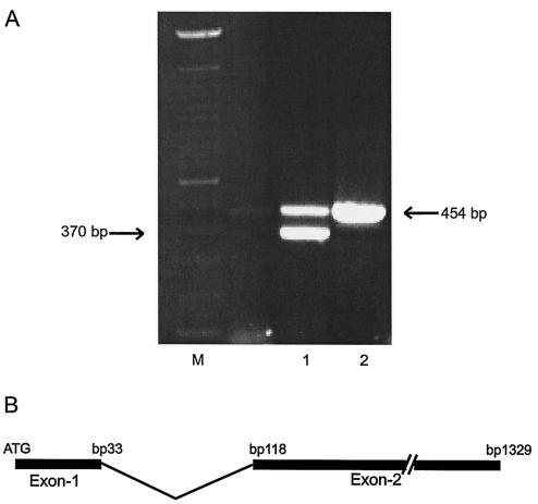
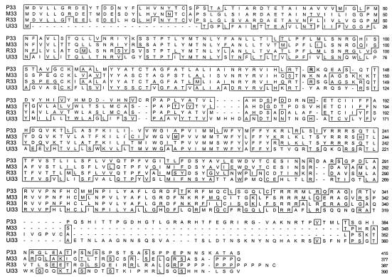
To confirm if the virus isolated was a CMV, we examined total DNA isolated from infected cells for the presence of homologs to the HCMV DNA polymerase and the HCMV UL33 gene. These genes were selected because the polymerase gene is well conserved among herpesviruses (6, 7, 15, 24), and a homologous form of the human CMV UL33 gene has been reported to be present in many CMVs. UL33 is nonessential for virus replication in cell culture (13, 33), making it a possible site for insertion of foreign genes. PCR analysis and sequencing of the UL33 homolog yielded preliminary sequence information, which was used to synthesize a small 29-mer probe for screening a series of plasmid clones of virus genomic DNA. The sequence obtained from a 3-kb genomic BamHI fragment identified a CMV UL33 homolog. The sequence revealed a high level of homology to MCMV M33 and rat CMV (RCMV) R33. Whereas UL33 and the RCMV homolog R33 are the products of unspliced RNA, the MCMV homolog, M33, is the product of a single splicing event (11). The P33 sequence of PCMV is contiguous, suggesting a splicing event. RT-PCR was performed with primers on either side of the putative splice-donor and -acceptor sequences. This experiment confirmed the presence of an 85-bp intron within the 1,333-bp gene, consisting of 33- and 1,215-bp exons (Fig. 1). When total RNA from PCMV-infected cells was used as a template, two bands were detected on the gel representing unspliced genomic copy of P33 and spliced P33 mRNA. M33, R33, and UL33 genes are homologous to G protein-coupled receptor (amino acid sequence similarities of 62.5, 59.9, and 39.1%, respectively) (Fig. 2). Transcriptional analysis in the presence of 50 μg of phosphonoformic acid (PFA)/ml inhibited P33 transcription confirming its identity as a true late gene (data not shown).
FIG. 1.
Analysis of RNA and DNA from Peromyscus cells infected with PCMV. (A) Demonstration of a presence of both genomic and spliced version of P33 gene in infected cells. Lanes: 1, P33-specific RT-PCR with RNA from PCMV-infected cells as a template; 2, P33-specific PCR with purified genomic PCMV DNA as a template; M, 100-bp ladder (Gibco-BRL). (B) Genomic arrangement of coding region of P33. The first exon, encoding the first 11 amino acids, is separated by an 85-bp intron from the second exon encoding an additional 404 amino acids.
FIG. 2.
Jotun-Hein alignment of the P33 protein with the homologous UL33 proteins from MCMV (M33), RCMV (R33), and HCMV (UL33). The consensus residues are boxed. The homologies are 62.5, 59.9, and 39.1%, respectively.
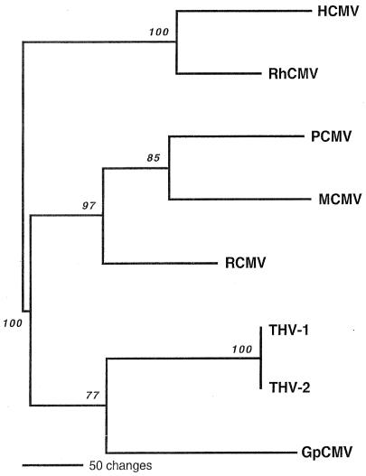
The DNA sequence of the DNA polymerase gene was compared to the corresponding regions of characterized DNA polymerases of other species-specific CMVs. As expected, the DNA polymerase ORF aligned most closely with those of previously characterized rodent CMVs, with the MCMV DNA polymerase homolog ORF being the most closely related, followed by the DNA polymerase of RCMV (Fig. 3). Other DNA polymerase homologs are grouped in different clades with HCMV and rhesus CMV (RhCMV) occupying one clade and guinea pig CMV (GpCMV) being grouped with two different tupaia herpesvirus DNA polymerases. This alignment and the fact that PCMV did not replicate in NIH 3T3 cells (data not shown) illustrate that PCMV is a novel CMV, one closely related to MCMV and RCMV.
FIG. 3.
Phylogenetic relationships of PCMV to representative CMVs isolated from rodents and primates. A single most parsimonious phylogenetic tree was generated on the basis of amino acid sequence differences of the 480-amino-acid fragment of the viral DNA polymerase by using PAUP (version 4b10). Maximum-parsimony analysis was conducted by using the “branch-and-bound” search option and a “protpars” weighting matrix. Gaps were treated as “missing data.” Lengths of the horizontal branches are proportional to the amino acid step differences between corresponding taxa (see bar scale). Vertical branches are for visual clarity only. Bootstrap values obtained from 1,000 replicates of the heuristic maximum-parsimony analysis are shown in italics at the appropriate branch points. Previously published CMV DNA polymerase sequences used in the analysis include the following: HCMV strain AD169 (GenBank accession number X17403), RhCMV strain 68-1 (AF033184), MCMV strain Smith (M73549), RCMV strain Maastricht (AF232689), GpCMV strain 22122 (L25706), and tupaia herpesvirus strains 1 (THV-1; AF074327) and 2 (THV-2; AF281817).
Insertion of SNV-G1 into PCMV.
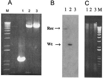
Although rat and mouse M33 is not essential for the viral replication cycle in tissue culture, recombinant RCMV and MCMV lacking the UL33 homolog displayed an attenuated phenotype, as demonstrated by a reduced tropism for the salivary gland (11) and an increased 50% lethal dose (2), respectively. The P33 gene was therefore targeted to be replaced with an expression cassette encoding a fusion gene comprised of the entire SNV-G1 (plus the nucleotide sequence for the first 50 amino acids of G2) placed in frame with the EGFP gene at the C-terminal end. The P33 gene, situated in the middle of a 3-kb BamHI genomic DNA fragment, contains two convenient XhoI sites that are 575 bp apart, with the translation start site 378 bp upstream of the first site and the stop codon 376 bp downstream of the second site. The 575-bp XhoI fragment was replaced with a plasmid that contains the fusion gene described above by homologous recombination. The recombinant virus was purified by using G418 selection and fluorescence-activated cell sorting (FACS). PCR analysis across the insertion site demonstrated no evidence of contaminating wild-type sequences (Fig. 1A). Southern blot analysis (Fig. 4) with a P33 specific probe demonstrated that there is a shift in size from 3 kb in the wt-PCMV to ca. 10 kb in the recombinant PCMV genomic fragment. It was therefore concluded that the recombinant virus was pure, and it was designated PCMV(ΔP33:G1EGFP).
FIG. 4.
Southern blot of recombinant and wt-PCMV DNA demonstrating insertion of SNV-G1 into P33. (A) PCR amplification with P33-specific primers with wt-PCMV DNA (lane 1), recombinant PCMV DNA (before FACS) (lane 2), and recombinant DNA (after FACS) (lane 3). (B) Southern blot of total DNA isolated from PMEC infected with PCMV(ΔP33:G1EGFP) (lane 1), wt-PCMV (lane 2), or uninfected control cells (lane 3). The DNAs were cleaved with BamHI, and the blot was probed with the 1.2-kb AgeI fragment containing the P33 gene. (C) Agarose gel, used for the Southern blot presented in panel B. Lane labeling is the same in panels B and C.
Expression of EGFP-G1.
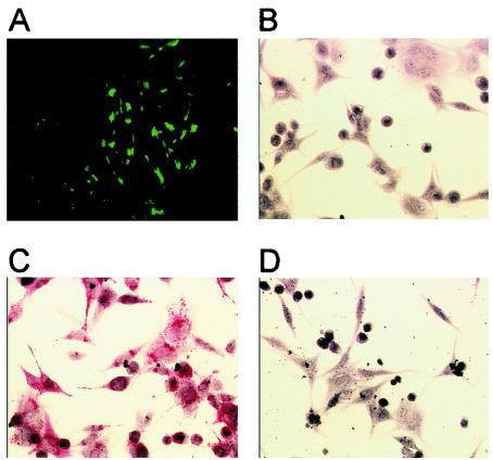
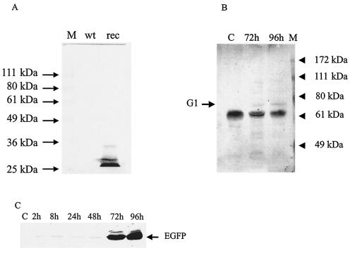
Cells infected with recombinant PCMV expressed EGFP, which was detected by using fluorescence microscopy (Fig. 5A). In order to determine whether G1 could be detected in infected cells, we stained cells with anti-G1 antibodies. Recombinant PCMV-infected PMEC strongly reacted with anti-SNV-G1 recombinant human antibody on immunohistochemistry slides (Fig. 5B to D). Protein lysates of PMEC infected with PCMV(ΔP33:G1EGFP) were examined by Western blot analysis with a monoclonal antibody to EGFP. Interestingly, Western blot analysis showed an abundant band corresponding to the molecular mass expected for EGFP (ca. 27 kDa) (Fig. 6A). The observed molecular mass of the protein band was less than the predicted molecular mass of the G1-EGFP fusion protein (>106 kDa). An infection time course of recombinant PCMV-infected PMEC showed the expression of the low-molecular-weight EGFP protein was detectable 72 h postinfection by Western blot analysis (Fig. 6C). Rabbit serum, generated against a peptide based on the amino acid sequence of SNV-G1, homologous to immunodominant domain of Pumaala G1, showed a band of ca. 75 kDa at late times postinfection (Fig. 6B). The G1-EGFP expression cassette is under the transcriptional control of the HCMV immediate-early promoter; however, there is no detectable signal for EGFP until 48 h postinfection.
FIG. 5.
Fluorescence and immunohistochemistry analysis of wt-PCMV and recombinant PCMV infection of PMEC. (A) Peromyscus cells 5 days after infection with recombinant PCMV(ΔP33:G1EGFP). Magnification, ×320. (B) PMEC infected with recombinant PCMV probed with nonspecific negative antibody control. (C) PMEC infected with recombinant PCMV probed with recombinant anti-SNV human antibody. (D) PMEC infected with wt-PCMV probed with recombinant anti-SNV human antibody. In panels B to D, primary antibody incubations were followed by incubation with alkaline phosphatase-conjugated secondary antibody-colorimetric development. Magnification, ×640.
FIG. 6.
Western analysis of recombinant proteins expressed with recombinant PCMV virus. (A) Western blot of wt-PCMV-infected PMEC and PCMV(ΔP33:G1EGFP)-infected PMEC at 96 h postinfection with a monoclonal antibody to EGFP. The predominant band on the blot is EGFP protein with a molecular mass of ca. 27 kDa. (B) Western blot of SNV expression in PCMV(ΔP33:G1EGFP)-infected PMEC at 72 and 96 h postinfection with a rabbit anti-peptide antibody raised against an epitope of SNV-G1. The minor band at ca. 72 kDa corresponds to SNV-G1. (C) Western blot of time course expression of EGFP in PCMV(ΔP33:G1EGFP)-infected PMEC.
Immunization of deer mice with PCMV(ΔP33:G1EGFP).
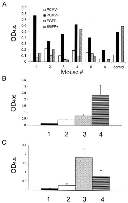
To determine whether PCMV(ΔP33:G1EGFP) can infect deer mice and evoke an immune response against SNV-G1 and EGFP, deer mice were immunized with 10 PFU of the recombinant PCMV or wt-PCMV and monitored for antibodies to SNV, EGFP, or PCMV. After a year, these mice were boosted with 10,000 PFU of the recombinant PCMV, and their serology for PCMV and SNV was monitored. Figure 7A demonstrates an early immune response (15 days postinfection) against PCMV and EGFP in deer mice infected with recombinant PCMV. Deer mice infected with recombinant PCMV showed a weak but statistically significant (P = 0.018) immune response against PCMV 4 weeks postinfection (Fig. 7B). The immune response against PCMV increased over a 12-month period. Reinfection of recombinant PCMV-infected mice with recombinant PCMV caused a dramatic increase in anti-PCMV and anti-SNV antibody levels (Fig. 7B and C). Therefore, it seems likely that the levels of G1 expression in the recombinant virus were sufficient to induce a significant humoral immune response. In addition, infection of wt-PCMV-infected mice with recombinant PCMV resulted in significant anti-SNV antibody levels by 3 weeks postinfection (P = 0.003).
FIG. 7.
Antibody response as measured by ELISA in deer mice infected with recombinant or wt-PCMV. (A) Antibody response to PCMV and EGFP in six deer mice infected with recombinant PCMV at 15 days postinfection. Column groups 1 to 6 represent individual mice. Control, pooled serum containing anti-GFP mouse monoclonal antibody against wt-PCMV from naturally infected wild mice. (B) Antibody response against PCMV antigens. Mouse serum dilution, 1:50. Columns: 1, uninfected control (three mice); 2, recombinant PCMV-infected mice 4 weeks postinfection (eight mice); 3, recombinant PCMV-infected mice 12 weeks postinfection (six mice); 4, wt-PCMV-infected mice 12 weeks postinfection (five mice). (C) Antibody response to SNV-G1. Mouse serum dilution, 1:100. Columns: 1, uninfected control (three mice); 2, recombinant PCMV-infected mice 12 weeks postinfection (six mice); 3, recombinant PCMV-infected mice 3 weeks after reinfection with recombinant PCMV (six mice); 4, wt-PCMV-infected mice 3 weeks after secondary infection with recombinant PCMV (three mice).
DISCUSSION
In this study we describe a CMV from P. maniculatis that has not been previously characterized. The virus was classified as a CMV based on the following criteria: (i) the cytopathology was typical of a CMV; (ii) electron microscopy indicated a morphology typical of a CMV (Jay Brown, unpublished data); (iii) DNA polymerase and UL33 homologs shared sequence homology with MCMV; and (iv) replication occurred exclusively in cells derived from P. maniculatus. Phylogenetic analysis places this virus in the same clade as MCMV and RCMV. The DNA polymerase fragment shows similarities, at the amino acid level, of 60.2 and 56.9% with the corresponding regions of the DNA polymerases of MCMV and RCMV, respectively.
The evolutionary relatedness to MCMV is also demonstrated by the fact that the UL33 homolog, P33, contains an 85-bp intron that separates the 33-bp 5′ end of the coding region from the remaining 1,215-bp second exon. In the M33 gene, the splice site is at the identical position in the gene. This finding is in contrast to the UL33 homologs in RCMV (R33) and HCMV, for which the gene is encoded on a contiguous ORF. The P33 gene is a true late gene in that no transcripts were detected in the presence of PFA, in contrast to transcription for the DNA polymerase, which represents an early cytomegalovirus gene. Although these G protein-coupled receptor homologs are dispensable for virus replication in tissue culture, they clearly play an essential role in viral pathogenesis, as is evident by a decreased tropism for the salivary glands (2, 11) and an increased 50% lethal dose of UL33-null CMVs.
We generated a recombinant PCMV by inserting an expression cassette for SNV-G1 fused to EGFP into the P33 gene. Fusion protein was designed with a preserved G1-G2 cleavage site between the G1 and EGFP coding regions. This was done to achieve expression of G1 in its natural form, which could be important for the generation of a correct immune response against G1, while providing easy EGFP detection for monitoring of recombinant protein expression and selection of recombinant virus. Expression of EGFP was confirmed by observing green fluorescence. Immunohistochemistry with specific anti-SNV-G1 recombinant human antibody confirmed the expression of SNV-G1 in PMEC infected with recombinant PCMV. Western blot analysis of infected cell lysates indicated that an abundant protein of 27 kDa, presumably EGFP, was present at early times in infection. Western blot analysis of these same lysates with a rabbit antiserum against a G1 epitope of SNV indicated a 75-kDa G1 protein was detected at late times in infection. These results confirm expression and cleavage of G1 and EGFP from their fusion protein precursors. Efficient expression from the G1-EGFP cassette occurred only late in infection and is also consistent with inefficient expression of the EGFP-G1 plasmid in transient transfections (data not shown). Although the HCMV promoter is an immediate-early promoter, this is in the context of the CMV virion and its position in the virus DNA. It is likely that when the promoter is present in the PCMV genome and in P. maniculatus cells that its temporal regulation is different. It appears that G1 and EGFP proteins were both expressed as individual polypeptides, since a G1-EGFP fusion protein is not detectable in infected cells. This is probably caused by the cleavage site between G1 and G2 in the SNV glycoprotein, which was retained in the G1-EGFP fusion construct.
When deer mice were challenged with PCMV (ΔP33:G1EGFP), they could be infected with as little as 10 PFU per animal. When the animals were challenged with recombinant virus, an antibody response to SNV-G1 and EGFP was detected. This is a strong indication that the expressed forms of G1 and EGFP are capable of inducing antibodies that recognize SNV-G1 and EGFP in Vero-E6 infected cell lysates. Interestingly, preexisting immunity to wt-PCMV does not interfere with the induction of an immune response against an immunogenic transgene expressed in this viral vector. This is in accordance with studies that show the presence of multiple strains of MCMV, resulting from subsequent infections, in wild mice (19). This is in contrast to HCMV infection in humans where, in healthy individuals, prior infection with either wild-type HCMV or a vaccine strain confers at least partial protection (1, 25, 26). This could prove to be an important advantage over other, more frequently used virus gene delivery systems in the deer mouse model, in which an immune response to the viral vector can interfere with the efficiency of transgene expression in vivo (12, 30). The fact that an antibody response against PCMV antigens is more profound in wt-PCMV-infected mice suggests that recombinant PCMV might display an attenuated phenotype. It is interesting that, although the virus is attenuated after insertion of a foreign gene into P33, it is still able to establish an infection with as little as 10 PFU per animal, as demonstrated by the observation that challenged animals produced antibodies to PCMV, SNV, and EGFP. However, the antibody response to PCMV in mice infected with recombinant PCMV was lower than in wt-PCMV-infected mice (data not shown).
There are other examples of the successful utilization of herpesviruses to induce an immune response through heterologous expression. The canine herpesvirus has been used to induce a protective immune response against rabies by expression of the rabies virus G protein (35) in dogs. Also, Marek's disease virus, expressing IBDV-VP2, was efficacious in protecting chickens from very virulent infectious bursal disease (34). Although the results presented here are encouraging, further studies need to be conducted to determine the efficacy of a PCMV vector in protecting deer mice from SNV infection. At this time little is known about the significance of antibody versus cell-mediated response in protection against SNV infection. Furthermore, the role of G1, G2, or nucleocapsid protein in a protective immune response against SNV is unclear. The data from our laboratory indicate that maternal antibody may protect young animals against infection with SNV (5).
We intend to explore the recombinant PCMV system further to identify the importance of G1, G2, and the nucleocapsid protein in protection against SNV. It is our intent to challenge animals immunized with the PCMV vector with wild-type virus to determine whether prior humoral immunity can protect against the animals developing a persistent infection or after the length of time the viremic phase of the disease occurs. These studies will be done in the near future when biocontainment level 4 space is available since animal studies require BSL-4 containment and the space in the United States for this type of containment is extremely limited.
Acknowledgments
A.A.R. and A.G.M.V.G. contributed equally to this study.
This project was supported by NIH grants AI36418 and HL63470.
REFERENCES
- 1.Adler, S. P., S. H. Hempfling, S. E. Starr, S. A. Plotkin, and S. Riddell. 1998. Safety and immunogenicity of the Towne strain cytomegalovirus vaccine. Pediatr. Infect. Dis. J. 17:200-206. [DOI] [PubMed] [Google Scholar]
- 2.Beisser, P. S., C. Vink, J. G. Van Dam, G. Grauls, S. J. Vanherle, and C. A. Bruggeman. 1998. The R33 G protein-coupled receptor gene of rat cytomegalovirus plays an essential role in the pathogenesis of viral infection. J. Virol. 72:2352-2363. [DOI] [PMC free article] [PubMed] [Google Scholar]
- 3.Berezesky, I. K., P. M. Grimley, S. A. Tyrrell, and A. S. Rabson. 1971. Ultrastructure of a rat cytomegalovirus. Exp. Mol. Pathol. 14:337-349. [DOI] [PubMed] [Google Scholar]
- 4.Borst, E., and M. Messerle. 2000. Development of a cytomegalovirus vector for somatic gene therapy. Bone Marrow Transplant. 25(Suppl. 2):S80-S82. [DOI] [PubMed] [Google Scholar]
- 5.Borucki, M. K., J. D. Boone, J. E. Rowe, M. C. Bohlman, E. A. Kuhn, R. DeBaca, and S. C. St. Jeor. 2000. Role of maternal antibody in natural infection of Peromyscus maniculatus with Sin Nombre virus. J. Virol. 74:2426-2429. [DOI] [PMC free article] [PubMed] [Google Scholar]
- 6.Capps, G. G., and M. C. Zuniga. 1990. A double-labeling method for measuring induction of protein phosphorylation. BioTechniques 8:62-69. [PubMed] [Google Scholar]
- 7.Chadwick, E. G., R. Yogev, S. Kwok, J. J. Sninsky, D. E. Kellogg, and S. M. Wolinsky. 1989. Enzymatic amplification of the human immunodeficiency virus in peripheral blood mononuclear cells from pediatric patients. J. Infect. Dis. 160:954-959. [DOI] [PubMed] [Google Scholar]
- 8.Childs, J. E., T. G. Ksiazek, C. F. Spiropoulou, J. W. Krebs, S. Morzunov, G. O. Maupin, K. L. Gage, P. E. Rollin, J. Sarisky, and R. E. Enscore. 1994. Serologic and genetic identification of Peromyscus maniculatus as the primary rodent reservoir for a new hantavirus in the southwestern United States. J. Infect. Dis. 169:1271-1280. [DOI] [PubMed] [Google Scholar]
- 9.Chu, Y. K., G. B. Jennings, and C. S. Schmaljohn. 1995. A vaccinia virus-vectored Hantaan virus vaccine protects hamsters from challenge with Hantaan and Seoul viruses but not Puumala virus. J. Virol. 69:6417-6423. [DOI] [PMC free article] [PubMed] [Google Scholar]
- 10.Dantas, J. R., Jr., Y. Okuno, H. Asada, M. Tamura, M. Takahashi, O. Tanishita, Y. Takahashi, T. Kurata, and K. Yamanishi. 1986. Characterization of glycoproteins of viruses causing hemorrhagic fever with renal syndrome (HFRS) using monoclonal antibodies. Virology 151:379-384. [DOI] [PubMed] [Google Scholar]
- 11.Davis-Poynter, N. J., D. M. Lynch, H. Vally, G. R. Shellam, W. D. Rawlinson, B. G. Barrell, and H. E. Farrell. 1997. Identification and characterization of a G protein-coupled receptor homolog encoded by murine cytomegalovirus. J. Virol. 71:1521-1529. [DOI] [PMC free article] [PubMed] [Google Scholar]
- 12.Engelhardt, J. F., L. Litzky, and J. M. Wilson. 1994. Prolonged transgene expression in cotton rat lung with recombinant adenoviruses defective in E2a. Hum. Gene Ther. 5:1217-1229. [DOI] [PubMed] [Google Scholar]
- 13.Hartwell, L. H., and T. A. Weiner. 1989. Checkpoints: controls that ensure the order of cell cycle events. Science 246:629-634. [DOI] [PubMed] [Google Scholar]
- 14.Heiskanen, T., A. Lundkvist, R. Soliymani, E. Koivunen, A. Vaheri, and H. Lankinen. 1999. Phage-displayed peptides mimicking the discontinuous neutralization sites of puumala Hantavirus envelope glycoproteins. Virology 262:321-332. [DOI] [PubMed] [Google Scholar]
- 15.Langan, T. A., J. Gautier, M. Lohka, R. Hollingsworth, S. Moreno, P. Nurse, J. Maller, and R. A. Sclafani. 1989. Mammalian growth-associated H1 histone kinase: a homolog of cdc2+/CDC28 protein kinases controlling mitotic entry in yeast and frog cells. Mol. Cell. Biol. 9:3860-3868. [DOI] [PMC free article] [PubMed] [Google Scholar]
- 16.Lyubsky, S., I. Gavrilovskaya, B. Luft, and E. Mackow. 1996. Histopathology of Peromyscus leucopus naturally infected with pathogenic NY-1 hantaviruses: pathologic markers of HPS viral infection in mice. Lab. Investig. 74:627-633. [PubMed] [Google Scholar]
- 17.Maciejewski, J., E. Bruening, R. Donahue, E. Mocarski, N. Young, and S. St. Jeor. 1992. Infection of hematopoietic progenitor cells by human cytomegalovirus. Blood 80:170-178. [PubMed] [Google Scholar]
- 18.McAllister, R. M., J. E. Filbert, and C. R. Goodheart. 1967. Human cytomegalovirus. Studies on the mechanism of viral cytopathology and inclusion body formation. Proc. Soc. Exp. Biol. Med. 124:932-937. [DOI] [PubMed] [Google Scholar]
- 19.Moro, D., M. L. Lloyd, A. L. Smith, G. R. Shellam, and M. A. Lawson. 1999. Murine viruses in an island population of introduced house mice and endemic short-tailed mice in Western Australia. J. Wildl. Dis. 35:301-310. [DOI] [PubMed] [Google Scholar]
- 20.Netski, D., B. H. Thran, and S. C. St Jeor. 1999. Sin Nombre virus pathogenesis in Peromyscus maniculatus. J. Virol. 73:585-591. [DOI] [PMC free article] [PubMed] [Google Scholar]
- 21.Nichol, S. T., C. F. Spiropoulou, S. Morzunov, P. E. Rollin, T. G. Ksiazek, H. Feldmann, A. Sanchez, J. Childs, S. Zaki, and C. J. Peters. 1993. Genetic identification of a hantavirus associated with an outbreak of acute respiratory illness. Science 262:914-917. [DOI] [PubMed] [Google Scholar]
- 22.Reference removed.
- 23.Pai, R. K., M. Bharadwaj, H. Levy, G. Overturf, D. Goade, I. A. Wortman, R. Nofchissey, and B. Hjelle. 1999. Absence of infection in a neonate after possible exposure to sin nombre hantavirus in breast milk. Clin. Infect. Dis. 29:1577-1579. [DOI] [PubMed] [Google Scholar]
- 24.Pardee, A. B. 1989. G1 events and regulation of cell proliferation. Science 246:603-608. [DOI] [PubMed] [Google Scholar]
- 25.Plotkin, S. A. 1999. Cytomegalovirus vaccine. Am. Heart J. 138:S484-S487. [DOI] [PubMed] [Google Scholar]
- 26.Plotkin, S. A. 1999. Vaccination against cytomegalovirus, the changeling demon. Pediatr. Infect. Dis. J. 18:313-325. [DOI] [PubMed] [Google Scholar]
- 27.Rowe, J. E., S. C. St. Jeor, J. Riolo, E. W. Otteson, M. C. Monroe, W. W. Henderson, T. G. Ksiazek, P. E. Rollin, and S. T. Nichol. 1995. Coexistence of several novel hantaviruses in rodents indigenous to North America. Virology 213:122-130. [DOI] [PubMed] [Google Scholar]
- 28.Schmaljohn, C. S., Y. Chu, A. L. Schmaljohn, and J. M. Dalrymple. 1990. Antigenic subunits of Hantaan virus expressed by baculovirus and vaccinia virus recombinants. J. Virol. 64:3162-3170. [DOI] [PMC free article] [PubMed] [Google Scholar]
- 29.Schmaljohn, C. S., S. E. Hasty, and J. M. Dalrymple. 1992. Preparation of candidate vaccinia-vectored vaccines for haemorrhagic fever with renal syndrome. Vaccine 10:10-13. [DOI] [PubMed] [Google Scholar]
- 30.Smith, T. A., B. D. White, J. M. Gardner, M. Kaleko, and A. McClelland. 1996. Transient immunosuppression permits successful repetitive intravenous administration of an adenovirus vector. Gene Ther. 3:496-502. [PubMed] [Google Scholar]
- 31.Spaete, R. R., and E. S. Mocarski. 1987. Insertion and deletion mutagenesis of the human cytomegalovirus genome. Proc. Natl. Acad. Sci. USA 84:7213-7217. [DOI] [PMC free article] [PubMed] [Google Scholar]
- 32.Takekoshi, M., F. Maeda-Takekoshi, S. Ihara, S. Sakuma, and Y. Watanabe. 1993. Inducible expression of a foreign gene inserted into the human cytomegalovirus genome. J. Gen. Virol. 74(Pt. 8):1649-1652. [DOI] [PubMed] [Google Scholar]
- 33.Taylor, G. R., J. S. Noble, J. L. Hall, P. Quirke, A. D. Stewart, and R. F. Mueller. 1989. Rapid screening for delta F508 deletion in cystic fibrosis. Lancet 8675:1345-1346. [PubMed] [Google Scholar]
- 34.Tsukamoto, K., C. Kojima, Y. Komori, N. Tanimura, M. Mase, and S. Yamaguchi. 1999. Protection of chickens against very virulent infectious bursal disease virus (IBDV) and Marek's disease virus (MDV) with a recombinant MDV expressing IBDV VP2. Virology 257:352-362. [DOI] [PubMed] [Google Scholar]
- 35.Xuan, X., K. Tuchiya, I. Sato, Y. Nishikawa, Y. Onoderaz, Y. Takashima, A. Yamamoto, A. Katsumata, A. Iwata, S. Ueda, T. Mikami, and H. Otsuka. 1998. Biological and immunogenic properties of rabies virus glycoprotein expressed by canine herpesvirus vector. Vaccine 16:969-976. [DOI] [PubMed] [Google Scholar]


