Abstract
Denaturing high-performance liquid chromatography (DHPLC) was evaluated as a method for identifying Bacillus anthracis by analyzing two chromosomal targets, the 16S-23S intergenic spacer region (ISR) and the gyrA gene. The 16S-23S ISR was analyzed by this method with 42 strains of B. anthracis, 36 strains of Bacillus cereus, and 12 strains of Bacillus thuringiensis; the gyrA gene was analyzed by this method with 33 strains of B. anthracis, 27 strains of B. cereus, and 9 strains of B. thuringiensis. Two blind panels of 45 samples each were analyzed to evaluate the potential diagnostic capability of this method. Our results show that DHPLC is an efficient method for the identification of B. anthracis.
Bacillus anthracis, Bacillus cereus, and Bacillus thuringiensis belong to the Bacillus cereus group of bacilli (22). The taxonomy of this group of organisms is questionable. Comparisons of the B. anthracis and B. cereus rRNA genes showed very little sequence variation between the two organisms, if any at all. In fact, these organisms are so closely related that there is no difference in the sequences of the 16S rRNA genes between some strains of B. anthracis and some strains of B. cereus (1). Some investigators have suggested that these closely related organisms should all be grouped as B. cereus (14, 17).
There are few techniques to distinguish B. anthracis from the other members of the B. cereus group (8). Traditionally, they have been differentiated from each other on the basis of colony morphology, penicillin susceptibility, gamma phage susceptibility, lack of hemolysis, and motility (18). The traditional methods of identifying these organisms are giving way to more quantifiable and interpretable nucleic acid-based assays (4, 9, 15). Selection of the appropriate set of genes is necessary for nucleic acid-based analysis. Virulent strains of B. anthracis have two plasmids that contain the genes that produce toxins and a capsule. The larger pXO1 plasmid contains the genes lef, pag, and cya, which encode the toxins lethal factor, protective antigen, and edema factor, respectively (3, 20, 26). The smaller pXO2 plasmid contains the genes capA, capB, and capC, which encode the information needed to produce a poly-d-glutamic acid capsule (11, 24). These two plasmids are the primary markers used to differentiate B. anthracis from closely related species. Some B. anthracis strains carry only one plasmid, and strains that lack both plasmids have been isolated (23). Plasmid-cured strains of B. anthracis can be differentiated from B. cereus only if they retain penicillin and gamma phage sensitivities and are weakly hemolytic. An assay based on a chromosomal target is needed to accurately differentiate members of the B. cereus group. The 16S-23S intergenic spacer region (ISR) of the rRNA operon and the gyrA gene are two such targets. These two areas of the bacterial genome have been used extensively to identify bacteria.
The genes that encode rRNA are usually in the order 16S-23S-5S in the bacterial genome (5). Between these genes are short ISRs containing tRNA genes as well as target sequences for RNase III and recognition signals needed for processing of the transcript. Also present are stretches of what is believed to be nonfunctional DNA. The nonfunctional DNA sequences found in the ISR often vary between species due to genetic drift.
Prokaryotic DNA gyrase introduces negative supercoils into DNA in an ATP-dependent reaction (19). The enzyme is a heterotetramer consisting of two A peptides and two B peptides that are products of the gyrA and gyrB genes, respectively. Alignment of their amino acid sequences from different organisms reveals regions of conservation separated by regions of variability (25). The variable regions of these peptides make the gyrA and gyrB genes suitable for use for bacterial identification.
Denaturing high-performance liquid chromatography (DHPLC) is used in a variety of genetic applications. Recently, DHPLC has been used to analyze the bacterial genomes of antibiotic-resistant strains of Staphylococcus aureus, Mycobacterium tuberculosis, and Neisseria meningitidis (6, 12, 21). We recently published a report demonstrating how DHPLC may be used to identify bacteria by analyzing a segment of the 16S rRNA gene (16).
The technique has four parts: amplification by PCR, quantification, hybridization, and analysis of the hybridized product. Once the PCR product is quantified, heteroduplexes are formed with driver and experimental amplicons during hybridization. Heteroduplexes are analyzed under partially denaturing conditions. In this study, DHPLC was used to identify B. anthracis by analyzing two chromosomal targets, the 16S-23S rRNA ISR and a segment of the gyrA gene.
MATERIALS AND METHODS
Bacterial strains.
The organisms used in this study were from reference material obtained from the American Type Culture Collection (ATCC; Manassas, Va.), clinics, or entries from previous United States Army Medical Research Institute of Infectious Diseases (USAMRIID; Fort Detrick, Md.) collections. The DNA was extracted by using either Bactozol kits (Molecular Research Center, Inc., Cincinnati, Ohio) or QIAamp DNA mini kits (Qiagen, Valencia, Calif.). The Bactozol kits were used according to the recommendations of the manufacturer. QIAmp kits were used as follows. Cells were pelleted and resuspended in 180 μl of Dulbecco's phosphate-buffered saline (Gibco BRL, Rockville, Md.). Twenty microliters of proteinase K and 200 μl of AL buffer were added, and the mixture was mixed by vortexing. The mixture was incubated for 60 min at 55°C to lyse the cells. After incubation, 210 μl of 100% ethanol was added to the sample. The mixture was transferred to a QIAamp spin column and centrifuged at 6,000 × g for 2 min. Following centrifugation, 500 μl of AW1 buffer was added and the sample was centrifuged for 2 min at 6,000 × g. Following this centrifugation step, 500 μl of AW2 buffer was added to the column and the sample was centrifuged at 6,000 × g for 2 min. After centrifugation, 100 μl of AE buffer preheated to 70°C was applied to the column, and the sample was centrifuged at 6,000 × g for 1 min to elute the DNA.
PCR amplification of the 16S-23S rRNA ISR.
PCR amplification of the 16S-23S rRNA ISR was performed with primer set Ec16S.1390p (5′-TTG TAC ACA CCG CCC GTC A-3) and Mb23S.44n (5′-TCT CGA TGC CAA GGC ATC CAC C-3′), described by Frothingham and Wilson (10). Each strain and driver were amplified in 100-μl reaction mixtures containing 1.0 μM each primer, 40 μM each deoxynucleoside triphosphate (dNTP), 10 μl of 10× PCR buffer II, 5.0 U of AmliTaq Gold (Applied Biosystems, Foster City, Calif.), and 16.0 μl of 25 mM MgCl2 in molecular biology-grade water. Cycling conditions were a 10-min preincubation at 95°C to activate the AmpiTaq Gold, followed by 35 cycles of 1 min at 95°C, 1 min at 50°C, and 1 min at 72°C, with a final extension at 72°C for 10 min. All PCRs were performed on a PTC-100 thermocycler (MJ Research, Waltham, Mass.).
DHPLC analysis of the 16S-23S rRNA ISR to identify B. anthracis.
Prior to hybridization, the PCR products were quantified by reverse-phase high-performance liquid chromatography (RP-HPLC) with Transgenomic (Omaha, Nebr.) WAVE software and a Transgenomic DNA fragment analysis system. The mobile phase was composed of buffer A (0.1 M triethylammonium acetate [pH 7.0], 0.025% acetonitrile) and buffer B (0.1 M triethylammonium acetate [pH 7.0], 25% acetonitrile). The analytical gradient used for quantification was as follows: 0.0 min, 44.0% buffer A and 56.0% buffer B; 0.5 min, 39.0% buffer A and 61.0% buffer B; 5.0 min, 30.0% buffer A and 70.0% buffer B; 5.1 min, 0.0% buffer A and 100.0% buffer B; 5.7 min, 44.0% buffer A and 56.0% buffer B; and 6.6 min, 44.0% buffer A and 56.0% buffer B. The buffers were added at 0.9 ml/min and 50°C. The columns used for analysis were DNASep cartridges (50 by 4.4 mm [inner diameter]) packed with nonporous polystyrenedivinylbenzene copolymer particles (diameter, 2.0 to 3.0 μm). Fifteen microliters of crude PCR product from each sample was injected onto the column.
The hybridization reaction mixture volumes were 200 μl. The mixtures contained 10 mM EDTA and equimolar amounts of the B. anthracis Sterne strain PCR product and the experimental PCR product in molecular biology-grade water. The quantity of crude PCR product was standardized to 200,000 units, as determined with Transgenomic software. Hybridization conditions were a 4-min preincubation at 95°C, followed by a period of cooling to 25°C over 45 min at −1.5°C/min.
DHPLC analysis of the 16S-23S ISR was performed on the Transgenomic WAVE system described above. Fifteen microliters of hybridized sample was run with the following analytical gradient: 0.0 min, 46.0% buffer A and 54.0% buffer B; 0.5 min, 41.0% buffer A and 59.0% buffer B; 5.0 min, 32.0% buffer A and 68.0% buffer B; 5.1 min, 0.0% buffer A and 100.0% buffer B; 5.7 min, 46.0% buffer A and 54.0% buffer B; and 6.6 min, 46.0% buffer A and 54.0% buffer B. The buffers were added at 0.9 ml/min and 55.5°C.
Analysis of 5′ end of gyrA gene to identify organisms for assay development.
The 5′ ends of the gyrA genes of 8 strains of B. anthracis, 33 strains of B. cereus, and 10 strains of B. thuringiensis were screened by DHPLC to identify candidate strains for sequencing. The B. anthracis Sterne strain was used as the driver.
DHPLC of the 5′ end of the gyrA gene was performed by using the primer set GYRAF1 (5′-ATG TCA GAC AAT CAA CAA CAA GC-3′) and GYRAR3 (5′-ACA TTC TTG CTT CTG TAT AAC GC-3′). Each strain and driver were amplified in 100-μl reaction mixtures containing 1.0 μM each primer, 40 μM each dNTP, 10 μl of 10× PCR buffer II, 5.0 U of AmliTaq Gold (Applied Biosystems), and 8.0 μl of 25 mM MgCl2 in molecular biology-grade water. Cycling conditions were a 10-min preincubation at 95°C to activate the AmpiTaq Gold, followed by 35 cycles of 1 min at 95°C, 1 min at 55°C, and 1 min at 72°C, with a final extension at 72°C for 10 min. All PCRs were performed on an MJ Research PTC-100 thermocycler.
The resulting 364-bp product was quantified with the following analytical gradient: 0.0 min, 47.0% buffer A and 53.0% buffer B; 0.5 min, 42.0% buffer A and 58.0% B; 5.0 min, 33.0% buffer A and 67.0% buffer B; 5.1 min, 0.0% A and 100.0% buffer B; 5.7 min, 47.0% buffer A and 53.0% buffer B; and 6.6 min, 47.0% buffer A and 53.0% buffer B. The buffers were added at 0.9 ml/min and 50.0°C. The hybridization reactions were set up as described above.
Mutation analysis was performed with the following analytical gradient: 0.0 min, 49.0% buffer A and 51.0% buffer B; 0.5 min, 44.0% buffer A and 56.0% buffer B; 5.0 min, 35.0% buffer A and 65.0% buffer B; 5.1 min, 0.0% buffer A and 100.0% buffer B; 5.7 min, 49.0% buffer A and 51.0% buffer B; 6.6 min, 49.0% buffer A and 51.0% buffer B. The buffers were added at 0.9 ml/min and 59.5°C.
Sequencing of gyrA gene.
The gyrA genes of three strains of B. anthracis and two strains of B. cereus were sequenced. Primers GYRAF1 and GYRAR1 (5′-CTG TAA TAT TAC AAG TCT TCA GAC CTT-3′) were designed to amplify the entire gyrA gene. These primers gave poor results. A new reverse primer, GYRAR2 (5′-TAT TAC AAG TCT TCA GAC CTT TAC CAC-3′), that hybridized 6 bp upstream from the stop codon was designed. This primer gave better results. The PCR conditions for this primer set were a 10-min preincubation at 95°C, followed by 30 cycles of 1 min at 95°C, 1 min at 55°C, and 1 min at 72°C and then 15 cycles at 1 min at 95°C, 1 min at 50°C, and 1 min at 72°C, with a final extension at 72°C for 10 min. All PCRs were performed on an MJ Research PTC-100 thermocycler.
The PCR product was purified with QIAQuick spin columns (Qiagen). The PCR product was sequenced with primers GYRAF1 (5′-ATG TCA GAC AAT CAA CAA CAA GC-3′), GYRAF480 (5′-ATT ACC AGC GCG TTT TCC TAA C-3′), GYRAF547 (5′-AAT ATT CCG CCG CAT CAA CT-3′), GYRAF1009 (5′-TCT CTTGTAAATGGA GAG CCG C-3′), GYRAF1153 (5′-CGA ATT GCC TTA GAC CAT TTG G-3′), GYRAF1440 (5′-CAA TGA TAA GAG ACG CAC GGA-3′), GYRAF1571 (5′-CGT ACA AAA CAC AGA ACC GTG G-3′), GYRAF1 (5′-ATG TCA GAC AAT CAA CAA CAA GC-3′), GYRAR3 (5′-ACA TTC TTG CTT CTG TAT AAC GC-3′), GYRAR474 (5′-ATT GGC TCC CTT TCA GAA CCA-3′), GYRAR503R (5′-GCG GCT CTC CAT TTA CAA GAG A-3′), GYRAR1898 (5′-TTC GCA AAT GAT GAA AGC GG-3′), and GYRAR2042 (5′-GAA CGC ACA TCT TGC TCG TTA A-3′).
The sequencing reaction mixture volumes were 20 μl, and the mixtures contained 2.5 μM primer, 45 ng of PCR product, 8 μl of Big Dye (Applied Biosystems), and molecular grade-biology water. The sequence cycling conditions were a 30-s preincubation at 85°C, followed by 25 cycles of 10 s at 96°C, 5 s at 50°C, and 4 min at 60°C, with a final extension at 60°C for 10 min. The sequencing reaction mixtures were purified on Centri Sep columns (Princeton Separations, Adelphia, N.J.).
DHPLC analysis of the gyrA gene to identify B. anthracis.
Two primer sets, primers GYRA1542S (5′-TGG TTA TAT TAA GAG GTT GCC AGC-3′) and GYRA1904A (5′-CGT ATA TTC GCA AAT GAT GAA AGT G-3′) and primers GYRA535F (5′-GGT ATG GCA ACA AAT ATT CCG C-3′) and GYRA956R (5′-TTT AAT AAT ACA TTA GCA TTG GCA TCA-3′), were designed from areas of the B. cereus gyrA gene that contained base pair substitutions compared to the sequence of B. anthracis. The PCR mixtures contained 1.0 μM each primer, 40 μM each dNTP, 10 μl of 10× PCR buffer II, 5.0 U of AmliTaq Gold (Applied Biosystems), and 8.0 μl of 25 mM MgCl2 in molecular biology-grade water. Cycling conditions were a 10-min preincubation at 95°C to activate the AmpiTaq Gold, followed by 35 cycles of 1 min at 95°C, 1 min at 60°C, and 1 min at 72°C, with a final extension at 72°C for 10 min. All PCRs were performed on an MJ Research PTC-100 thermocycler.
Quantification and the DHPLC conditions for the GYRA1542S and GYRA1904A primer set were as follows. Quantification was performed with the following analytical gradient: 0.0 min, 47.0% buffer A and 53.0% buffer B; 0.5 min, 42.0% buffer A and 58.0% buffer B; 5.0 min, 33.0% buffer A and 67.0% buffer B; 5.1 min, 0.0% buffer A and 100.0% buffer B; 5.7 min, 47.0% buffer A and 53.0% buffer B; and 6.6 min, 47.0% buffer A and 53.0% buffer B. The buffers were added at 0.9 ml/min and 50.0°C. The hybridization reactions were set up as described above. The DHPLC conditions for the identification of B. anthracis were as follows: 0.0 min, 49.0% buffer A and 51.0% buffer B; 0.5 min, 44.0% buffer A and 56.0% buffer B; 5.0 min, 35.0% buffer A and 65.0% buffer B; 5.1 min, 0.0% buffer A and 100.0% buffer B; 5.7 min, 49.0% buffer A and 51.0% buffer B; and 6.6 min, 49.0% buffer A and 51.0% buffer B. The buffers were added at 0.9 ml/min and 58.5°C.
Quantification and the DHPLC conditions for the GYRA535F and GYRA956R primer set were as follows. Quantification was performed with the following analytical gradient: 0.0 min, 46.0% buffer A and 54.0% buffer B; 0.5 min, 41.0% buffer A and 59.0% buffer B; 5.0 min, 32.0% buffer A and 68.0% buffer B; 5.1 min, 0.0% buffer A and 100.0% buffer B; 5.7 min, 46.0% buffer A and 54.0% buffer B; and 6.6 min, 46.0% A and 54.0% buffer B. The buffers were added at 0.9 ml/min and 50.0°C. The hybridization reactions were set up as described above. The DHPLC conditions for the identification of B. anthracis were as follows: 0.0 min, 48.0% buffer A and 52.0% buffer B; 0.5 min, 43.0% buffer A and 57.0% buffer B; 5.0 min, 34.0% buffer A and 66.0% buffer B; 5.1 min, 0.0% buffer A and 100.0% buffer B; 5.7 min, 48.0% buffer A and 52.0% buffer B; and 6.6 min, 48.0% buffer A and 52.0% buffer B. The buffers were added at 0.9 ml/min and 58.0°C.
Nucleotide sequence accession numbers.
The sequences of B. cereus BACI170 and BACI180 are identical. The sequence of B. cereus BACI170 has been submitted to GenBank, and its accession number is AY291535. The accession number of B. anthracis dANR is AY291534.
RESULTS
DHPLC analysis of the 16S-23S rRNA ISR to identify B. anthracis.
To determine if DHPLC could be used in conjunction with the 16S-23S ISR to identify B. anthracis, we formed heteroduplexes with B. anthracis Sterne as our driver DNA and hybridized it to 42 strains of B. anthracis, 36 strains of B. cereus, and 12 strains of B. thuringiensis (Table 1).
TABLE 1.
Bacillus strains used for 16S-23S ISR and gyrA analysis by DHPLC
| Species | USAMRIID acquisition no. and/or strain | ATCC no. | Hybridization with:
|
Species | USAMRIID acquisition no. and/or strain | ATCC no. | Hybridization with:
|
|||
|---|---|---|---|---|---|---|---|---|---|---|
| 16S-23S ISR | gyrA | 16S-23S ISR | gyrA | |||||||
| B. anthracis | BA0018 | X | X | |||||||
| B. anthracis | BA002 | X | X | |||||||
| B. anthracis | BA0065 | X | X | |||||||
| B. anthracis | BA0068 | X | X | |||||||
| B. anthracis | BA0070 | X | ||||||||
| B. anthracis | BA0074 | X | ||||||||
| B. anthracis | BA0076, Delta Ames | X | X | |||||||
| B. anthracis | BA0078 | X | X | |||||||
| B. anthracis | BA1000 | X | X | |||||||
| B. anthracis | BA1001 | X | ||||||||
| B. anthracis | BA1002 | X | ||||||||
| B. anthracis | BA1003 | X | ||||||||
| B. anthracis | BA1004, Ames | X | ||||||||
| B. anthracis | BA1007 | X | ||||||||
| B. anthracis | BA1008 | X | X | |||||||
| B. anthracis | BA1010 | X | X | |||||||
| B. anthracis | BA1011 | X | ||||||||
| B. anthracis | BA1013 | X | X | |||||||
| B. anthracis | BA1014 | X | X | |||||||
| B. anthracis | BA1015 | X | X | |||||||
| B. anthracis | BA1016 | X | X | |||||||
| B. anthracis | BA1017 | X | X | |||||||
| B. anthracis | BA1018 | X | X | |||||||
| B. anthracis | BA1019 | X | X | |||||||
| B. anthracis | BA1020 | X | X | |||||||
| B. anthracis | BA1021 | X | ||||||||
| B. anthracis | BA1022 | X | X | |||||||
| B. anthracis | BA1023 | X | X | |||||||
| B. anthracis | BA1024 | X | X | |||||||
| B. anthracis | BA1025 | X | X | |||||||
| B. anthracis | BA1026 | X | X | |||||||
| B. anthracis | BA1027 | X | X | |||||||
| B. anthracis | BA1030 | X | X | |||||||
| B. anthracis | BA1031 | X | X | |||||||
| B. anthracis | BA1032 | X | X | |||||||
| B. anthracis | BA1033 | X | X | |||||||
| B. anthracis | BA1036, Sterne | X | X | |||||||
| B. anthracis | BA1037 | X | X | |||||||
| B. anthracis | BA1175 | X | X | |||||||
| B. anthracis | BA1176, SPS 97.13.079 | X | ||||||||
| B. anthracis | Delta ANR | X | X | |||||||
| B. anthracis | Delta Sterne | X | ||||||||
| B. anthracis | SPS | X | ||||||||
| B. cereus | BACI015 | 10876 | X | |||||||
| B. cereus | BACI016 | 13061 | X | X | ||||||
| B. cereus | BACI017 | 14579 | X | |||||||
| B. cereus | BACI164 | 7039 | X | X | ||||||
| B. cereus | BACI165 | 12480 | X | X | ||||||
| B. cereus | BACI166 | 13472 | X | X | ||||||
| B. cereus | BACI167 | 13824 | X | X | ||||||
| B. cereus | BACI168 | 14603 | X | X | ||||||
| B. cereus | BACI169 | 14893 | X | X | ||||||
| B. cereus | BACI170 | 15816 | X | X | ||||||
| B. cereus | BACI171 | 19625 | X | X | ||||||
| B. cereus | BACI172 | 19637 | X | X | ||||||
| B. cereus | BACI173 | 21182 | X | X | ||||||
| B. cereus | BACI174 | 21366 | X | X | ||||||
| B. cereus | BACI175 | 21634 | X | X | ||||||
| B. cereus | BACI176 | 21768 | X | X | ||||||
| B. cereus | BACI177 | 21769 | X | X | ||||||
| B. cereus | BACI178 | 21770 | X | X | ||||||
| B. cereus | BACI179 | 21771 | X | |||||||
| B. cereus | BACI180 | 21772 | X | X | ||||||
| B. cereus | BACI181 | 21928 | X | X | ||||||
| B. cereus | BACI182 | 25621 | X | X | ||||||
| B. cereus | BACI183 | 27348 | X | X | ||||||
| B. cereus | BACI184 | 27522 | X | X | ||||||
| B. cereus | BACI185 | 27877 | X | X | ||||||
| B. cereus | BACI186 | 31293 | X | X | ||||||
| B. cereus | BACI187 | 31429 | X | X | ||||||
| B. cereus | BACI188 | 31430 | X | X | ||||||
| B. cereus | BACI189 | 33018 | X | X | ||||||
| B. cereus | BACI190 | 33019 | X | X | ||||||
| B. cereus | BACI191 | 43881 | X | X | ||||||
| B. cereus | BACI192 | 53522 | X | X | ||||||
| B. cereus | BACI193 | 55055 | X | X | ||||||
| B. cereus | BACI194 | 700282 | X | |||||||
| B. cereus | BACI205 | 9139 | X | X | ||||||
| B. cereus | BACI206 | 9818 | X | X | ||||||
| B. thuringiensis | BACI037 | 39152 | X | |||||||
| B. thuringiensis | BACI052 | X | X | |||||||
| B. thuringiensis | BACI195 | 10792 | X | X | ||||||
| B. thuringiensis | BACI196 | 13366 | X | X | ||||||
| B. thuringiensis | BACI197 | 13367 | X | X | ||||||
| B. thuringiensis | BACI198 | 19266 | X | |||||||
| B. thuringiensis | BACI199 | 19267 | X | X | ||||||
| B. thuringiensis | BACI200 | 19268 | X | X | ||||||
| B. thuringiensis | BACI201 | 19269 | X | X | ||||||
| B. thuringiensis | BACI202 | 19270 | X | X | ||||||
| B. thuringiensis | BACI203 | 29730 | X | X | ||||||
| B. thuringiensis | BACI204 | 33679 | X | |||||||
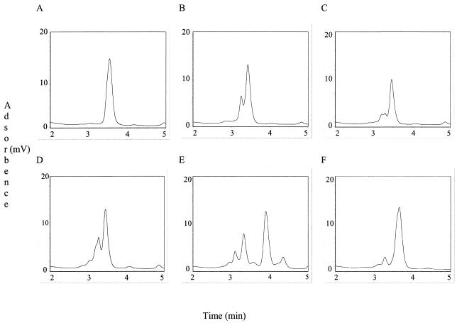
RP-HPLC chromatograms for quantification of the 16S-23S ISR showed various numbers of peaks between organisms. Some Bacillus strains had one peak, whereas others had multiple peaks (Fig. 1). For all strains of B. anthracis, the peaks eluted between 3 and 4 min. When the amount of PCR product to be added to the hybridization reactions was determined, the only peaks that were counted were those that eluted between 3 and 4 min.
FIG. 1.
Examples of RP-HPLC chromatograms used for quantification of the 16S-23S PCR product. Peaks that eluted between 3 and 4 min were used for DHPLC analysis. (A) B. thuringiensis ATCC 19269; (B) B. thuringiensis ATCC 19266; (C) B. anthracis BA1033; (D) B. anthracis Sterne; (E) B. cereus ATCC 21770; (F) B. cereus ATCC 33019. Some strains of B. anthracis had RP-HPLC peak profiles that were similar or identical to those of some strains of B. thuringiensis.
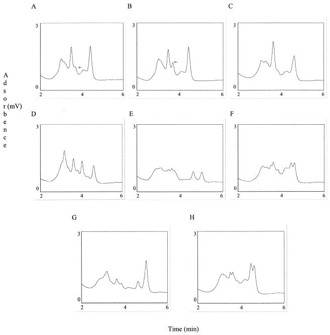
DHPLC analysis of the B. anthracis strains revealed two peak profile types (Fig. 2A and B). These two peak profiles differed from all of the peak profiles obtained for the strains of B. cereus and B. thuringiensis (Fig. 2C to H).
FIG. 2.
Peak profiles of 16S-23S heteroduplexes and homoduplex determined by DHPLC. (A) B. anthracis BA1018; (B) B. anthracis Sterne homoduplex. Two strains of B. anthracis had peak profiles similar to that of strain BA1018. All of the other strains tested had peak profiles similar to that of the Sterne strain. The arrows indicate the differences between the two peak profiles from all the B. anthracis strains tested. Examples of the DHPLC peak profiles of the 16S-23S ISR heteroduplexes of B. thuringiensis ATCC 19269 (C), B. thuringiensis ATCC 19266 (D), B. cereus ATCC 21770 (E), B. cereus ATCC 33019 (F), B. cereus ATCC 14579 (G), and B. thuringiensis ATCC 33679 (H) are shown. All strains of B. cereus and B. thuringiensis had peak profiles different from that of B. anthracis.
DHPLC analysis of the gyrA gene to identify B. anthracis.
Given the highly conserved nature of the B. cereus group genome, we decided to screen our organisms by DHPLC to identify organisms whose gyrA genes should be sequenced. The amino acids at the amino termini of gyrase A peptides are more conserved than the amino acids at the carboxyl termini (25). We screened the 5′ ends of the gyrA genes by DHPLC to identify organisms suitable for assay development.
Eight strains of B. anthracis, 33 strains of B. cereus, and 10 strains of B. thuringiensis were analyzed (Table 2). All of the B. anthracis strains analyzed had similar peak profiles, indicating that their sequences were identical. Two strains of B. cereus, BACI177 and BACI180, had peak profiles identical to the B. anthracis peak profiles. Sequencing of the gyrA genes of these organisms and three strains of B. anthracis revealed an allele with changes at 6 base pairs (Table 3). Primer sets spanning these DNA differences were tested by DHPLC to determine if any of them could be exploited for identification purposes.
TABLE 2.
Bacillus strains screened by DHPLC to identify candidate organisms for gyrA sequencing
| Species | USAMRIID acquisition no. | ATCC no. |
|---|---|---|
| B. anthracis | BA1015 | |
| B. anthracis | BA1019 | |
| B. anthracis | BA1022 | |
| B. anthracis | BA1023 | |
| B. anthracis | BA1024 | |
| B. anthracis | BA1025 | |
| B. anthracis | SPS | |
| B. anthracis | Sterne | |
| B. cereus | BACI164 | 7039 |
| B. cereus | BACI165 | 12480 |
| B. cereus | BACI166 | 13472 |
| B. cereus | BACI167 | 13824 |
| B. cereus | BACI168 | 14603 |
| B. cereus | BACI169 | 14893 |
| B. cereus | BACI170 | 15816 |
| B. cereus | BACI171 | 19625 |
| B. cereus | BACI172 | 19637 |
| B. cereus | BACI173 | 21182 |
| B. cereus | BACI174 | 21366 |
| B. cereus | BACI175 | 21634 |
| B. cereus | BACI176 | 21768 |
| B. cereus | BACI177 | 21769 |
| B. cereus | BACI178 | 21770 |
| B. cereus | BACI179 | 21771 |
| B. cereus | BACI180 | 21772 |
| B. cereus | BACI181 | 21928 |
| B. cereus | BACI182 | 25621 |
| B. cereus | BACI183 | 27348 |
| B. cereus | BACI184 | 27522 |
| B. cereus | BACI185 | 27877 |
| B. cereus | BACI186 | 31293 |
| B. cereus | BACI187 | 31429 |
| B. cereus | BACI188 | 31430 |
| B. cereus | BACI189 | 33018 |
| B. cereus | BACI190 | 33019 |
| B. cereus | BACI191 | 43881 |
| B. cereus | BACI192 | 53522 |
| B. cereus | BACI193 | 55055 |
| B. cereus | BACI194 | 70282 |
| B. cereus | BACI205 | 9139 |
| B. cereus | BACI206 | 9818 |
| B. thuringiensis | BACI196 | 13366 |
| B. thuringiensis | BACI197 | 13367 |
| B. thuringiensis | BACI198 | 19266 |
| B. thuringiensis | BACI199 | 19267 |
| B. thuringiensis | BACI200 | 19268 |
| B. thuringiensis | BACI201 | 19269 |
| B. thuringiensis | BACI202 | 19270 |
| B. thuringiensis | BACI203 | 29730 |
| B. thuringiensis | BACI204 | 33679 |
| B. thuringiensis | BACI037 | 39152 |
TABLE 3.
gyrA nucleotide differences between B. anthracis and B. cereus strains BACI170 and BACI180
| Codon change | Base position | Amino acid change |
|---|---|---|
| TTT→TTC | 656 | Phe→Phe |
| TTA→CTA | 792 | Leu→Leu |
| TTG→CTG | 1065 | Leu→Leu |
| GAC→GAT | 1667 | Asp→Asp |
| AAA→AAG | 1692 | Lys→Lys |
| ATC→ATT | 1926 | Ile→Ile |
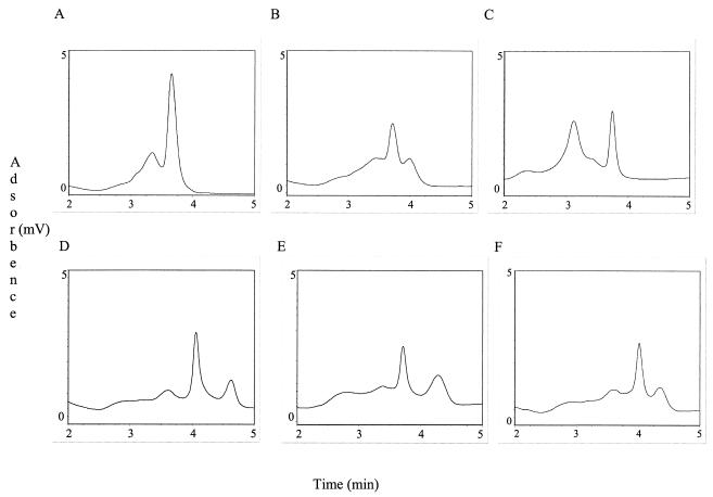
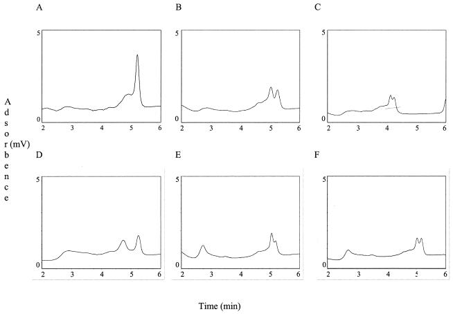
Two primer sets gave promising results. Primer set GYRA1542S and GYRA1904A produced one peak profile type for all of the B. anthracis strains tested which differed from those of all of the B. cereus and B. thuringiensis strains tested (Table 1; Fig. 3). Primer set GYRA535F and GYRA956R produced two peak profile types for all of the B. anthracis strains tested. Three strains of B. anthracis had peak profiles that differed from those of the other strains of B. anthracis tested. Interestingly, these strains were the same ones that produced different peak profiles with the 16S-23S ISR primer set. None of the B. cereus strains or B. thuringiensis strains had peak profiles similar to either B. anthracis peak profile type (Fig. 4).
FIG. 3.
Examples of DHPLC peak profiles of the homoduplex and heteroduplexes obtained with primers GYRA1542S and GYRA1904A. (A) B. anthracis Sterne homoduplex; (B) B. cereus ATCC 33019; (C) B. cereus ATCC 21769; (D) B. thuringiensis ATCC 19268; (E) B. cereus ATCC 27522; (F) B. thuringiensis ATCC 19267.
FIG. 4.
Examples of DHPLC peak profiles of the homoduplex and heteroduplexes obtained with primers GYRA535F and GYRA956R. (A) B. anthracis Sterne homoduplex; (B) B. cereus ATCC 33019; (C) B. cereus ATCC 21769; (D) B. thuringiensis ATCC 19268; (E) B. cereus ATCC 27522; (F) B. thuringiensis ATCC 19267.
Blind panel analysis to identify B. anthracis.
To evaluate DHPLC as a method of identifying B. anthracis, we tested the 16S-23S ISR primer set that gave two peak profiles with a blind panel of 45 Bacillus organisms and the gyrA primer set that gave one peak profile with a panel of 45 Bacillus organisms (Table 4). Of the 24 B. anthracis strains in the blind panel tested with the 16S-23S ISR primer set, all were correctly identified; of the 16 B. anthracis strains in the blind panel tested with the gyrA primer set, all were correctly identified.
TABLE 4.
Bacillus strains used in blind panels
| Primer set and sample no. | Species | USAMRIID acquisition no. | Primer set and sample no. | Species | USAMRIID acquisition no. | |
|---|---|---|---|---|---|---|
| 16S-23S primer set | ||||||
| 1 | B. anthracis | BA1022 | ||||
| 2 | B. anthracis | BA1012 | ||||
| 3 | B. anthracis | BA1001 | ||||
| 4 | B. anthracis | BA1026 | ||||
| 5 | B. anthracis | BA0076 | ||||
| 6 | B. cereus | BACI164 | ||||
| 7 | B. cereus | BACI178 | ||||
| 8 | B. anthracis | BA1023 | ||||
| 9 | B. anthracis | BA1001 | ||||
| 10 | B. cereus | BACI184 | ||||
| 11 | B. cereus | BACI183 | ||||
| 12 | B. anthracis | BA1016 | ||||
| 13 | B. anthracis | BA1036 | ||||
| 14 | B. anthracis | BA1100 | ||||
| 15 | B. cereus | BACI188 | ||||
| 16 | B. cereus | BACI165 | ||||
| 17 | B. thuringiensis | BACI200 | ||||
| 18 | B. anthracis | BA1024 | ||||
| 19 | B. cereus | BACI172 | ||||
| 20 | B. thuringiensis | BACI203 | ||||
| 21 | B. thuringiensis | BACI199 | ||||
| 22 | B. anthracis | BA1014 | ||||
| 23 | B. anthracis | BA1008 | ||||
| 24 | B. thuringiensis | BACI198 | ||||
| 25 | B. anthracis | BA1002 | ||||
| 26 | B. anthracis | BA1032 | ||||
| 27 | B. cereus | BACI177 | ||||
| 28 | B. anthracis | BA0076 | ||||
| 29 | B. cereus | BACI179 | ||||
| 30 | B. thuringiensis | BACI196 | ||||
| 31 | B. cereus | BACI189 | ||||
| 32 | B. anthracis | BA1003 | ||||
| 33 | B. anthracis | BA1015 | ||||
| 34 | B. anthracis | BA1023 | ||||
| 35 | B. anthracis | BA1019 | ||||
| 36 | B. cereus | BACI182 | ||||
| 37 | B. anthracis | BA1027 | ||||
| 38 | B. thuringiensis | BACI201 | ||||
| 39 | B. cereus | BACI181 | ||||
| 40 | B. cereus | BACI180 | ||||
| 41 | B. anthracis | BA1021 | ||||
| 42 | B. thuringiensis | BACI197 | ||||
| 43 | B. cereus | BACI191 | ||||
| 44 | B. anthracis | BA002 | ||||
| 45 | B. anthracis | BA1033 | ||||
| gyrA primer set blind panel | ||||||
| 1 | B. anthracis | BA0018 | ||||
| 2 | B. anthracis | BA002 | ||||
| 3 | B. thuringiensis | BACI202 | ||||
| 4 | B. thuringiensis | BACI199 | ||||
| 5 | B. cereus | BACI181 | ||||
| 6 | B. anthracis | BA1022 | ||||
| 7 | B. anthracis | BA1027 | ||||
| 8 | B. cereus | BACI166 | ||||
| 9 | B. cereus | BACI171 | ||||
| 10 | B. cereus | BACI177 | ||||
| 11 | B. cereus | BACI164 | ||||
| 12 | B. cereus | BACI174 | ||||
| 13 | B. anthracis | BA1019 | ||||
| 14 | B. thuringiensis | BACI201 | ||||
| 15 | B. thuringiensis | BACI200 | ||||
| 16 | B. anthracis | BA1018 | ||||
| 17 | B. cereus | BACI184 | ||||
| 18 | B. cereus | BACI196 | ||||
| 19 | B. anthracis | BA1036 | ||||
| 20 | B. anthracis | BA0065 | ||||
| 21 | B. anthracis | BA1031 | ||||
| 22 | B. cereus | BACI176 | ||||
| 23 | B. cereus | BACI197 | ||||
| 24 | B. cereus | BACI205 | ||||
| 25 | B. cereus | BACI192 | ||||
| 26 | B. cereus | BACI173 | ||||
| 27 | B. cereus | BACI206 | ||||
| 28 | B. anthracis | BA1021 | ||||
| 29 | B. anthracis | BA1030 | ||||
| 30 | B. cereus | BACI169 | ||||
| 31 | B. anthracis | BA1025 | ||||
| 32 | B. thuringiensis | BACI052 | ||||
| 33 | B. anthracis | BA1015 | ||||
| 34 | B. cereus | BACI170 | ||||
| 35 | B. thuringiensis | BACI203 | ||||
| 36 | B. cereus | BACI166 | ||||
| 37 | B. cereus | BACI174 | ||||
| 38 | B. cereus | BACI185 | ||||
| 39 | B. cereus | BACI172 | ||||
| 40 | B. anthracis | BA1021 | ||||
| 41 | B. anthracis | BA1023 | ||||
| 42 | B. cereus | BACI164 | ||||
| 43 | B. cereus | BACI167 | ||||
| 44 | B. cereus | BACI016 | ||||
| 45 | B. anthracis | BA1018 |
DISCUSSION
The two closest relatives of B. anthracis are B. cereus and B. thuringiensis. Studies have shown that these organisms are genetically indistinguishable (14). The primary characteristics used to distinguish these organisms from each other are the genes carried on plasmids. The existence of plasmid-free B. anthracis strains and B. anthracis-like B. cereus strains can complicate the identification process. We analyzed the 16S-23S ISR and the gyrA gene by DHPLC to evaluate this method as a means of differentiating B. anthracis from B. cereus and B. thuringiensis.
The 16S-23S ISR is used extensively to identify bacteria. Harrell et al. (13) reported that there is a 1-nucleotide difference between B. anthracis Ames, B. anthracis Vollum, and B. cereus ATCC 14579 (BACI017). In another study, Bourque et al. (2) analyzed the 16S-23S ISRs of a number of strains of B. thuringiensis as well as those of strains of B. cereus and B. anthracis. Their data showed little sequence divergence among species and strains. DHPLC is capable of detecting these subtle variations in DNA. Recently, we showed how this method may be used to identify bacteria (16).
Before hybridization, the crude PCR product was quantified by RP-HPLC. All chromatograms from the 16S-23S ISR primer set for B. anthracis quantification had more than one peak, indicating that there is more than one 16S-23S ISR type in the B. anthracis chromosome. Recent submissions to GenBank confirm this finding. GenBank submissions by Daffonchio et al. (7) indicate that different tRNAs are encoded in this region. Some strains of B. anthracis and B. cereus had similar profiles on RP-HPLC chromatograms. However, their DHPLC peak profiles differed. Analysis of the 16S-23S ISR by DHPLC revealed two peak profile types for B. anthracis. These two-peak profile types differed from the peak profile types obtained for all of the B. cereus and B. thuringiensis strains tested.
In addition to the 16S-23S ISR, we also analyzed the gyrA gene. Similar to the 16S-23S ISR, this gene is also used extensively for bacterial identification. The 5′ end of this gene is more conserved than the 3′ end. We screened the 5′ end of this gene by DHPLC to determine if it was possible to use it to differentiate B. anthracis from B. cereus and B. thuringiensis. All of the B. anthracis strains tested had similar peak profiles. Two strains of B. cereus had peak profiles similar to those of the B. anthracis strains tested. Sequencing of the gyrA genes of these two strains revealed an allele with 6 base pair substitutions. Several base pair substitutions were analyzed to determine if unique chromatograms could be obtained for B. anthracis. We were able to differentiate B. anthracis from B. cereus and B. thuringiensis with two primer sets: GYRA1542S and GYRA1904A, which spanned 2 base pair substitutions at positions 1667 and 1692 of the gyrA gene, and GYRA535F and GYRA956R, which spanned the base pair substitutions at positions 656 and 792.
To evaluate DHPLC as a method of identifying B. anthracis, we tested the 16S-23S-specific and gyrA-specific primers, using the B. anthracis reference strains BA1036 and BA1016 with two blind panels of 45 organisms each. Of the 24 B. anthracis strains in the panel tested with the 16S-23S ISR-specific primer set, all were correctly identified. Of the 16 B. anthracis strains in the panel tested with both gyrA-specific primer sets, all strains were correctly identified. Given the high degree of specificity, these results indicate that DHPLC can be used to screen large numbers of samples for B. anthracis.
In summary, we tested a total of 73 members of the B. cereus group to determine if DHPLC could be used to differentiate B. anthracis from B. cereus and B. thuringiensis by analyzing the 16S-23S ISR and a portion of the gyrA gene. In addition, blind panels of 45 samples were used to investigate the capability of DHPLC to identify B. anthracis. We were successful in identifying all the B. anthracis strains in each panel.
Acknowledgments
This work was supported by an In-House Laboratory Independent Research Reports grant from the Medical, Chemical, Biological, and Defense Research Program of the United States Army Medical Research and Materiel Command, Fort Detrick, Md.
REFERENCES
- 1.Ash, C., J. A. Farrow, M. Dorsch, E. Stackebrandt, and M. D. Collins. 1991. Comparative analysis of Bacillus anthracis, Bacillus cereus, and related species on the basis of reverse transcriptase sequencing of 16S rRNA. Int. J. Syst. Bacteriol. 41:343-346. [DOI] [PubMed] [Google Scholar]
- 2.Bourque, S. N., J. R. Valero, M. C. Lavoie, and R. C. Levesque. 1995. Comparative analysis of the 16S to 23S ribosomal intergenic spacer sequences of Bacillus thuringiensis strains and subspecies and of closely related species. Appl. Environ. Microbiol. 61:1623-1626. [DOI] [PMC free article] [PubMed] [Google Scholar]
- 3.Bragg, T. S., and D. L. Robertson. 1989. Nucleotide sequence and analysis of the lethal factor gene (lef) from Bacillus anthracis. Gene 81:45-54. [DOI] [PubMed] [Google Scholar]
- 4.Carl, M., R. Hawkins, N. Coulson, J. Lowe, D. L. Robertson, W. M. Nelson, R. W. Titball, and J. N. Woody. 1992. Detection of spores of Bacillus anthracis using the polymerase chain reaction. J. Infect. Dis. 165:1145-1148. [DOI] [PubMed] [Google Scholar]
- 5.Christensen, H., K. Jorgensen, and J. E. Olsen. 1999. Differentiation of Campylobacter coli and C. jejuni by length and DNA sequence of the 16S-23S rRNA internal spacer region. Microbiology 145(Pt. 1):99-105. [DOI] [PubMed] [Google Scholar]
- 6.Cooksey, R. C., G. P. Morlock, B. P. Holloway, J. Limor, and M. Hepburn. 2002. Temperature-mediated heteroduplex analysis performed by using denaturing high-performance liquid chromatography to identify sequence polymorphisms in Mycobacterium tuberculosis complex organisms. J. Clin. Microbiol. 40:1610-1616. [DOI] [PMC free article] [PubMed] [Google Scholar]
- 7.Daffonchio, D., A. Cherif, and S. Borin. 2000. Homoduplex and heteroduplex polymorphisms of the amplified ribosomal 16S-23S internal transcribed spacers describe genetic relationships in the “Bacillus cereus group.” Appl. Environ. Microbiol. 66:5460-5468. [DOI] [PMC free article] [PubMed] [Google Scholar]
- 8.Dixon, T. C., M. Meselson, J. Guillemin, and P. C. Hanna. 1999. Anthrax. N. Engl. J. Med. 341:815-826. [DOI] [PubMed] [Google Scholar]
- 9.Ellerbrok, H., H. Nattermann, M. Ozel, L. Beutin, B. Appel, and G. Pauli. 2002. Rapid and sensitive identification of pathogenic and apathogenic Bacillus anthracis by real-time PCR. FEMS Microbiol. Lett. 214:51-59. [DOI] [PubMed] [Google Scholar]
- 10.Frothingham, R., and K. H. Wilson. 1993. Sequence-based differentiation of strains in the Mycobacterium avium complex. J. Bacteriol. 175:2818-2825. [DOI] [PMC free article] [PubMed] [Google Scholar]
- 11.Green, B. D., L. Battisti, T. M. Koehler, C. B. Thorne, and B. E. Ivins. 1985. Demonstration of a capsule plasmid in Bacillus anthracis. Infect. Immun. 49:291-297. [DOI] [PMC free article] [PubMed] [Google Scholar]
- 12.Hannachi-M'Zali, F., J. E. Ambler, C. F. Taylor, and P. M. Hawkey. 2002. Examination of single and multiple mutations involved in resistance to quinolones in Staphylococcus aureus by a combination of PCR and denaturing high-performance liquid chromatography (DHPLC). J. Antimicrob. Chemother. 50:649-655. [DOI] [PubMed] [Google Scholar]
- 13.Harrell, L. J., G. L. Andersen, and K. H. Wilson. 1995. Genetic variability of Bacillus anthracis and related species. J. Clin. Microbiol. 33:1847-1850. [DOI] [PMC free article] [PubMed] [Google Scholar]
- 14.Helgason, E., O. A. Okstad, D. A. Caugant, H. A. Johansen, A. Fouet, M. Mock, I. Hegna, and A.-B. Kolsto. 2000. Bacillus anthracis, Bacillus cereus, and Bacillus thuringiensis—one species on the basis of genetic evidence. Appl. Environ. Microbiol. 66:2627-2630. [DOI] [PMC free article] [PubMed] [Google Scholar]
- 15.Henderson, I., C. J. Duggleby, and P. C. Turnbull. 1994. Differentiation of Bacillus anthracis from other Bacillus cereus group bacteria with the polymerase chain reaction. Int. J. Syst. Bacteriol. 44:99-105. [DOI] [PubMed] [Google Scholar]
- 16.Hurtle, W., D. Shoemaker, E. Henchal, and D. Norwood. 2002. Denaturing HPLC for identifying bacteria. BioTechniques 33:386-388, 390-391. [DOI] [PubMed] [Google Scholar]
- 17.Kaneko, T., R. Nozaki, and K. Aizawa. 1978. Deoxyribonucleic acid relatedness between Bacillus anthracis, Bacillus cereus and Bacillus thuringiensis. Microbiol. Immunol. 22:639-641. [DOI] [PubMed] [Google Scholar]
- 18.Logan, N. A., J. A. Carman, J. Melling, and R. C. Berkeley. 1985. Identification of Bacillus anthracis by API tests. J. Med. Microbiol. 20:75-85. [DOI] [PubMed] [Google Scholar]
- 19.Maxwell, A., and M. Gellert. 1984. The DNA dependence of the ATPase activity of DNA gyrase. J. Biol. Chem. 259:14472-14480. [PubMed] [Google Scholar]
- 20.Robertson, D. L., M. T. Tippetts, and S. H. Leppla. 1988. Nucleotide sequence of the Bacillus anthracis edema factor gene (cya): a calmodulin-dependent adenylate cyclase. Gene 73:363-371. [DOI] [PubMed] [Google Scholar]
- 21.Shlush, L. I., D. M. Behar, A. Zelazny, N. Keller, J. R. Lupski, A. L. Beaudet, and D. Bercovich. 2002. Molecular epidemiological analysis of the changing nature of a meningococcal outbreak following a vaccination campaign. J. Clin. Microbiol. 40:3565-3571. [DOI] [PMC free article] [PubMed] [Google Scholar]
- 22.Somerville, H. J., and M. L. Jones. 1971. Genetic relatedness within the Bacillus cereus group of bacilli. J. Gen. Microbiol. 69:ix. [PubMed] [Google Scholar]
- 23.Turnbull, P. C., R. A. Hutson, M. J. Ward, M. N. Jones, C. P. Quinn, N. J. Finnie, C. J. Duggleby, J. M. Kramer, and J. Melling. 1992. Bacillus anthracis but not always anthrax. J. Appl. Bacteriol. 72:21-28. [DOI] [PubMed] [Google Scholar]
- 24.Uchida, I., T. Sekizaki, K. Hashimoto, and N. Terakado. 1985. Association of the encapsulation of Bacillus anthracis with a 60 megadalton plasmid. J. Gen. Microbiol. 131(Pt. 2):363-367. [DOI] [PubMed] [Google Scholar]
- 25.Wang, J. C. 1996. DNA topoisomerases. Annu. Rev. Biochem. 65:635-692. [DOI] [PubMed] [Google Scholar]
- 26.Welkos, S. L., J. R. Lowe, F. Eden-McCutchan, M. Vodkin, S. H. Leppla, and J. J. Schmidt. 1988. Sequence and analysis of the DNA encoding protective antigen of Bacillus anthracis. Gene 69:287-300. [DOI] [PubMed] [Google Scholar]


