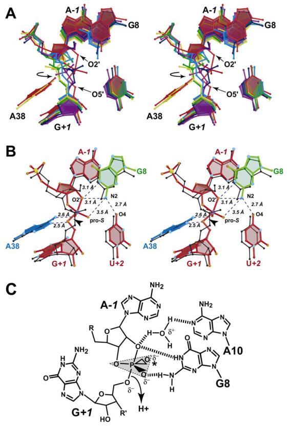
FIGURE 6.

Superposition ensemble of structures representing the native G8 structure, the four purine variants at position 8, and the 4WJ hairpin ribozyme-vanadate complex (10), as well as a proposed role for water. (A) Stereo diagram for the least squares superposition ensemble The structures are: G8A (yellow), G8AP (green), G8DAP (blue), G8I (purple), G8 native (dark blue) and the hairpin ribozyme-vanadate complex (red). A circular arrow indicates the dihedral angle rotation required to produce a more in-line angle. (B) Stereo diagram showing a superposition between the hairpin ribozyme-vanadate complex (ball-and-stick model) with the native G8 structure (black). Atoms O4 of U+2, N2, of G8, O2' of A−1 and the pro-S oxygen of G+1 are labeled for clarity. (C) Schematic diagram indicating a proposed role for water in the cleavage reaction Dashed lines indicate possible H-bonds. The asterisk indicates the location of the pro-R oxygen, which coordinates to the exocyclic amines of A9 and A38 in the transition-state.
