Abstract
Antigenicities of several formalin-detoxified botulinum neurotoxin preparations were measured by inhibition and sandwich enzyme-linked immunosorbent assay (ELISA), and immunogenicity was studied in mice. The toxoids were derived primarily from the serotype A 150-kDa neurotoxin protein, while one toxoid was derived from the naturally occurring 900-kDa toxin-hemagglutinin complex. Antigenicity was severely compromised in two commercially available toxoids. A variety of new toxoids were synthesized in-house by optimizing formaldehyde reaction conditions. Three of the resulting toxoids were found to be antigenically identical to the native toxin, as measured by inhibition ELISA, in spite of showing a reduction of toxicity by more than 100,000-fold. Sandwich ELISAs indicated that the in-house toxoids were two- to threefold less antigenic than the neurotoxin compared to commercial toxoids, which were about 100-fold less antigenic. Mice were immunized twice, on day 0 and day 14. By day 28, relatively high toxin-specific immunoglobulin G (IgG) titers were detected in animals that had received any of the in-house toxoids, with greater than 99% being IgG1 and the remainder being IgG2. These immunized mice remained asymptomatic after being challenged with 50 to 1,000,000 50% lethal dose (LD50) units of the 900-kDa neurotoxin. In contrast, animals immunized with several different batches of commercially available toxoids did not develop measurable toxin-specific antibody titers. However, these mice survived neurotoxin challenges with 2 LD50 units but died when challenged with 6 LD50 units. Neutralizing titers measured from pools of sera generated with the in-house toxoid preparations ranged from 2.5 to 5 U/ml. In terms of predicting immunogenicity, inhibition ELISAs comparing each formalin toxoid to the parent toxin provided good insight for screening the new toxoids as well as for estimating their relative in vivo potencies. Inhibition ELISA data indicate that those toxoids that most closely resemble the native toxin are highly immunogenic and protective. The superior quality of these new toxoids makes them useful tools for continued use in ELISA development and for antitoxin production.
Isolates of Clostridium botulinum tend to produce one of seven structurally and genetically similar neurotoxins (13). Although intoxication may occur if the microorganisms colonize the human intestine, botulism is considered a toxemia rather than an infection because the botulinum neurotoxin (BoNT) alone will cause the disease. BoNT serotypes A (BoNT/A), -B, and -E cause most human cases of botulism, while serotype F has caused only a few confirmed cases (30, 31). BoNTs are bound to many nontoxic clostridial proteins, forming heterogeneous complexes of up to 900 kDa in mass. These complexes consist of one 150-kDa neurotoxin molecule, hemagglutinins (Hmgs) and a variety of other proteins (32). The number and types of proteins associated with the neurotoxin vary with the C. botulinum strain (24). Antibodies that recognize the Hmg and other nontoxic clostridial proteins do not prevent illness (16, 18). Protective immunity is derived solely from antibodies that bind to the neurotoxin molecule itself (8).
Vaccination is the only approach that can be used to prevent botulism. A pentavalent botulinum toxoid comprised of formalin-detoxified BoNT/A, -B, -C, -D, and -E Hmg complexes has been used to immunize laboratory and military personnel since 1961, but this has never been licensed by the U.S. FDA (8, 12, 27). Outside of the United States, a collaboration among three major laboratories in Japan has recently led to the manufacture of a tetravalent botulinum toxoid for serotypes A, B, E, and F that is being used to immunize personnel who are at high risk of exposure to BoNTs (35).
Vaccination immediately after toxin exposure has no protective benefit because the immune response is relatively slow compared to the rate of intoxication. The only drug treatment available after intoxication is antibody therapy, which entails the injection of equine-derived botulinum antitoxin (BAT) or human-derived botulinum immunoglobulin (Ig) to remove the toxin from the blood circulation (1, 3, 34). Antibody therapy does not directly alleviate symptoms of botulism but can limit the amount of toxin that enters nerve terminals and thus may lessen the severity and shorten the duration of paralysis.
Since a vaccine can be used to either protect a human population or produce a BAT or human-derived botulinum Ig product, it is important to have reliable methods of evaluating the antigenic integrity of botulinum vaccines. An in vitro assay that can serve in this capacity could evaluate the consistency of the antigen throughout the manufacturing process, as well as generate correlate potency data that may reduce in vivo testing. This study was designed to characterize several formalin toxoids that were synthesized in-house to support the development of two enzyme-linked immunosorbent assay (ELISA) methods to characterize BoNT antigens. Data for several in-house C. botulinum toxoids in comparison with commercially available toxoids are reported here.
MATERIALS AND METHODS
Antibodies.
Equine BAT, which is the potency reference standard maintained under the U.S. Code of Federal Regulations [section 610.20(a)], was obtained from the U.S. FDA/CBER. BAT was prepared as a glycerol stock with a potency of 25 IU per ml and stored at −80°C. Rabbit anti-botulinum serotype A IgG was purchased from Metabiologics, Inc. (Madison, WI). Rabbit IgG was biotinylated with N-hydroxysuccinimidobiotin (Sigma-Aldrich, St. Louis, MO). Rabbit anti-horse, goat anti-mouse, avidin-alkaline phosphatase conjugates, p-formaldehyde, and buffer reagents were obtained from Sigma-Aldrich. Goat anti-mouse IgG1 and IgG2 were purchased from Jackson ImmunoResearch, Inc. (West Grove, PA).
Toxins and toxoids.
Research-grade BoNT/A and BoNT/A formalin toxoids (150-kDa and 900-kDa forms) were purchased from Metabiologics, Inc. The 150-kDa neurotoxin had a specific toxicity of (2.6 to 3.0) × 108 50% lethal dose (LD50) units per mg of protein, whereas the 900-kDa complex had 3.0 × 10⁷ LD50 units per mg of protein. The commercial toxoids were prepared in 0.5% formalin. For botulinum toxoid synthesis, BoNT/A (150 kDa) was diluted in phosphate-buffered saline (PBS) to a protein concentration of 0.16 mg/ml. In some cases, l-lysine ranging from 2 to 20 mM was added to PBS-toxin solutions. Buffered formalin was prepared by dissolving 60 mg of p-formaldehyde per ml of PBS and heating at 60°C for 45 min and was diluted into toxin-PBS solutions to produce a final formalin concentration of 0.08 to 0.50% (wt/vol). Reaction mixtures proceeded at 37°C without agitation until detoxification was complete. This typically required 1 (toxoid 0) to 5 (toxoid 3) weeks, depending on the formaldehyde concentration. Detoxification was semiquantified by an incremental process of injecting (intraperitoneally) 0.1 μg of toxoid into a naïve mouse (∼25,000 LD50 equivalents). If the animal remained asymptomatic, then another animal was injected with a higher toxoid dose. Several iterations of this process were performed to determine the level of active neurotoxin within a preparation of toxoid. The optimized toxoid preparation was 100,000 times less toxic than the parent toxin.
ELISA methods.
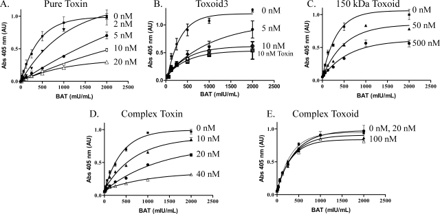
The inhibition ELISA (see Fig. 1) was performed by coating 96-well plates (Immulon 1B) with the 150-kDa neurotoxin (1 μg/ml) overnight at room temperature. Plates were washed several times and blocked for at least 30 min using Tris-buffered saline buffer (0.5 M NaCl, 10 mM Tris, pH 7.4, 0.05% Triton X-100, 0.3% bovine serum albumin [BSA], and 0.02% sodium azide). Serial twofold dilutions of equine BAT were prepared by using PBS containing 0.3% BSA. Each dilution was divided into four aliquots. The neurotoxin or toxoid was added to each of three sets of BAT dilutions. Neither the neurotoxin nor toxoid was added to control (BAT only) dilutions. After a 60-min incubation at room temperature, 50 μl of each aliquot was transferred to neurotoxin-coated wells, followed by a 60-min incubation at room temperature. Wells were washed with Tris-buffered saline buffer, and then 50 μl of anti-horse IgG conjugated to alkaline phosphatase was deposited in each well. After 30 min, plates were washed and an alkaline phosphatase substrate, p-nitrophenyl phosphate (Sigma-Aldrich), was added. Color development proceeded until a maximum optical density at 405 nm of around 1.0 was achieved for the wells, defining the upper plateau of the standard toxin curve.
FIG. 1.
Inhibition ELISA with variable BAT concentrations. Soluble toxins or toxoids were used to inhibit equine BAT binding to pure neurotoxin adsorbed to 96-well plates. (A) Inhibition by soluble 150-kDa neurotoxin occurred in the range of 2 to 20 nM neurotoxin. (B) Toxoid 3 inhibited BAT binding with similar effectiveness as the parent toxin. (C) A commercial toxoid derived from the pure neurotoxin required at least 10-fold higher concentrations of antigen to partially prevent BAT binding to the plate-bound neurotoxin. (D and E) The inhibition profile produced by the Hmg toxin complex (D) and its corresponding toxoid (E). Each data point is the average absorbance from four wells on a single ELISA plate. Error bars are one standard deviation. Abs, absorbance.
Sandwich ELISA.
Wells of microtiter plates were coated with 0.1 ml of purified rabbit anti-BoNT/A IgG at 2.5 μg/ml in PBS (overnight at 37°C). Serial dilutions of neurotoxin or toxoids were applied to the IgG-coated wells. In one variation of the assay, biotinylated rabbit IgG and avidin conjugated to alkaline phosphatase were used to quantify the neurotoxin or toxoid bound to the ELISA plate. In a second variation of the assay, mouse antitoxin generated with toxoid 7 and anti-mouse alkaline phosphatase were used in place of biotinylated IgG and avidin. Both assay methods produced similar results.
Indirect ELISA for antibody titer determination.
IgG, IgG1, and IgG2 were measured by coating microtiter plates with neurotoxin and applying serial twofold dilutions of mouse antiserum across the plate. Goat anti-mouse alkaline phosphatase was used to detect toxin-specific mouse IgG. ELISA titers are presented as the reciprocal of the dilution that produces 0.1 absorbance units (AU).
In vivo survival and immunogenicity.
BALB/CAnNCr male mice (NCT, Frederick, MD) were used for all in vivo tests. All aspects of the experiments were conducted in accordance with the National Institutes of Health Guide for the Care and Use of Laboratory Animals (22). The research had prior approval from the CBER Animal Care and Use Committee. Mice were injected twice (on days 0 and 14) by the subcutaneous route with 75 ng of antigen in 50 μl PBS containing 0.2% alhydrogel (Accurate Chemical & Scientific Corp., Westbury, NY). Antigen was mixed with the adjuvant and incubated at room temperature for 60 min prior to injection. Serum was collected on day 28. Toxin-specific antibody titers were measured using an indirect ELISA, and passive immunization of mice was used to quantify neutralizing antibody titers. Protective immunity was assessed by directly challenging immunized mice with neurotoxin, using two or three mice per toxin challenge dose.
The mouse neutralization assay remains the standard assay used by the WHO to quantify functional antibody titers to BoNT (4, 14). Serum collected from immunized mice was diluted into PBS and 0.2% BSA and combined with 3 ng or about 100 mouse LD50s of 900-kDa BoNT/A. The antiserum and toxin mixtures were incubated at room temperature for 1 to 2 h before being administered to mice (0.4 ml, intraperitoneally). The amount of immune serum that fully protected mice was used to calculate the neutralizing titer of the test serum.
Direct toxin challenge was performed on immunized mice by injecting measured quantities of 900-kDa neurotoxin in a volume of 0.4 ml PBS/BSA. For the first 8 hours, mice were observed hourly to identify early symptoms of botulism, such as narrowing of the waist, unusual gait, and rapid, shallow breathing. If symptoms did not develop during this initial time frame, mice were observed three times daily for up to 7 days. None of the botulinum toxoid-immunized mice developed symptoms following challenges of up to 1 million mouse LD50 units. The potency of the toxin material used in both the neutralization assay and in the direct challenge assay was determined by injecting 0.5 LD50 units and 2.0 LD50 units of neurotoxin into two naïve mice. Mice that were exposed to 0.5 LD50 units routinely developed minor, nonlethal symptoms of botulism, whereas the 2.0 LD50 group died within 24 h.
Data analysis.
Optical density was measured using a GENios microplate reader and Magellan 4 software (Tecan U.S., Inc., Research Triangle Park, NC). Nonlinear regression analysis of data was performed using GraphPad Prism 5 (GraphPad Software, Inc., San Diego, CA). Error bars represent the standard deviation of triplicate values.
RESULTS
Toxoid preparation.
BoNT/A (150 kDa) was subjected to different concentrations of formaldehyde with or without lysine (Table 1). Toxoid 0 was generated by adding formaldehyde at a final concentration of 61 mM (0.5% formalin) with the toxin at 1 mg/ml. In order to reduce the severity of the detoxification reaction, subsequent botulinum toxoid preparations used less toxin protein (0.15 to 0.17 mg/ml) and lower concentrations of formaldehyde. Formaldehyde at 10 mM (toxoid 3) inactivated the neurotoxin over 4 to 6 weeks, reducing neurotoxicity by about 5 orders of magnitude. Formaldehyde at 20 mM (toxoid 7) or 40 mM (toxoid 8) inactivated the neurotoxin relatively rapidly, requiring only 11 days to reduce toxicity by about 100,000- and 200,000-fold, respectively. After 3 to 4 weeks, toxoid 7 and toxoid 8 were detoxified by more than 600,000-fold. The extent of detoxification was diminished when 5 mM of formaldehyde or 10 mM of formaldehyde in combination with 5 mM of lysine was used in the reaction. Even after 8 weeks of incubation at 37°C, BoNT/A was detoxified by only 2 to 3 orders of magnitude by these lower levels of free formaldehyde (toxoid 4 and toxoid 5).
TABLE 1.
Detoxification and antitoxin determination
| Toxoid | Formalin (%) | No. of days at 37°C | Detoxification (fold) | Approx. neutralizing titer of antitoxin (U/ml)a |
|---|---|---|---|---|
| 0 | 0.5 | 7 | >600,000 | ND |
| 1 | 0.5 | 7 | >600,000 | ND |
| 2 | 0.5b | 7 | >600,000 | ND |
| 3 | 0.08 | 28-42 | 100,000 | 5.0 |
| 4 | 0.04 | 56 | 100-200 | ND |
| 5 | 0.08b | 56 | 100-400 | ND |
| 6 | 0.16b | 56 | 100-1000 | ND |
| 7 | 0.16 | 21-28 | >600,000 | 2.5-3.1 |
| 8 | 0.33 | 21-28 | >600,000 | 3.75-5.0 |
ND, values were not determined.
Reaction mixture contained 5 mM of lysine.
In vitro analyses.
Using equine BAT, a semiquantitative inhibition ELISA was developed to assess the antigenic similarity between neurotoxin and formalin toxoids. In all cases regarding Fig. 1, the native 150-kDa neurotoxin was adsorbed to 96-well plates and was detected using serial dilutions of equine BAT from 30 to 2,000 mU/ml. Inhibition analysis was performed by incubating BAT dilutions with known concentrations of soluble neurotoxin to define a standard titration curve or with formalin toxoid. The standard curve routinely produced a 50% effective concentration (EC50) of approximately 290 mU/ml. Soluble neurotoxin in the range of 2 to 20 nM was required to incrementally inhibit BAT binding to the plate-adsorbed neurotoxin (Fig. 1A). When the same experiment was performed using toxoid 3, the inhibition pattern was analogous to inhibition caused by the native neurotoxin (Fig. 1B). Based on these results, toxoid 3 was antigenically identical to the native neurotoxin in spite of the formalin treatment (Table 1). Toxoid 4 to toxoid 8 performed the same as toxoid 3 (data not shown). In contrast, a commercially available 150-kDa formalin toxoid at 50 to 500 nM generated partial inhibition of BAT binding (Fig. 1C), which indicated that this toxoid retained 2% to 10% antigenic similarity to the native neurotoxin; i.e., epitopes common to the native neurotoxin are 10- to 50-fold less abundant on this commercial toxoid than on the new toxoids.
Inhibition ELISA analysis was repeated using commercially available 900-kDa Hmg toxin and Hmg toxoid (Fig. 1D and 1E). The results show that while the biologically active Hmg toxin was antigenically very similar to the biologically active 150-kDa neurotoxin, the corresponding formalin Hmg toxoid was ineffective at blocking BAT binding. The most likely explanation is that the antigenicities of the commercially available 150-kDa toxoid and 900-kDa Hmg complex toxoid were severely compromised by the formalin detoxification process (Fig. 1B and 1E). The abilities of polyclonal antibodies to recognize toxin-specific epitopes on the 900-kDa complex is not dramatically altered by the nontoxic proteins associated with the neurotoxin; however, formaldehyde treatment of the 900-kDa complex drastically diminished the abilities of antibodies to recognize these epitopes.
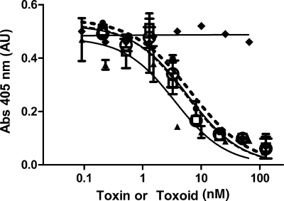
The inhibition ELISA was further refined by choosing a fixed concentration of equine BAT (Fig. 2). The 150-kDa neurotoxin inhibited BAT binding to the plate-adsorbed neurotoxin, with a 50% inhibitory concentration (IC50) of 3.5 ± 2.0 nM and a slope of 1.0 ± 0.4. The inhibition curves for toxoid 3, toxoid 7, and toxoid 8 are overlapping, with IC50s ranging between 5.3 to 6.6 nM and with slopes ranging from 0.88 to 0.94. The parallelism of all four curves indicates that toxin-specific epitopes are retained in the toxoids. The commercially available formalin toxoid has lost all toxin-specific epitopes when evaluated with this assay.
FIG. 2.
Inhibition ELISA with fixed BAT concentration. The same ELISA methodology was performed as shown in Fig. 1 except that BAT was maintained at 500 mU/ml. Soluble BoNT/A (▴) inhibited BAT binding with an IC50 of 3.5 ± 2.0 nM. Toxoid 3 (○), toxoid 7 (•), and toxoid 8 (□) inhibited BAT binding in a manner that is parallel to the native BoNT/A, indicating that the majority of toxin-specific epitopes are retained on each botulinum toxoid. Inhibitory IC50s for these botulinum toxoids were between 5.3 to 6.6 nM. A commercial botulinum toxoid (⧫) did not inhibit BAT binding. Abs, absorbance.
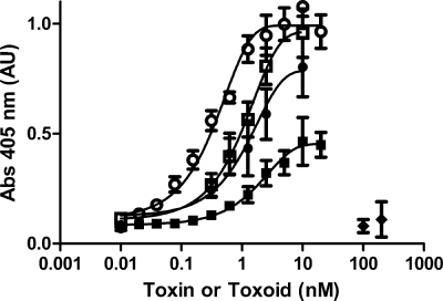
A sandwich ELISA was devised as an alternative to the inhibitory ELISA to assess the antigen quality (Fig. 3). Biotinylated and nonbiotinylated rabbit anti-BoNT/A IgG were used to trap and detect the toxin and toxoids. BoNT/A was used to generate the standard curve, which had an EC50 of 0.37 nM. The 150-kDa commercial toxoid at very high concentrations elicited only a modest rise in AU compared to the full titration curve generated by the native neurotoxin. Toxoid 0 did not produce a detectable signal over the background. The degree of disparity between these toxoids and the native BoNT/A could not be quantified other than to say that the commercial toxoid and toxoid 0 are several orders of magnitude less antigenic than the parent BoNT/A, using this assay. In contrast, toxoid 2 resembled BoNT/A, with an EC50 of 1.9 nM. Toxoid 4 most closely resembled the parent toxin, with an EC50 of 1.1 nM. However, toxoid 4 was detoxified by only about 99%, which was inadequate for follow-up animal immunization studies. Toxoid 3 had an EC50 of 1.2 nM but was detoxified by about 105-fold. Further improvement was achieved with toxoid 7 and toxoid 8, which had sandwich ELISA profiles identical to those of toxoid 3 but were each detoxified by greater than 600,000-fold (Table 1). These in vitro results suggested that toxoid 3, toxoid 7, and toxoid 8 would be the most efficacious toxoids in vivo.
FIG. 3.
Sandwich ELISA of toxins and toxoids. Biotinylated anti-BoNT/A IgG was used to detect toxin or toxoid applied to 96-well plates. Separate dilutions of BoNT/A (○), toxoid 4 (□), toxoid 3 (•), toxoid 2 (▪), and toxoid 0 (♦) were applied to 96-well plates that were coated with rabbit anti-BoNT/A IgG. Each symbol is the average of a minimum of three data points. Error bars represent one standard deviation unit. Abs, absorbance.
In vivo analyses.
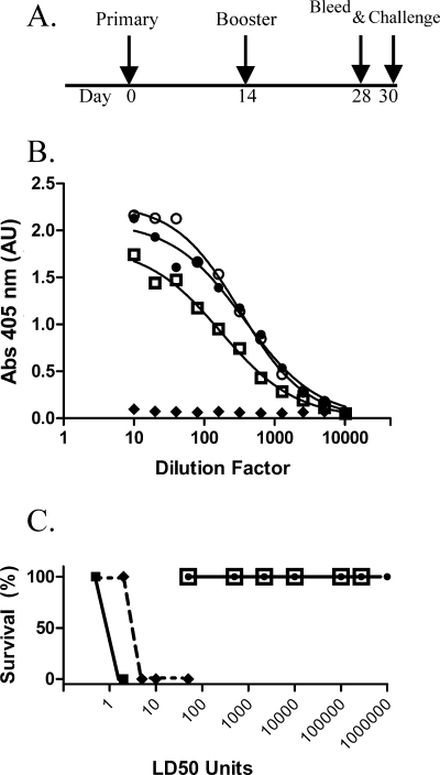
Immunogenicity was assessed for the different commercial toxoids, as well as for toxoid 3, toxoid 7, and toxoid 8 (Fig. 4B) (Table 1). Interestingly, at 75 ng of antigen per immunization, the commercial toxoid failed to elicit measurable antibody titers (Fig. 4B), whereas all of the in-house toxoids elicited elevated toxin-specific titers by day 28. The antibody pools generated by toxoid 3, toxoid 7, and toxoid 8 had ELISA titers of 8,900, 13,200, and 5,600, respectively (Fig. 4B).
FIG. 4.
Immunogenicity of toxoids in mice. (A) Depiction of the immunization schedule. (B) Toxin-specific IgG response measured on serum pools collected on day 28. Toxoid 3 (○), toxoid 7 (•), and toxoid 8 (□) elicited antitoxin titers that were approximately the same regardless of the chemistry used to generate the toxoid. The commercial 150-kDa toxoid (♦) did not elicit detectable toxin-specific antibodies. (C) Direct toxin challenge and the resulting survival of immunized animals between days 30 and 37. Nonimmunized mice (▪) survived 0.5 LD50 but died from 2 LD50. The commercial toxoid (♦) elicited little protection, whereas mice immunized with toxoid 3 or toxoid 8 (□) were fully protected from the highest challenge dose of 275,000 LD50 units, and two mice immunized with toxoid 7 (•) were fully protected by as much as 1 million LD50 units. Abs, absorbance.
Several days after serum collection, the same animals were challenged with biologically active Hmg-BoNT/A complex. Mice immunized with the commercially available 150-kDa toxoid were able to survive a challenge of 2 LD50 units of toxin but died when challenged with 6 LD50 units (Fig. 4C). Because of this result, two lots of the commercial 150-kDa toxoid and one lot of the 900-kDa Hmg toxoid were examined in a side-by-side immunogenicity trial, and none of these toxoids conferred protection when challenged with 5 LD50 units. In stark contrast, toxoid 3- and toxoid 8-immunized mice survived all levels of toxin challenge, which were as high as 275,000 LD50 units, and toxoid 7 mice survived 1 million LD50 units (Fig. 4C). Higher challenges were not performed. This experiment was later repeated by immunizing new groups of mice with the same botulinum toxoid batches. All botulinum toxoid-immunized mice remained healthy 7 days following toxin challenge.
Neutralizing antibody titers were measured for each of the 4-week serum pools using the standard mouse neutralization assay as described by the WHO (4). Serum pools generated using the commercially available toxoids could not be analyzed by this approach because antibody titers were too low to neutralize the test dose of toxin. Neutralization titers for the toxoids are summarized in Table 1. Toxoid 7 generated the lowest protective titers, between 2.5 and 3.1 U/ml, whereas toxoid 3 elicited neutralizing titers of around 5 U/ml. Toxoid 8 titers were intermediate between toxoid 3 and toxoid 7. Subsequent immunogenicity trials have routinely elicited neutralizing titers between 2.5 and 5.5 U/ml 4 weeks after the primary immunization. For toxoid 3, toxoid 7, and toxoid 8, the IgG isotype content regularly showed that IgG1 was the predominant isotype, about 3 orders of magnitude greater than IgG2ab.
DISCUSSION
This study has demonstrated that formalin toxoids derived from the purified 150-kDa BoNT protein can elicit protective IgG titers that vary greatly depending on the reaction conditions used during detoxification. This effect was first demonstrated for tetanus and diphtheria toxoids and later with pertussis toxin (11, 21, 23, 25). However, a systematic investigation to assess and optimize the quality of botulinum formalin toxoids has never been reported. In vitro and in vivo tests performed in this laboratory have shown that several new toxoids are nearly identical to the native toxin and appear to be immunogenically superior to other botulinum vaccines. The combined in vitro and in vivo results demonstrate that when a toxoid resembles the native toxin, it elicits very high protective antibody titers that effectively cross-recognize the native neurotoxin. On the other hand, if a toxoid does not resemble the native toxin by in vitro analysis, such as when formalin conditions are too harsh, then the toxoid elicits little, if any, protection in vivo. Toxin-specific immunogenicity, in fact, is quite poor in such instances.
The commercially available formalin botulinum toxoids tested were made by methods that are generally consistent with the manufacture of the pentavalent botulinum toxoid (7, 36). The pentavalent toxoid consists of Hmg-BoNT complexes from five different BoNT serotypes, and consequently, less than 30% of the protein content within the original formulation is from neurotoxin protein, with the remaining 70% being comprised of contaminating nontoxic clostridial proteins. Only about 2 to 6% of the total protein in the pentavalent toxoid is from BoNT/A. Therefore, within the context of the present study, it was difficult to establish experimental conditions to directly compare the original pentavalent toxoid with the new toxoids.
Research-grade 900-kDa Hmg toxin was evaluated by inhibition ELISA and was found to be antigenically very similar to the 150-kDa neurotoxin. In contrast, the commercially available toxoids were found to be nearly devoid of toxin-specific epitopes. This is not surprising, since the Hmg proteins surround the neurotoxin molecule and protect BoNT from proteases in the gastrointestinal tract (24, 33). Also, it is well known that formaldehyde cross-linking creates rigid covalent protein structures by both inter- and intramolecular bonds (9, 10, 20). Therefore, detoxification with formalin most likely covalently linked the nontoxic clostridial Hmg proteins to the neurotoxin molecule and physically blocked toxin epitopes. Regardless of whether the antigen is derived from the Hmg toxin or the highly purified 150-kDa neurotoxin, inappropriate formaldehyde reaction conditions can chemically alter epitopes on proteins, exacerbating the degree of antigenic dissimilarity between toxoid and neurotoxin (19, 23, 25, 29).
A review of the literature suggests that toxoid 3, toxoid 7, and toxoid 8 are superior to the pentavalent toxoid and a more recently described recombinant (C-fragment) botulinum vaccine (5, 6, 17). Clayton et al. (6) reported the only direct comparison between the BoNT/A component in the pentavalent toxoid and that of the recombinant BoNT/A C-fragment vaccine. The authors showed that immunization with about 3 μg of the A antigen, regardless of whether it was from the C fragment or the pentavalent toxoid, protected mice from a challenge with 100,000 LD50 of toxin, which is about 3 μg of 900-kDa Hmg toxin. Byrne et al. (5) tested a refined C-fragment vaccine and found that 1.5 to 2 μg of antigen was required to generate protective titers to a challenge of about 3 μg of active toxin. Other researchers have investigated whether the C fragment can act as a multivalent vaccine by various routes of administration, and more recently, a recombinant 150-kDa holotoxoid has been reported along with an alkylated neurotoxoid (2, 15, 26, 28). However, in all of these cases, mice were immunized with relatively high doses of antigen, e.g., >4 μg, over a protracted immunization schedule, and protective titers were assessed with relatively low challenge doses of toxin.
In spite of the difficulties in comparing the botulinum toxoid results with previously published immunization studies, the new botulinum toxoids appear to have outperformed the pentavalent and C-fragment antigen preparations. Immunization with a botulinum toxoid antigen dose of 0.15 μg (75 ng × two immunizations) elicited antibody titers that neutralized more than 100,000 LD50 units. In fact, the 275,000 LD50 challenge reported here is 9.1 μg of Hmg toxin. The ratio of the toxin challenge dose to the vaccine dose (9.1 to 0.15) is 60-fold higher for the botulinum toxoids compared to either the C-fragment or pentavalent vaccine. In addition to the high protective activity elicited by the relatively low dose of botulinum toxoid, the protective antibody titers developed more rapidly than the literature reports for the pentavalent and C-fragment vaccines. All of the results consistently indicate that the botulinum toxoids are antigenically very similar to the native toxin, and as a result, the polyclonal response elicited by toxoid 3, toxoid 7, or toxoid 8 effectively neutralizes the native toxin. This also suggests that the C fragment and pentavalent toxoids elicit antibodies that do not cross-recognize the native neurotoxin as well.
The inhibition and sandwich ELISAs were used here to measure the antigenic content of several botulinum toxoids. The assays were originally developed to measure the relative similarity between the antigen and parent toxin as a quality control test for new botulinum vaccines intended for human use. In the current research, both ELISAs were critical research tools used to monitor the development and refinement of the botulinum toxoids. Prior to having these ELISAs, the only method available to assess new botulinum antigens was in vivo immunogenicity testing, which requires 1 month to immunize mice and more time to measure neutralizing antibody titers. In comparison, each ELISA analysis requires 6 h, and the resulting data approximate the in vivo potency of the various toxoids. The botulinum toxoids have been used to generate polyclonal antiserum that is now a standard assay reagent for the inhibition and sandwich ELISAs. In the future, we hope to use toxoid 7 to replace the active neurotoxin used in each ELISA. Aside from assay development, the new botulinum toxoids can be used in other capacities as well. For example, new epitopes previously missed due to poor or limited antigenicity of the original formalin toxoids might be identified. And a botulinum toxoid might prove to be safe and useful for creating a new equine- or human-derived BAT or as a next-generation botulism vaccine for general human use.
Acknowledgments
Much of the ELISA analysis was performed by Pravina Mattoo (FDA/CBER) and Adaku Iwueze. Dan Li (FDA/CBER) assisted with determining mouse neutralization titers. Christine Anderson (FDA/CBER) provided the standard equine antitoxin, and Juan Arciniega (FDA/CBER) provided valuable discussion and advice during the writing of this paper. D. Sesardic (NIBSC, United Kingdom) and R. Jones (NIBSC, United Kingdom) provided valued assistance to confirm some of the neutralizing titer data. Michael Goodnough and Carl Malizio (Metabiologics, Inc.) provided very helpful information on the preparation of the commercial toxoid and subsequently performed confirmatory testing by immunizing mice with botulinum toxoids and challenging with botulinum toxin.
This work was supported by intramural FDA/CBER funding and by FDA/CBER-NIAID/NIH interagency agreement no. Y1-AI-6153-01/no. 224-06-1322.
The findings and conclusions in this article have not been formally disseminated by the Food and Drug Administration and should not be construed to represent any agency determination or policy.
Footnotes
Published ahead of print on 30 July 2008.
REFERENCES
- 1.Arnon, S. S., R. Schechter, S. E. Maslanka, N. P. Jewell, and C. L. Hatheway. 2006. Human botulism immune globulin for the treatment of infant botulism. N. Engl. J. Med. 354:462-471. [DOI] [PubMed] [Google Scholar]
- 2.Baldwin, M. R., W. H. Tepp, A. Przedpelski, C. L. Pier, M. Bradshaw, E. A. Johnson, and J. T. Barbieri. 2008. Subunit vaccine against the seven serotypes of botulism. Infect. Immun. 76:1314-1318. [DOI] [PMC free article] [PubMed] [Google Scholar]
- 3.Black, R. E., and R. A. Gunn. 1980. Hypersensitivity reactions associated with botulinal antitoxin. Am. J. Med. 69:567-570. [DOI] [PubMed] [Google Scholar]
- 4.Bowmer, E. J. 1963. Preparation and assay of the international standards for Clostridium botulinum types A, B, C, D and E antitoxins. Bull. World Health Organ. 29:701-709. [PMC free article] [PubMed] [Google Scholar]
- 5.Byrne, M. P., T. J. Smith, V. A. Montgomery, and L. A. Smith. 1998. Purification, potency, and efficacy of the botulinum neurotoxin type A binding domain from Pichia pastoris as a recombinant vaccine candidate. Infect. Immun. 66:4817-4822. [DOI] [PMC free article] [PubMed] [Google Scholar]
- 6.Clayton, M. A., J. M. Clayton, D. R. Brown, and J. L. Middlebrook. 1995. Protective vaccination with a recombinant fragment of Clostridium botulinum neurotoxin serotype A expressed from a synthetic gene in Escherichia coli. Infect. Immun. 63:2738-2742. [DOI] [PMC free article] [PubMed] [Google Scholar]
- 7.Fiock, M. A., M. A. Cardella, and N. F. Gearinger. 1963. Studies on immunity to toxins of Clostridium botulinum. IX. Immunologic response of man to purified pentavalent ABCDE botulinum toxiod. J. Immunol. 90:697-702. [PubMed] [Google Scholar]
- 8.Fiock, M. A., L. F. Devine, N. F. Gearinger, J. T. Duff, G. G. Wright, and P. J. Kadull. 1962. Studies on immunity to toxins of Clostridium botulinum. VIII. Immunological response of man to purified bivalent AB botulinum toxoid. J. Immunol. 88:277-283. [PubMed] [Google Scholar]
- 9.Fraenkel-Conrat, H. 1954. Reaction of nucleic acid with formaldehyde. Biochim. Biophys. Acta 15:307-309. [DOI] [PubMed] [Google Scholar]
- 10.Fraenkel-Conrat, H. O., and H. S. Olcott. 1948. Reaction of formaldehyde with proteins. VI. Cross-linking of amino groups with phenol, imidazole, or indole groups. J. Biol. Chem. 174:827-843. [PubMed] [Google Scholar]
- 11.Fulthorpe, A. J. 1958. Estimation of tetanus toxoid by different methods, including haemagglutination inhibition. Immunology 1:365-372. [PMC free article] [PubMed] [Google Scholar]
- 12.Gelzleichter, T. R., M. A. Myers, R. G. Menton, N. A. Niemuth, M. C. Matthews, and M. J. Langford. 1999. Protection against botulinum toxins provided by passive immunization with botulinum human immune globulin: evaluation using an inhalation model. J. Appl. Toxicol. 19(Suppl. 1):S35-S38. [DOI] [PubMed] [Google Scholar]
- 13.Hill, K. K., T. J. Smith, C. H. Helma, L. O. Ticknor, B. T. Foley, R. T. Svensson, J. L. Brown, E. A. Johnson, L. A. Smith, R. T. Okinaka, P. J. Jackson, and J. D. Marks. 2007. Genetic diversity among botulinum neurotoxin-producing clostridial strains. J. Bacteriol. 189:818-832. [DOI] [PMC free article] [PubMed] [Google Scholar]
- 14.Jones, R. G., M. J. Corbel, and D. Sesardic. 2006. A review of WHO International Standards for botulinum antitoxins. Biologicals 34:223-226. [DOI] [PubMed] [Google Scholar]
- 15.Jones, R. G., Y. Liu, P. Rigsby, and D. Sesardic. 2008. An improved method for development of toxoid vaccines and antitoxins. J. Immunol. Methods 337:42-48. [DOI] [PubMed] [Google Scholar]
- 16.Lamanna, C., and J. P. Lowenthal. 1951. The lack of identity between hemagglutinin and the toxin of type A botulinal organism. J. Bacteriol. 61:751-752. [DOI] [PMC free article] [PubMed] [Google Scholar]
- 17.LaPenotiere, H. F., M. A. Clayton, and J. L. Middlebrook. 1995. Expression of a large, nontoxic fragment of botulinum neurotoxin serotype A and its use as an immunogen. Toxicon 33:1383-1386. [DOI] [PubMed] [Google Scholar]
- 18.Lowenthal, J. P., and C. Lamanna. 1951. Factors affecting the botulinal hemagglutination reaction, and the relationship between hemagglutinating activity and toxicity of toxin preparations. Am. J. Hyg. 54:342-353. [DOI] [PubMed] [Google Scholar]
- 19.Metz, B., G. F. Kersten, P. Hoogerhout, H. F. Brugghe, H. A. Timmermans, A. de Jong, H. Meiring, J. ten Hove, W. E. Hennink, D. J. Crommelin, and W. Jiskoot. 2004. Identification of formaldehyde-induced modifications in proteins: reactions with model peptides. J. Biol. Chem. 279:6235-6243. [DOI] [PubMed] [Google Scholar]
- 20.Mohammad, A., H. S. Olcott, and H. Fraenkel-Conrat. 1949. The reaction of proteins with acetaldehyde. Arch. Biochem. 24:270-280. [PubMed] [Google Scholar]
- 21.Moloney, P. J. 1926. The preparation and testing of diphtheria toxoid (Anatoxine-Ramon). Am. J. Public Health (NY) 16:1208-1210. [DOI] [PMC free article] [PubMed] [Google Scholar]
- 22.National Research Council. 1996. Guide for the care and use of laboratory animals. National Academy Press, Washington, DC.
- 23.Nencioni, L., G. Volpini, S. Peppoloni, M. Bugnoli, T. De Magistris, I. Marsili, and R. Rappuoli. 1991. Properties of pertussis toxin mutant PT-9K/129G after formaldehyde treatment. Infect. Immun. 59:625-630. [DOI] [PMC free article] [PubMed] [Google Scholar]
- 24.Ohishi, I., S. Sugii, and G. Sakaguchi. 1977. Oral toxicities of Clostridium botulinum toxins in response to molecular size. Infect. Immun. 16:107-109. [DOI] [PMC free article] [PubMed] [Google Scholar]
- 25.Petre, J., M. Pizza, L. Nencioni, A. Podda, M. T. De Magistris, and R. Rappuoli. 1996. The reaction of bacterial toxins with formaldehyde and its use for antigen stabilization. Dev. Biol. Stand. 87:125-134. [PubMed] [Google Scholar]
- 26.Pier, C. L., W. H. Tepp, M. Bradshaw, E. A. Johnson, J. T. Barbieri, and M. R. Baldwin. 2008. Recombinant holotoxoid vaccine against botulism. Infect. Immun. 76:437-442. [DOI] [PMC free article] [PubMed] [Google Scholar]
- 27.Pittman, P. R., D. Hack, J. Mangiafico, P. Gibbs, K. T. McKee, Jr., A. M. Friedlander, and M. H. Sjogren. 2002. Antibody response to a delayed booster dose of anthrax vaccine and botulinum toxoid. Vaccine 20:2107-2115. [DOI] [PubMed] [Google Scholar]
- 28.Ravichandran, E., F. H. Al-Saleem, D. M. Ancharski, M. D. Elias, A. K. Singh, M. Shamim, Y. Gong, and L. L. Simpson. 2007. Trivalent vaccine against botulinum toxin serotypes A, B, and E that can be administered by the mucosal route. Infect. Immun. 75:3043-3054. [DOI] [PMC free article] [PubMed] [Google Scholar]
- 29.Robinson, J. P., J. B. Picklesimer, and D. Puett. 1975. Tetanus toxin. The effect of chemical modifications on toxicity, immunogenicity, and conformation. J. Biol. Chem. 250:7435-7442. [PubMed] [Google Scholar]
- 30.Shapiro, R. L., C. Hatheway, J. Becher, and D. L. Swerdlow. 1997. Botulism surveillance and emergency response. A public health strategy for a global challenge. JAMA 278:433-435. [PubMed] [Google Scholar]
- 31.Shapiro, R. L., C. Hatheway, and D. L. Swerdlow. 1998. Botulism in the United States: a clinical and epidemiologic review. Ann. Intern. Med. 129:221-228. [DOI] [PubMed] [Google Scholar]
- 32.Somers, E., and B. R. DasGupta. 1991. Clostridium botulinum types A, B, C1, and E produce proteins with or without hemagglutinating activity: do they share common amino acid sequences and genes? J. Protein Chem. 10:415-425. [DOI] [PubMed] [Google Scholar]
- 33.Sugii, S., I. Ohishi, and G. Sakaguchi. 1977. Correlation between oral toxicity and in vitro stability of Clostridium botulinum type A and B toxins of different molecular sizes. Infect. Immun. 16:910-914. [DOI] [PMC free article] [PubMed] [Google Scholar]
- 34.Tacket, C. O., W. X. Shandera, J. M. Mann, N. T. Hargrett, and P. A. Blake. 1984. Equine antitoxin use and other factors that predict outcome in type A foodborne botulism. Am. J. Med. 76:794-798. [DOI] [PubMed] [Google Scholar]
- 35.Torii, Y., Y. Tokumaru, S. Kawaguchi, N. Izumi, S. Maruyama, M. Mukamoto, S. Kozaki, and M. Takahashi. 2002. Production and immunogenic efficacy of botulinum tetravalent (A, B, E, F) toxoid. Vaccine 20:2556-2561. [DOI] [PubMed] [Google Scholar]
- 36.Wright, G. G., J. T. Duff, M. A. Fiock, H. B. Devlin, and R. L. Soderstrom. 1960. Studies on immunity to toxins of Clostridium botulinum. V. Detoxification of purified type A and type B toxins, and the antigenicity of univalent and bivalent aluminium phosphate adsorbed toxoids. J. Immunol. 84:384-389. [PubMed] [Google Scholar]


