



Abstract
The T cell stimulatory activity of peptides is known to be associated with the cell surface stability and lifetime of the peptide-MHC (pepMHC) complex. In this report, soluble high-affinity T cell receptors (TCRs) that are specific for pepMHC complexes recognized by the mouse CD8+ clone 2C were used to monitor the cell surface lifetimes of synthetic agonist peptides. In the 2C system, Ld-binding peptide p2Ca (LSPFPFDL) has up to 10,000-fold lower activity than peptide QL9 (QLSPFPFDL) even though the 2C TCR binds to p2Ca-Ld and QL9-Ld complexes with similar affinities. Unexpectedly, p2Ca-Ld complexes were found to have a longer cell surface lifetime than QL9-Ld complexes. However, the strong agonist activity of QL9 correlated with its ability to participate in efficient intracellular delivery followed by cell surface expression of the peptide, resulting in high and persistent surface levels of QL9-Ld. The ability of target cells to take up and present QL9 was observed with TAP-deficient cells and TAP-positive cells, including dendritic cells. The process was brefeldin A-sensitive, indicating a requirement for transport of the pepMHC through the ER and/or golgi. Thus, strong T cell stimulatory activity of some pepMHC complexes can be accomplished not only through long cell surface lifetimes of the ligand, but through a mechanism that leads to delayed presentation of the exogenous antigen after intracellular uptake.
Keywords: MHC, Antigen Presentation, T Cells
Introduction
Many factors contribute to the ability of pepMHC complexes to stimulate T cells. These include the affinity of the peptide for the MHC product, the cell surface stability (i.e. lifetime) of the pepMHC complex, and the affinity or dissociation constant of the T cell receptor for the pepMHC (Baker et al., 2000; Holler and Kranz, 2003). A biochemical analysis of several pep-Kb, pep-Kbm1, and pep-Kbm8 ligands suggested that cell surface stability of the pepMHC correlated more with T cell activity than the affinity of the peptide for the MHC (Rudolph et al., 2001). Another study suggested that heat capacity of the TCR:pepMHC interaction, and hence conformational rearrangements, may also influence T cell stimulatory capacity (Krogsgaard et al., 2003).
Numerous studies have shown that cell surface stabilities (i.e. lifetimes) of pepMHC ligands have very significant impacts on peptide activity. The conventional process for antigen presentation by class I MHC products involves the endogenous pathway of protein degradation by proteosomes, TAP-dependent transport of peptides to the ER, tapasin-mediated association of peptide with class I/β2m, and delivery of the pepMHC class I complex to the cell surface (Rock and Goldberg, 1999). An alternative process involving the cross-presentation of proteins that are acquired from the extracellular environment has been described (Bevan, 1976), reviewed in (Ackerman and Cresswell, 2004). While there is some debate about the relative contributions of pepMHC cell surface expression through the conventional endogenous pathway and the exogenous pathway, there is no doubt that cross-presentation can efficiently stimulate a T cell response (Freigang et al., 2003) (Palliser et al., 2005). Furthermore, recent experiments with the 2C system showed that cross-presentation of SIYR antigen by tumor stroma was very important in the ability of 2C CTLs to destroy large, established tumor burdens (Spiotto et al., 2004).
Notwithstanding the normal in vivo mechanisms of antigen processing and presentation, the standard approach to examine antigen potency in T cell immunity has been to use synthetic peptides in the presence of antigen presenting cells (or target cells). Exogenous, synthetic peptides can be presented by exchange with peptides that are bound to cell surface MHC molecules of the antigen presenting cell (APC). Addition of peptides to TAP-deficient cell lines leads to an increase in total pepMHC levels due to the retention of stabilized pepMHC on the surface. Thus, relative pepMHC stabilities can be judged by determining the amount of peptide required for half-maximal pepMHC upregulation (Tallquist et al., 1998). Similarly, the potency of peptides in T cell activation is typically evaluated as the concentration of peptide that elicits half-maximal stimulation of a T cell. It has been shown that exogenous peptides can also be taken up by cells and delivered to the endoplasmic reticulum (Day et al., 1997) or other compartments (Reits et al., 2003), but the possible role of the intracellular peptides in class I presentation and T cell activation is not clear.
Measurement of the cell surface lifetime of a specific pepMHC complex requires a peptide-specific, MHC-restricted probe such as a monoclonal antibody. Several antibodies with pepMHC specificity have been identified (Andersen et al., 1996; Dadaglio et al., 1997; Porgador et al., 1997), and recent developments in the screening of antibody libraries displayed on the surface of phage may provide a useful source of such probes (Cohen et al., 2003; Denkberg and Reiter, 2006; Krogsgaard et al., 2000). Alternatively, soluble TCRs, the natural ligand of pepMHC complexes, could in principle provide a source of specific reagents but their low intrinsic affinity (micromolar range) and problems with expression have hindered their use. Our lab has overcome these problems by engineering high-affinity TCRs (nanomolar range) that retain a high degree of peptide specificity (Chlewicki et al., 2005; Holler et al., 2000). Soluble forms of these TCRs have been expressed in insect cells, and their binding and specificity have been characterized in various biochemical assays (Holler et al., 2003; Holler et al., 2000; Peng et al., 2004). In the present study, cell surface lifetimes of pepMHC complexes were evaluated using these soluble high affinity probes.
In the 2C CTL system, the pepMHC complex LSPFPFDL-Ld (p2CA-Ld) is a weak agonist whereas QLSPFPFDL-Ld (QL9-Ld) is a strong agonist, with up to 104 fold difference in biological activity (on a peptide concentration basis) (Holler and Kranz, 2003; Sykulev et al., 1994b; Udaka et al., 1992). Clone 2C also recognizes the pepMHC complex EQYKFYSV-Kb (dEV8-Kb) as a weak agonist (Tallquist et al., 1996) and SIYRYYGL-Kb (SIYR-Kb) as a strong agonist (Udaka et al., 1996), with greater than 105 fold difference in biological activity (Degano et al., 2000). In both of these weak/strong agonist pairs, the affinity of the 2C TCR for the weak and strong agonist pepMHC is relatively similar and thus does not explain the large difference in biological function (Garcia et al., 1997). Furthermore, T cells that express the higher affinity mutants of the 2C TCR retain these differences in activity between the p2Ca/QL9 and dEV8/SIYR pairs (Holler et al., 2003; Holler and Kranz, 2003; Holler et al., 2001).
In this report we used the soluble high-affinity TCRs m6α and m67α (Holler et al., 2003; Holler et al., 2000) to examine the cell surface lifetimes of the pepMHC molecules recognized by CTL 2C. The dEV8/SIYR-Kb and the p2Ca/QL9-Ld and complexes had different explanations for their differences in biological activity. The strong agonist (SIYR-Kb) exhibited a much longer cell surface lifetime than the weak agonist (dEV8-Kb), thereby accounting for their dramatic difference in activity, as previously suggested (Holler and Kranz, 2003). Unexpectedly, the strong agonist QL9-Ld had a slightly shorter cell surface lifetime than the weak agonist p2Ca-Ld. The stronger activity of QL9 appears to be associated with its ability to participate in a pathway that involves uptake of the peptide, followed by delayed presentation of the QL9-Ld complex. This pathway was found to operate in Ld-positive tumor cell lines and in dendritic cells (DCs). Our results indicate that the biological potency of strong agonists can be attained through different mechanisms, and that the process of peptide internalization can contribute significantly to the cell surface density and persistence of some pepMHC complexes.
Materials and Methods
Peptides and Antibodies
Peptides that bind to Kb (SIYR, SIYRYYGL; dEV8, EQYKFYSV; OVA, SIINFEKL) and peptides that bind to Ld (p2CA LSPFPFPDL, QL9 QLSPFPFDL, and MCMV, YPHFMPTNL) were synthesized by standard F-moc chemistry at the Protein Science Facility at the University of Illinois (Urbana, IL) or Protein Chemistry Laboratory at Texas A&M University (College Station, TX). Peptides were purified by C-18 reverse phase HPLC with a linear acetonitrile gradient. Peptide purity and concentration were determined by mass spectrometry and quantitative amino acid analysis. The following monoclonal antibodies were used: biotinylated anti-TCR Cβ, H57-597 (Pharmingen), anti-Ld, 28.14.8 (Pharmingen), anti-Kb, B.8.24.3, anti-OVA (SIINFEKL)-Kb, 25.D1-16, and anti-CD11c (Pharmingen). Polyclonal, biotinylated goat anti-mouse IgG (Pierce) was used as a secondary detecting agent.
Soluble T Cell Receptors
Soluble high affinity TCRs specific for QL9-Ld and p2Ca-Ld (m6α) or SIYR-Kb (m67α), derived from the 2C TCR, were produced as described previously (Holler et al., 2003). Briefly, full-length, soluble TCRs that contained the extracellular domains (VαCα and VβCβ) were cloned into an insect expression vector and produced in secreted form as an αβ heterodimer (Garcia et al., 1997). Culture supernatants from these transfectants were used directly as a source of soluble, high-affinity TCRs. Bound TCRs were detected with biotinylated anti-TCR Cβ antibody H57-597 followed by strepavidin-phycoerythrin (SA-PE).
Cell Lines and Bone Marrow Cell Preparations
T2-Ld and T2-Kb, a TAP deficient human lymphoblastoid cell line transfected with the α chain of Ld or Kb respectively (Alexander et al., 1989), were maintained in RPMI 1640 media supplemented with 10% FCS, 5mM HEPES, 1.3mM L-glutamine, 50μM 2-mercaptoethanol, penicillin, streptomycin (supplemented media), and 0.5mg/ml geneticin. P815 is a DBA/2-derived H-2d mastocytoma tumor line that was maintained in supplemented RPMI media. Immature dendritic cells (DC) were obtained from the bone marrow of BALB/c (H-2d) mice. Cells were washed in supplemented RPMI media, harvested and suspended in red blood cell lysis buffer (150 mM NH4Cl, 10mM KHCO3, and 0.1mM Na2EDTA, pH 7.4). After 5 minutes at 37°C, bone marrow cells were washed twice and suspended in supplemented RPMI media containing GM-CSF from cell line J5 (Inaba et al., 1992). After two days, media and non-adherent cells were aspirated and replaced with fresh supplemented media containing GM-CSF. After six days, cells were harvested and washed in supplemented RPMI media for use in cell surface lifetime assays. These studies have been reviewed and approved by the Institutional Animal Care and Use Committee of the University of Illinois, Urbana-Champaign.
QL9-Ld and p2Ca-Ld Surface Lifetimes
QL9 and p2Ca peptides (10 μM) were incubated with 105 T2-Ld, P815, or DC for 2 hours at 37°C, washed twice with media, and suspended in media containing 1 μM of the null peptide, MCMV (YPHFMPTNL). Cells were further incubated at 37°C and, at various time intervals (0 to 24 hours), aliquots of the cells were harvested, washed with PBS/BSA, and incubated with m6α TCR on ice. After 30 minutes, cells were washed twice, and incubated with biotinylated anti-TCR Cβ followed by SA-PE. Flow cytometry was performed on a Coulter Epics XL flow cytometer. Cell surface half-life (t1/2= ln2/koff) was calculated using the koff determined from the linear portion of the plot of percent maximal pepMHC versus time. Percent maximal pepMHC was calculated by normalization with the highest MFUs for the assay:
where MFUtime x is the mean fluorescence units (MFU) of bound TCR measured at time x and MFUmax is the maximum MFU of bound TCR measured in the experiment.
In assays using brefeldin A (BFA, ICN Biomedicals), cells were pretreated with BFA (10 μg/ml) for 30 minutes before the addition of exogenous peptide QL9. After the wash step, BFA (5 μg/ml) was added to the media and for the longer term assays, BFA was added again at 4 hours and 8 hours. BFA inhibition of QL9-Ld expression was assessed relative to the total level of Ld on the cell surface. For P815 and T2-Ld cells, the amount of Ld on the surface was determined during the time course assay. QL9 pulsed cells were harvested at 0, 3, or 24 hours, washed with PBS/BSA and incubated on ice with anti-Ld antibody 28.14.8. Cells were washed, incubated with biotinylated goat anti-mouse IgG followed by SA-PE, and analyzed by flow cytometry. The percent QL9-Ld relative to total Ld was determined as follows:
where MFUQL9-Ld is the mean fluorescence units of bound TCR and MFULd is the mean fluorescence units of bound anti-Ld antibody measured at 0, 3 or 24 hours in the presence and absence of BFA. These %QL9-Ld values were normalized relative to the maximum %QL9-Ld values in the absence of BFA. The percent inhibition of QL9-Ld complexes by BFA at 0, 3, and 24 hours was determined as follows:
SIYR-Kb, dEV8-Kb, and OVA-Kb Surface Lifetimes
To determine the cell surface lifetime of SIYR (SIYRYYGL)-Kb complexes, experiments were performed as described above except that SIYR peptide, and null peptide OVA (SIINFEKL) were used. SIYR-Kb complexes were detected with soluble m67α TCR, a high affinity TCR for the SIYR-Kb complex (Holler et al., 2003), biotinylated anti-TCR Cβ, and SA-PE. Cell surface half-life (t1/2 = ln2/koff) was calculated as described above. In addition, total Kb complexes at each time (x) were detected with anti-Kb mAb B.8.24.3, biotinylated goat anti-mouse IgG and SA-PE. For comparison, the cell surface lifetime of OVA (SIINFEKL)-Kb complexes was determined. Experiments were performed as described for the SIYR-Kb complexes except that the detecting reagent was anti-OVA-Kb (25.D1-16), biotinylated goat anti mouse IgG, and SA-PE (Porgador et al., 1997) and the null peptide was SIYR.
The affinity of m67α TCR for dEV8-Kb is too low to use m67α as a detecting agent. Thus, an indirect approach was used to estimate the relative lifetime of the dEV8 (EQYKFYSV)-Kb complex. T2-Kb cells were incubated for two hours with 10μM dEV8, 10μM OVA (control), or no peptide, cells were harvested and suspended in media containing the SIYR peptide (10 μM). This allowed exchange of dissociated dEV8 or OVA with SIYR, which could be detected with the m67α TCR. Flow cytometry was performed with the soluble m67α TCR, biotinylated anti-TCR Cβ, and SA-PE. The percent maximal dEV8-Kb or OVA-Kb complex on the cell surface was calculated as follows:
where MFUmax is the mean fluorescence units for the maximal amount of SIYR-Kb detected in the assay and MFUx is the mean fluorescence units detected at time x after incubation of the dEV8 or OVA loaded cells with excess SIYR. Since this assay is an indirect measure of peptide-Kb complexes, MFUx corresponds to the mean fluorescence units due to dEV8 or OVA peptide bound to Kb at time x.
Results
Cell Surface Lifetimes of QL9-Ld and p2Ca-Ld Complexes
Cell surface lifetimes are a key measure of the stability of pepMHC complexes, influencing their ability to activate T cells. In most cases, it has been impossible to assess lifetimes due to the lack of probes for specific pepMHC complexes. The isolation of high-affinity soluble TCRs allowed us to assess the lifetimes of pepMHC complexes in the 2C T cell system (Holler et al., 2003). Two pairs of pepMHC complexes, p2Ca-Ld/QL9-Ld and dEV8-Kb/SIYR-Kb were examined, each pair consisting of one weak and one strong agonist, respectively. The affinity of these peptides for Ld or Kb, TCR affinity for the pepMHC, and the biological activity of these pepMHC complexes have been determined previously and are shown in Table 1 (Garcia et al., 1997; Holler and Kranz, 2003; Hornell et al., 2003; Matsumura et al., 1992; Sykulev et al., 1994a; Sykulev et al., 1994b; Sykulev et al., 1998; Tallquist et al., 1998). Based on the observation that pepMHC stability can be more important than pepMHC affinity (Rudolph et al., 2001), we hypothesized that the strong agonists would have longer cell surface lifetimes than the weak agonists, especially given similar binding affinities of the TCR for the cognate pair of pep-MHC.
Table 1.
Binding parameters and T cell activity in the 2C system.
| Peptide | MHC | Peptide Affinitya Ka (M−1) | TCR Affinityb 2C TCR Kd (nM) | 2C T Cell Activityc SD50 (nM) | Fold Difference (relative to agonist) |
|---|---|---|---|---|---|
| SIYRYYGL (SIYR) | Kb | 1×10⁸ | 31,900 | 0.01 | 1 |
| EQYKFYSV (dEV8) | Kb | ~1×10⁶ | 84,100 | 1,000 | 100,000 |
| SIINFEKL (OVA) | Kb | 1.2×10⁸ | Null | ||
| QLSPFPFDL (QL9) | Ld | 2×10⁸ | 3,900 | 0.9 | 1 |
| LSPFPFDL (p2Ca) | Ld | 4×10⁶ | 3,300 | 2,000 | 2,200 |
| YPHFMPTNL (MCMV) | Ld | 2×10⁸ | Null |
Values from (Matsumura et al., 1992; Sykulev et al., 1994b; Sykulev et al., 1998)
Values from (Garcia et al., 1997)
Sensitization doses yielding one-half maximal stimulation (SD50 values) of the 2C cognate peptides have been measured for the original 2C CTL clone and various hybridomas or 2C αβ transfectants. For comparison purposes, the values for QL9 and p2Ca are from Holler et al. (Holler et al., 2001) and the values for SIYR and dEV8 are from Degano et al. (Degano et al., 2000). Other reported SD50 differences between the p2Ca/QL9 cognate peptide pair are shown in Tables 2 and Figure 6.
We first examined the weak agonist p2Ca-Ld and the strong agonist QL9-Ld. These peptides exhibited a 2,000-fold difference in potency for CD8-negative T cells transfected with wild type 2C TCR and a 10,000-fold difference for T cells transfected with high affinity m6α TCR (Holler et al., 2001). T2-Ld cells were pulsed with these two peptides for 2 hours, washed, and suspended in media containing the null peptide MCMV (YPHFMPTNL), an Ld binding peptide used to block rebinding of QL9 or p2Ca peptide. Cells were assayed at different time points for the levels of pep-Ld complexes remaining on the cell surface, using soluble m6α TCR and flow cytometry. This approach measures the loss of p2Ca-Ld or QL9-Ld due to dissociation of the peptide or internalization of the complex from the cell surface. Unexpectedly, the weak agonist p2Ca-Ld complex had a longer lifetime, 55 ± 9.4 minutes, than the strong agonist QL9-Ld complex, 13 ± 2.5 minutes (Figure 1A).
FIGURE 1. Cell surface lifetimes of p2Ca-Ld and QL9-Ld.
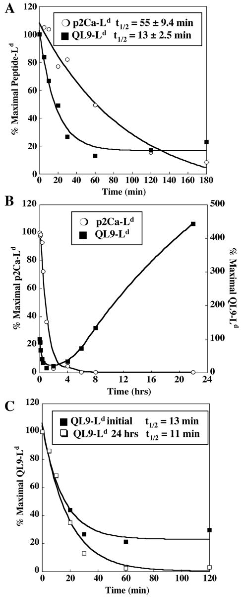
A. T2-Ld cells were incubated with p2Ca or QL9 peptide for 2 hours, washed, and suspended in media containing excess null peptide MCMV. Cells were incubated at 37°C and p2Ca-Ld and QL9-Ld levels were monitored at various time points by flow cytometry with m6α TCR. Standard deviation of cell surface lifetime was determined from four experiments. B. p2Ca-Ld and QL9-Ld levels were monitored over a 24 hour period, as described above. C. The initial QL9-Ld complexes (after the 2 hour incubation with peptide (see A above) and QL9-Ld complexes from the 24 hour time point in B were monitored in the presence of excess MCMV.
In monitoring the decrease in surface levels of QL9-Ld complexes over time, it was observed that the level of QL9-Ld complexes was not reduced to background levels (i.e. MFU in the absence of QL9). This property was not observed with p2Ca-Ld, in which the surface levels were reduced to background after a few hours (Figure 1A). In extending the assay with QL9/Ld for periods longer than three hours, the surface levels of QL9-Ld complexes increased such that by approximately 24 hours, the level of QL9-Ld complexes had increased to levels equal to or higher than those initially detected at the onset of the experiment (Figure 1B). The cell surface lifetime of these re-expressed QL9-Ld complexes (post 24 hours) was determined to be 11 minutes, about the same as the initial QL9-Ld complexes (Figure 1C). Thus, the initial QL9-Ld complexes and the re-expressed QL9-Ld complexes appear to be the same species and not a modified form of QL9-Ld. Furthermore, the level of re-expressed complexes reached background levels after several hours (Figure 1C), unlike the complexes detected in the first few hours of the assay. This observation shows that significant recycling did not occur after re-expression of the QL9-Ld and it suggests that the mechanism underlying delayed expression involves internalization of a QL9 peptide pool, rather than internalization and re-expression of the initial surface QL9-Ld complex. The ability of QL9, but not p2Ca, to be internalized and bound to internal stores of Ld likely contributes to the potency of QL9 as an agonist.
Cell Surface Lifetimes of SIYR-Kb and dEV8-Kb Complexes
In the 2C T cell system, the dEV8-Kb complex is a weak agonist and the SIYR-Kb complex is a strong agonist, with greater than 100,000-fold difference in potency between the two peptides (Table 1) (Degano et al., 2000; Sykulev et al., 1998). The cell surface lifetime of strong agonist SIYR-Kb was evaluated directly using the high affinity TCR m67α, but the affinity of m67α for dEV8-Kb is too low to be used as a direct probe for dEV8-Kb cell surface levels (Holler and Kranz, 2003). The surface half-life of the SIYR-Kb complex expressed on T2-Kb cells was 10 ± 1 hours (data not shown). The OVA peptide has been reported to have an affinity for Kb similar to SIYR peptide (KD value ~10 nM) (Matsumura et al., 1992; Sykulev et al., 1998) and OVA-Kb complexes have been shown to have a long surface lifetime (24 to 48 hours) on dendritic cells (Kukutsch et al., 2000). Consistent with these findings, the half-life of the OVA-Kb complex on T2-Kb cells was 23 ± 5 hours, as monitored with the anti-OVA-Kb specific antibody (data not shown).
Since the affinity of m67α TCR for the weak agonist dEV8-Kb complex is too low to allow direct detection, an indirect approach was used to estimate the dEV8-Kb lifetime. After a two-hour incubation with dEV8 peptide, OVA peptide, or media, cells were washed and suspended in media containing excess SIYR peptide. Upon dissociation of dEV8 or OVA peptides from Kb, SIYR peptide would compete for Kb binding and the SIYR-Kb complexes could be detected with the m67α TCR. The loss of dEV8 peptide bound to Kb complexes was plotted in comparison to OVA (Figure 2). In this indirect assay, OVA-Kb had a longer indirect cell surface lifetime than dEV8-Kb, 73 min compared to less than 5 minutes, respectively. Because the indirect assay monitors the presence of SIYR-Kb complexes, it is likely that empty Kb molecules associate with SIYR, leading to an underestimate of the true half-life of these complexes. Thus, while this represents a 20-fold difference in OVA-Kb lifetime compared to the direct measurement (23 ± 5 hours), it is clear that dEV8-Kb has a considerably more rapid dissociation rate. This observation is consistent with previous measurements showing the instability of dEV8-Kb compared to SIYR-Kb (Holler and Kranz, 2003; Krogsgaard et al., 2003; Tallquist et al., 1998).
FIGURE 2. Cell surface lifetimes of dEV8-Kb and OVA-Kb.
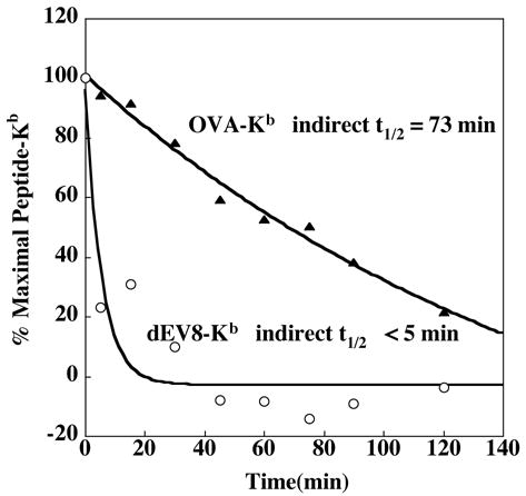
T2-Kb cells were incubated with dEV8, OVA, or without peptide for 2 hours. Cells were washed, resuspended in media containing excess SIYR peptide, and incubated at 37°C for various time periods. SIYR-Kb levels were detected by flow cytometry with m67α TCR. The amount of detectable SIYR-Kb complexes represented the loss of dEV8-Kb or OVA-Kb complexes. The percent maximal dEV8-Kb or OVA-Kb complex on the cell surface was calculated as described in Materials and Methods.
Delayed expression of QL9-Ld complexes in another tumor cell line
To ensure that the expression properties of the QL9 peptide were not an artifact due to peptide transport deficiency of the T2 cell line, QL9-Ld complexes were evaluated in a TAP-positive tumor cell line, P815. Cell surface expression levels of QL9-Ld complexes were monitored after 0 hrs, 3 hrs, and 24 hrs (Figure 3A). P815 cells, like T2-Ld cells, had high levels of QL9-Ld complexes at 0 hours, reduced levels at 3 hours and elevated levels after 24 hours. Thus, the mechanism of peptide recycling occurs either in the presence or absence of TAP.
FIGURE 3. Cell surface levels of QL9-Ld on P815 and T2-Ld cells at various times, in the presence and absence of Brefeldin.
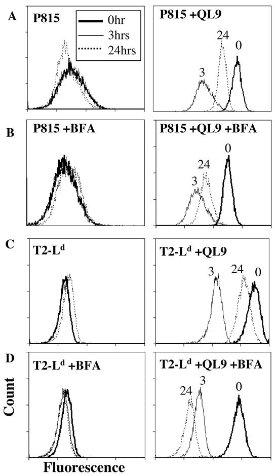
A. P815 cells incubated without (A) or with (B) Brefeldin A (BFA) were incubated without or with QL9 peptide for 2 hours, washed and maintained at 37°C for 0 hours, 3 hours, or 24 hours. The level of QL9-Ld complexes was measured at these times using m6α TCR by flow cytometry. For BFA experiments, cells were treated 30 minutes prior to peptide incubation, after the 2 hour peptide pulse, and at 4 hours and 8 hours. T2-Ld cells were also incubated without (C) or with (D) Brefeldin A (BFA) and assayed as described for P815 cells.
ER and golgi dependence of delayed pep-MHC expression
The studies described above suggested that the pepMHC complexes that were expressed after several hours were derived from peptides taken up by the cells and associated with intracellular Ld (rather than QL9-Ld recycled from the surface). To further elucidate the mechanism of delayed expression of QL9-Ld complexes, P815 and T2-Ld cells were treated with brefeldin A (BFA) and the levels of QL9-Ld were assessed compared to untreated cells (Figure 3). Brefeldin A inhibits the anterograde transport of vesicles from the ER to the golgi apparatus, thereby preventing pepMHC proteins from being transported to the cell surface. BFA treated P815 and T2-Ld cells were incubated with QL9 peptide and assayed for expression levels at 0, 3, and 24 hours (Figure 3B, D) compared to untreated P815 and T2-Ld cells (Figure 3A, C). Under these conditions, there was reduced expression of QL9-Ld complexes after 24 hours.
To quantitate the effects of BFA on re-expression of QL9-Ld complexes in T2-Ld and P815 cells, the percent inhibition of QL9-Ld in the presence of BFA was calculated (Figure 4). BFA also affected the total amount of detectable Ld complexes on the cell surface as determined by anti-Ld antibody 28.14.8 binding, therefore the percent of QL9-Ld detected by m6α TCR relative to total Ld is illustrated in Figure 4. Inhibition increased over time with nearly 100% inhibition observed for T2-Ld cells (Figure 4A, C) and 85% inhibition observed for P815 cells (Figure 4B, C) after 24 hours. In order to confirm that BFA acts on intracellular transport mechanisms, and does not simply inhibit the uptake of exogenous peptide, an additional experiment was done in which BFA was not added until after incubation with QL9 peptide. Again, 100% inhibition was observed (data not shown), suggesting that BFA effectively blocks re-expression of QL9-Ld complexes by interfering with trafficking of Ld through the ER and golgi apparatus.
FIGURE 4. Extent of inhibition of QL9-Ld surface levels by Brefeldin.
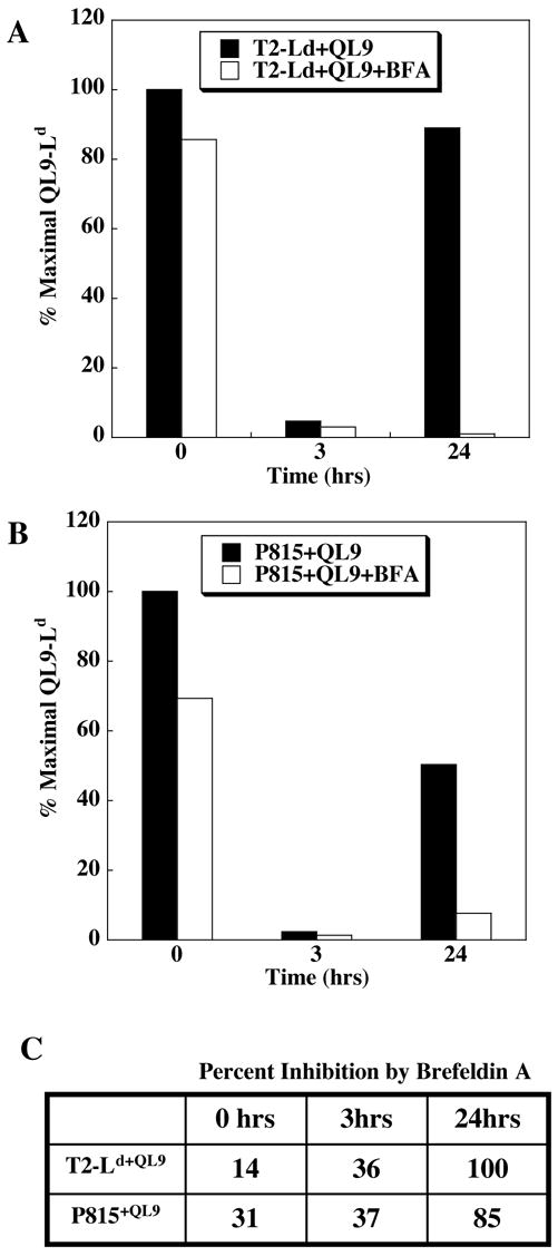
A. Results from experiments described in Figure 3, were used to calculate the level of QL9-Ld relative to total Ld levels at different times for T2-Ld
(A) or P815 (B). Bar graphs represent the percent QL9-Ld relative to the total amount of Ld on the cell surface at 0, 3, and 24 hours. The level of QL9-Ld and Ld on the cell surface was detected with m6α TCR and anti-Ld antibody 28.14.8, respectively. C. The percent inhibition of QL9-Ld complexes by BFA, relative to the absence of BFA, for T2-Ld and P815 cells pulsed with QL9 peptide.
Delayed expression of QL9-Ld complexes in dendritic cells
To determine whether the delayed expression of QL9-Ld complexes can occur in antigen presenting cells, dendritic cells isolated from the bone marrow of BALB/c mice (H-2d) were examined in vitro. Bone marrow cells were cultured with GM-CSF to generate immature DCs, and cells were gaited using an antibody to the surface marker CD11c. The level of QL9-Ld complexes on the CD11c-negative (Figure 5A) and CD11c-positive (Figure 5B) populations of peptide-pulsed DCs was evaluated at 0, 3, and 24 hours. For both populations of cells, the highest level of QL9-Ld was detected at 0 hours, the lowest level at 3 hours, and elevated expression at 24 hours. Thus, dendritic cells are also capable of this alternative pathway of peptide presentation.
FIGURE 5. Cell surface levels of QL9-Ld on bone marrow-derived dendritic cells at various times.
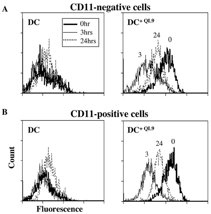
Bone marrow cells were cultured in GM-CSF. On day 6, cells were washed, incubated with or without QL9 peptide for 2 hours, washed, and maintained at 37°C for 0 hours (bold line), 3 hours (solid line), and 24 hours (dashed line). QL9-Ld complexes were monitored with m6α TCR, gaiting on the CD11c-negative (A) and CD11c-positive (B) cell populations.
Discussion
It has been shown previously in the 2C system that the ability of a specific peptide to activate T cells is dependent on several properties of the peptide: the affinity of the peptide for MHC, the stability of the pepMHC complex on the surface of the target cell, and the affinity of the TCR for the pepMHC complex (Holler and Kranz, 2003). Krogsgaard et al. showed that pepMHC stability and TCR:pepMHC half-lives are key factors in biological activity in class I and class II pepMHC systems, but they also observed that a change in specific heat capacity (i.e. possible conformational effects on TCR binding) may impact activity for some systems (Krogsgaard et al., 2003). By using high-affinity TCRs as probes for specific pepMHC, we were able to measure cell surface lifetimes in the SIYR-Kb/dEV8-Kb and QL9-Ld/p2Ca-Ld systems. In the Kb system, the weak agonist dEV8 has greater than 100,000 fold lower 2C T cell activity than SIYR and the affinities of the two peptides for Kb differ by ~100 fold (Holler and Kranz, 2003; Krogsgaard et al., 2003; Tallquist et al., 1998). Our indirect assay suggested that the lifetimes of the dEV8-Kb complex are indeed very short and thus the potency of SIYR, compared to dEV8, arises from the higher Kb-binding affinity of SIYR and its long cell surface lifetime.
In the Ld system however, the complexity of the molecular mechanisms that account for peptide potency in T cell immunity was highlighted by two unexpected observations. First, a strong agonist pepMHC complex (QL9-Ld) can have a shorter cell surface lifetime than a weak agonist pepMHC (p2Ca-Ld). Second, peptides provided to target cells exogenously could be taken up by the cell and presented several hours later. In fact, the potency of the peptide QL9 appears to be due at least in part to the ability of QL9 to be taken up by target cells, to associate with Ld, and to be transported to the cell surface hours after the original cell surface QL9-Ld has been lost. The delayed cell surface expression of these QL9-Ld complexes likely contributes to the stimulatory capacity of QL9 when assayed in vitro.
The re-expressed QL9-Ld complexes were shown to bind to the m6α TCR and have lifetimes identical to the initial QL9-Ld complexes, suggesting that the peptide or the complexes were not presented in an altered conformation as has been observed for endogenous processing of some class II MHC-binding peptides (Pu et al., 2004). In this regard, it is important to point out that many amino acid variants of QL9 affect either Ld binding or TCR binding (Kageyama et al., 2001; Schlueter et al., 1996), so that if side-chain modifications had occurred, it may have been identified by this approach. QL9 has been shown to undergo N-terminal cyclization to pyroglutamine and it is possible that this modification could stabilize the peptide from peptidases (Kageyama et al., 2001). Alternatively, cellular chaperones may protect some peptides from complete degradation (Rock et al., 2004).
The delayed expression of the pepMHC appeared to require transport through the ER or golgi since brefeldin A inhibited the process. As similar results were observed in TAP-negative or TAP-positive cell lines, the process does not require peptide transport through this mechanism. Alternative pathways that involve loading of exogenous antigens onto human and mouse class I proteins, as assessed in both in vitro and in vivo assays, have been identified (Ackerman and Cresswell, 2004; Chefalo et al., 2003; Chefalo and Harding, 2001; Gromme et al., 1999; Norbury et al., 2001) (Palliser et al., 2005). The post-golgi pathways are TAP independent and BFA insensitive, whereas the pathways that release peptides into the cytosol are TAP dependent and BFA sensitive. As QL9-Ld cross-presentation is BFA sensitive and TAP independent, the mechanism appears to involve yet another alternative pathway. One possible pathway for QL9-Ld complex re-expression involves endocytosis of QL9 peptides into endosomes or phagosomes. The QL9-loaded vesicles could fuse with the ER membrane, thereby releasing peptides that then bind to nascent MHC complexes. Several papers have shown that exogenous antigens taken up by dendritic cells can access the endoplasmic reticulum (Ackerman et al., 2005; Guermonprez et al., 2003; Houde et al., 2003), although some debate remains as to whether ER-phagosome fusion occurs (Touret et al., 2005). In addition, exogenous peptides that bind to class I proteins have been shown to be maintained in or transported among multiple cellular compartments, including the ER (Day et al., 1997; Reits et al., 2003). Our results indicate that such peptides could serve as a source for class I MHC protein binding and presentation.
Bone marrow derived immature DCs pulsed with QL9 peptide also exhibited re-expression of QL9-Ld complexes after 24 hours. In fact, both the CD11c-positive and the CD11c-negative populations were shown to re-express QL9-Ld complexes. The observation that the amount of re-expressed QL9-Ld was at least as high as the initial QL9-Ld suggests that the process is very efficient and it could have relevance in vivo. There are potentially important consequences of extending the high levels of QL9-Ld surface expression from minutes to over a day (see Figure 1B) through this pathway. T cells could be stimulated over a considerably longer period of time, yielding significant increases in peptide sensitivity. If long-term stimulatory potential makes a difference in this Ld-system, then it may be possible to distinguish a difference between the potency of p2Ca and QL9 peptides in short-term versus long-term T cell assays. To examine this possibility, we reviewed the published literature that has used 2C T cells in different types of assays, where both p2Ca and QL9 peptides were evaluated (Table 2). The reported SD50 values for p2Ca and QL9 peptides were used to calculate the ratio of p2Ca/QL9 potency in the different assays (Figure 6). For example, the agonist peptide QL9 has been reported to have up to ~240-fold more activity than the weak agonist p2Ca in a short-term (4 hour) cytotoxicity assay (Hornell et al., 2001). In assays that spanned longer periods of time (12–96 hours), the SD50 of both peptides was reduced, but this reduction in activity was less pronounced for QL9 (Table 2). Accordingly, a difference in activity of up to 10,000 fold in long-term assays was observed (Holler and Kranz, 2003), presumably due to the ability of QL9 peptide to be taken up by cells and presented hours later. These findings indicate that it is important to consider other features of peptides, in addition to their stability or cell surface lifetimes in complex with an MHC molecule, in order to interpret the basis of their biological activity. Studies involving comparisons of peptides for their T cell stimulatory activity must also consider the nature of the assay due to the possible role of peptide uptake and delayed presentation.
Table 2.
SD50 values for QL9 and p2Ca peptides in the 2C system.
| Reference | QL9 SD50 (M) | p2Ca SD50 (M) | Assay type | Assay time | Fold Difference (p2Ca/QL9) |
|---|---|---|---|---|---|
| (Sykulev et al., 1994b) | 5×10⁻¹²* | 1×10⁻¹⁰* | 51Cr release | 4 hours | 20 |
| (Sykulev et al., 1996) | 5×10⁻¹²* | 3×10⁻¹⁰* | 51Cr release | 4 hours | 60 |
| (Kageyama et al., 2001) | 3×10⁻¹¹ | 1×10⁻⁹ | 51Cr release | 4 hours | 33 |
| (Hornell et al., 2001) | 5.9×10⁻¹² | 1.4×10⁻⁹ | 51Cr release | 4 hours | 237 |
| (Schlueter et al., 1996) | 1.9×10⁻¹¹ | 1.6×10⁻¹⁰ | 51Cr release | 4 hours | 8 |
| (Cai et al., 1997) | 7×10⁻¹⁰ * | 7×10⁻⁷* | TCR downregulation | 12 hours | 1,000 |
| (Holler and Kranz, 2003) | 1×10⁻¹⁰* | 1×10⁻⁶* | IL-2 release | 30 hours | 10,000 |
| (Holler) (unpublished results) | 7.6×10⁻¹¹ | 2.1×10⁻⁷ | Proliferation | 72 hours | 2,763 |
| (Chambers et al., 1998) | 3×10⁻⁹* | 3×10⁻⁶* | Proliferation | 96 hours | 1,000 |
SD50 values were estimated, based on graphical representations.
FIGURE 6. Relative activity of QL9 and p2Ca peptides in short and long-term assays.
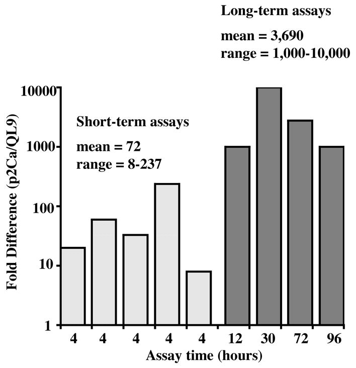
SD50 values from references listed in Table 2 were plotted as fold difference in SD50 values (p2Ca/QL9) versus assay time. Short-term assays (4 hours) are shown by the light grey bars and long-term assays (12–96 hours) are shown by the dark grey bars. Unpaired t-test (one-tailed) comparing the short-term and long-term values yielded a P value of 0.048.
It has been common practice to evaluate the potency of T cell ligands through the use of synthetic peptides cultured with target cells or APCs. In this approach, the sensitization dose that yields half maximal activity (SD50) is considered a measure of peptide activity. One of the major contributing factors to a peptide’s activity is the ability to form stable complexes with MHC molecules on the surface of APCs. The direct correlation of T cell activity with pepMHC stabilization has been demonstrated in many studies, including those with the 2C T cell system described in the present report. Experiments to assess pepMHC stability have frequently relied on MHC up-regulation assays, in which various concentrations of the peptide are incubated with cells that are deficient in TAP, and the increase in total pepMHC are measured several hours later. In contrast, T cell assays may be performed over a larger time frame, depending on the functional read-out. Examples of peptides whose T cell stimulatory activity does not correlate directly with pepMHC stability or TCR:pepMHC affinity have been described (e.g. (Baker et al., 2000)). While conformational changes upon TCR:pepMHC binding may account for some of these cases (Krogsgaard et al., 2003), the findings described here suggest that the potency of some peptides may need to take into account yet another property of the peptide, its ability to be taken up by a cell, to associate with intracellular MHC, and to be transported to the cell surface at a later time.
Finally, it is possible that different class I molecules will exhibit variations in the extent to which alternative peptide presentation pathways contribute to surface persistence (Hansen et al., 2000). The intracellular peptide pathway may be less critical for activity in cases where the cell surface lifetime of the peptide is very long (e.g. SIYR and OVA in the Kb system). In contrast, Ld, or Ld-like family members such as Db that have similar structures (Ciatto et al., 2001), the ability of cells to present antigenic peptides taken up from extracellular stores may be important for increasing the probability of a productive T cell response.
Acknowledgments
We thank Herman Eisen for comments on the manuscript and for the suggestion to analyze the published values for SD50 values of p2Ca and QL9. We also thank the staff of the University of Illinois Biotechnology Center Flow Cytometry Facility for their assistance.
Footnotes
This work was supported by NIH grant GM55767
Publisher's Disclaimer: This is a PDF file of an unedited manuscript that has been accepted for publication. As a service to our customers we are providing this early version of the manuscript. The manuscript will undergo copyediting, typesetting, and review of the resulting proof before it is published in its final citable form. Please note that during the production process errors may be discovered which could affect the content, and all legal disclaimers that apply to the journal pertain.
References
- Ackerman AL, Cresswell P. Cellular mechanisms governing cross-presentation of exogenous antigens. Nat Immunol. 2004;5:678–84. doi: 10.1038/ni1082. [DOI] [PubMed] [Google Scholar]
- Ackerman AL, Kyritsis C, Tampe R, Cresswell P. Access of soluble antigens to the endoplasmic reticulum can explain cross-presentation by dendritic cells. Nat Immunol. 2005;6:107–13. doi: 10.1038/ni1147. [DOI] [PubMed] [Google Scholar]
- Alexander J, Payne JA, Murray R, Frelinger JA, Cresswell P. Differential transport requirements of HLA and H-2 class I glycoproteins. Immunogenetics. 1989;29:380–399. doi: 10.1007/BF00375866. [DOI] [PubMed] [Google Scholar]
- Andersen PS, Stryhn A, Hansen BE, Fugger L, Engberg J, Buus S. A recombinant antibody with the antigen-specific, major histocompatibility complex-restricted specificity of T cells. Proc Natl Acad Sci. 1996;93:1820–24. doi: 10.1073/pnas.93.5.1820. [DOI] [PMC free article] [PubMed] [Google Scholar]
- Baker MB, Gagnon JS, Biddison EW, Wiley CD. Conversion of a T cell antagonist into an agonist by repairing a defect in the TCR/peptide/MHC interface: implications for TCR signaling. Immunity. 2000;13:475–84. doi: 10.1016/s1074-7613(00)00047-9. [DOI] [PubMed] [Google Scholar]
- Bevan MJ. Cross-priming for a secondary cytotoxic response to minor H antigens with H-2 congenic cells which do not cross-react in the cytotoxic assay. J Exp Med. 1976;143:1283–8. doi: 10.1084/jem.143.5.1283. [DOI] [PMC free article] [PubMed] [Google Scholar]
- Cai Z, Kishimoto H, Brunmark A, Jackson MR, Peterson PA, Sprent J. Requirements for peptide-induced T cell receptor downregulation on naive CD8+ T cells. J Exp Med. 1997;185:641–51. doi: 10.1084/jem.185.4.641. [DOI] [PMC free article] [PubMed] [Google Scholar]
- Chambers CA, Sullivan TJ, Truong T, Allison JP. Secondary but not primary T cell responses are enhanced in CTLA-4-deficient CD8+ T cells. Eur J Immunol. 1998;28:3137–43. doi: 10.1002/(SICI)1521-4141(199810)28:10<3137::AID-IMMU3137>3.0.CO;2-X. [DOI] [PubMed] [Google Scholar]
- Chefalo PJ, Grandea AG, 3rd, Van Kaer L, Harding CV. Tapasin−/− and TAP1−/−macrophages are deficient in vacuolar alternate class I MHC (MHC-I) processing due to decreased MHC-I stability at phagolysosomal pH. J Immunol. 2003;170:5825–33. doi: 10.4049/jimmunol.170.12.5825. [DOI] [PubMed] [Google Scholar]
- Chefalo PJ, Harding CV. Processing of exogenous antigens for presentation by class I MHC molecules involves post-Golgi peptide exchange influenced by peptide-MHC complex stability and acidic pH. J Immunol. 2001;167:1274–82. doi: 10.4049/jimmunol.167.3.1274. [DOI] [PubMed] [Google Scholar]
- Chlewicki LK, Holler PD, Monti BC, Clutter MA, Kranz DM. High-Affinity, Peptide-Specific T Cell Receptors Can Be Generated by Mutations in CDR1, CDR2 or CDR3. J Mol Biol. 2005;346:223–239. doi: 10.1016/j.jmb.2004.11.057. [DOI] [PubMed] [Google Scholar]
- Ciatto C, Tissot AC, Tschopp M, Capitani G, Pecorari F, Pluckthun A, Grutter MG. Zooming in on the hydrophobic ridge of H-2D(b): implications for the conformational variability of bound peptides. J Mol Biol. 2001;312:1059–71. doi: 10.1006/jmbi.2001.5016. [DOI] [PubMed] [Google Scholar]
- Cohen CJ, Sarig O, Yamano Y, Tomaru U, Jacobson S, Reiter Y. Direct phenotypic analysis of human MHC class I antigen presentation: visualization, quantitation, and in situ detection of human viral epitopes using peptide-specific, MHC-restricted human recombinant antibodies. J Immunol. 2003;170:4349–61. doi: 10.4049/jimmunol.170.8.4349. [DOI] [PubMed] [Google Scholar]
- Dadaglio G, Nelson CA, Deck MB, Petzold SJ, Unanue ER. Characterization and quantitation of peptide-MHC complexes produced from hen egg lysozyme using a monoclonal antibody. Immunity. 1997;6:727–38. doi: 10.1016/s1074-7613(00)80448-3. [DOI] [PubMed] [Google Scholar]
- Day PM, Yewdell JW, Porgador A, Germain RN, Bennink JR. Direct delivery of exogenous MHC class I molecule-binding oligopeptides to the endoplasmic reticulum of viable cells. Proc Natl Acad Sci U S A. 1997;94:8064–9. doi: 10.1073/pnas.94.15.8064. [DOI] [PMC free article] [PubMed] [Google Scholar]
- Degano M, Garcia KC, Apostolopoulos V, Rudolph MG, Teyton L, Wilson IA. A functional hot spot for antigen recognition in a superagonist TCR/MHC complex. Immunity. 2000;12:251–61. doi: 10.1016/s1074-7613(00)80178-8. [DOI] [PubMed] [Google Scholar]
- Denkberg G, Reiter Y. Recombinant antibodies with T-cell receptor-like specificity: novel tools to study MHC class I presentation. Autoimmun Rev. 2006;5:252–7. doi: 10.1016/j.autrev.2005.07.004. [DOI] [PubMed] [Google Scholar]
- Freigang S, Egger D, Bienz K, Hengartner H, Zinkernagel RM. Endogenous neosynthesis vs. cross-presentation of viral antigens for cytotoxic T cell priming. Proc Natl Acad Sci U S A. 2003;100:13477–82. doi: 10.1073/pnas.1835685100. [DOI] [PMC free article] [PubMed] [Google Scholar]
- Garcia KC, Tallquist MD, Pease LR, Brunmark A, Scott CA, Degano M, Stura EA, Peterson PA, Wilson IA, Teyton L. Alphabeta T cell receptor interactions with syngeneic and allogeneic ligands: affinity measurements and crystallization. Proc Natl Acad Sci U S A. 1997;94:13838–13843. doi: 10.1073/pnas.94.25.13838. [DOI] [PMC free article] [PubMed] [Google Scholar]
- Gromme M, Uytdehaag FG, Janssen H, Calafat J, van Binnendijk RS, Kenter MJ, Tulp A, Verwoerd D, Neefjes J. Recycling MHC class I molecules and endosomal peptide loading. Proc Natl Acad Sci U S A. 1999;96:10326–31. doi: 10.1073/pnas.96.18.10326. [DOI] [PMC free article] [PubMed] [Google Scholar]
- Guermonprez P, Saveanu L, Kleijmeer M, Davoust J, Van Endert P, Amigorena S. ER-phagosome fusion defines an MHC class I cross-presentation compartment in dendritic cells. Nature. 2003;425:397–402. doi: 10.1038/nature01911. [DOI] [PubMed] [Google Scholar]
- Hansen T, Balendiran G, Solheim J, Ostrov D, Nathenson S. Structural features of MHC class I molecules that might facilitate alternative pathways of presentation. Immunol Today. 2000;21:83–8. doi: 10.1016/s0167-5699(98)01426-1. [DOI] [PubMed] [Google Scholar]
- Holler PD, Chlewicki LK, Kranz DM. TCRs with high affinity for foreign pMHC show self-reactivity. Nat Immunol. 2003;4:55–62. doi: 10.1038/ni863. [DOI] [PubMed] [Google Scholar]
- Holler PD, Holman PO, Shusta EV, O’Herrin S, Wittrup KD, Kranz DM. In vitro evolution of a T cell receptor with high affinity for peptide/MHC. Proc Natl Acad Sci U S A. 2000;97:5387–92. doi: 10.1073/pnas.080078297. [DOI] [PMC free article] [PubMed] [Google Scholar]
- Holler PD, Kranz DM. Quantitative analysis of the contribution of TCR/pepMHC affinity and CD8 to T cell activation. Immunity. 2003;18:255–264. doi: 10.1016/s1074-7613(03)00019-0. [DOI] [PubMed] [Google Scholar]
- Holler PD, Lim AR, Cho BK, Rund LA, Kranz DM. CD8− T cell transfectants that express a high affinity T cell receptor exhibit enhanced peptide-dependent activation. J Exp Med. 2001;194(8):1043–1052. doi: 10.1084/jem.194.8.1043. [DOI] [PMC free article] [PubMed] [Google Scholar]
- Hornell TM, Martin SM, Myers NB, Connolly JM. Peptide length variants p2Ca and QL9 present distinct conformations to L(d)-specific T cells. J Immunol. 2001;167:4207–14. doi: 10.4049/jimmunol.167.8.4207. [DOI] [PubMed] [Google Scholar]
- Hornell TM, Myers N, Hansen TH, Connolly JM. Homology between an alloantigen and a self MHC allele calibrates the avidity of the alloreactive T cell repertoire independent of TCR affinity. J Immunol. 2003;170:4506–14. doi: 10.4049/jimmunol.170.9.4506. [DOI] [PubMed] [Google Scholar]
- Houde M, Bertholet S, Gagnon E, Brunet S, Goyette G, Laplante A, Princiotta MF, Thibault P, Sacks D, Desjardins M. Phagosomes are competent organelles for antigen cross-presentation. Nature. 2003;425:402–6. doi: 10.1038/nature01912. [DOI] [PubMed] [Google Scholar]
- Inaba K, Inaba M, Romani N, Aya H, Deguchi M, Ikehara S, Muramatsu S, Steinman RM. Generation of large numbers of dendritic cells from mouse bone marrow cultures supplemented with granulocyte/macrophage colony-stimulating factor. J Exp Med. 1992;176:1693–702. doi: 10.1084/jem.176.6.1693. [DOI] [PMC free article] [PubMed] [Google Scholar]
- Kageyama S, Tsomides TJ, Fukusen N, Papayannopoulos IA, Eisen HN, Sykulev Y. Potent cytolytic response by a CD8+ CTL clone to multiple peptides from the same protein in association with an allogeneic class I MHC molecule. J Immunol. 2001;166:3028–34. doi: 10.4049/jimmunol.166.5.3028. [DOI] [PubMed] [Google Scholar]
- Krogsgaard M, Prado N, Adams EJ, He XL, Chow DC, Wilson DB, Garcia KC, Davis MM. Evidence that structural rearrangements and/or flexibility during TCR binding can contribute to T cell activation. Mol Cell. 2003;12:1367–78. doi: 10.1016/s1097-2765(03)00474-x. [DOI] [PubMed] [Google Scholar]
- Krogsgaard M, Wucherpfennig KW, Canella B, Hansen BE, Svejgaard A, Pyrdol J, Ditzel H, Raine C, Engberg J, Fugger L. Visualization of myelin basic protein (MBP) T cell epitopes in multiple sclerosis lesions using a monoclonal antibody specific for the human histocompatibility leukocyte antigen (HLA)-DR2-MBP 85–99 complex. J Exp Med. 2000;191:1395–412. doi: 10.1084/jem.191.8.1395. [DOI] [PMC free article] [PubMed] [Google Scholar]
- Kukutsch NA, Rossner S, Austyn JM, Schuler G, Lutz MB. Formation and kinetics of MHC class I-ovalbumin peptide complexes on immature and mature murine dendritic cells. J Invest Dermatol. 2000;115:449–53. doi: 10.1046/j.1523-1747.2000.00084.x. [DOI] [PubMed] [Google Scholar]
- Matsumura M, Fremont DH, Peterson PA, Wilson IA. Emerging principles for the recognition of peptide antigens by MHC class I molecules. Science. 1992;257:927–934. doi: 10.1126/science.1323878. [DOI] [PubMed] [Google Scholar]
- Norbury CC, Princiotta MF, Bacik I, Brutkiewicz RR, Wood P, Elliott T, Bennink JR, Yewdell JW. Multiple antigen-specific processing pathways for activating naive CD8+ T cells in vivo. J Immunol. 2001;166:4355–62. doi: 10.4049/jimmunol.166.7.4355. [DOI] [PubMed] [Google Scholar]
- Palliser D, Guillen E, Ju M, Eisen HN. Multiple intracellular routes in the cross-presentation of a soluble protein by murine dendritic cells. J Immunol. 2005;174:1879–87. doi: 10.4049/jimmunol.174.4.1879. [DOI] [PubMed] [Google Scholar]
- Peng K-W, Holler PD, Orr BA, Kranz DM, Russell SJ. Targeting membrane fusion to specific peptide/MHC complexes through a high-affinity T-cell receptor. Gene Ther. 2004;11:1234–9. doi: 10.1038/sj.gt.3302286. [DOI] [PubMed] [Google Scholar]
- Porgador A, Yewdell JW, Deng Y, Bennink JR, Germain RN. Localization, quantitation, and in situ detection of specific peptide-MHC class I complexes using a monoclonal antibody. Immunity. 1997;6:715–26. doi: 10.1016/s1074-7613(00)80447-1. [DOI] [PubMed] [Google Scholar]
- Pu Z, Lovitch SB, Bikoff EK, Unanue ER. T cells distinguish MHC-peptide complexes formed in separate vesicles and edited by H2-DM. Immunity. 2004;20:467–76. doi: 10.1016/s1074-7613(04)00073-1. [DOI] [PubMed] [Google Scholar]
- Reits E, Griekspoor A, Neijssen J, Groothuis T, Jalink K, van Veelen P, Janssen H, Calafat J, Drijfhout JW, Neefjes J. Peptide diffusion, protection, and degradation in nuclear and cytoplasmic compartments before antigen presentation by MHC class I. Immunity. 2003;18:97–108. doi: 10.1016/s1074-7613(02)00511-3. [DOI] [PubMed] [Google Scholar]
- Rock KL, Goldberg AL. Degradation of cell proteins and the generation of MHC class I- presented peptides. Annu Rev Immunol. 1999;17:739–79. doi: 10.1146/annurev.immunol.17.1.739. [DOI] [PubMed] [Google Scholar]
- Rock KL, York IA, Goldberg AL. Post-proteasomal antigen processing for major histocompatibility complex class I presentation. Nat Immunol. 2004;5:670–7. doi: 10.1038/ni1089. [DOI] [PubMed] [Google Scholar]
- Rudolph MG, Speir JA, Brunmark A, Mattsson N, Jackson MR, Peterson PA, Teyton L, Wilson IA. The crystal structures of K(bm1) and K(bm8) reveal that subtle changes in the peptide environment impact thermostability and alloreactivity. Immunity. 2001;14:231–42. doi: 10.1016/s1074-7613(01)00105-4. [DOI] [PubMed] [Google Scholar]
- Schlueter CJ, Manning TC, Schodin BA, Kranz DM. A residue in the center of peptide QL9 affects binding to both Ld and the T cell receptor. J Immunol. 1996;157:4478–4485. [PubMed] [Google Scholar]
- Spiotto MT, Rowley DA, Schreiber H. Bystander elimination of antigen loss variants in established tumors. Nat Med. 2004;10:294–8. doi: 10.1038/nm999. [DOI] [PubMed] [Google Scholar]
- Sykulev Y, Brunmark A, Jackson M, Cohen RJ, Peterson PA, Eisen HN. Kinetics and affinity of reactions between an antigen-specific T cell receptor and peptide-MHC complexes. Immunity. 1994a;1:15–22. doi: 10.1016/1074-7613(94)90005-1. [DOI] [PubMed] [Google Scholar]
- Sykulev Y, Brunmark A, Tsomides TJ, Kageyama S, Jackson M, Peterson PA, Eisen HN. High-affinity reactions between antigen-specific T-cell receptors and peptides associated with allogeneic and syngeneic major histocompatibility complex class I proteins. Proc Natl Acad Sci USA. 1994b;91:11487–11491. doi: 10.1073/pnas.91.24.11487. [DOI] [PMC free article] [PubMed] [Google Scholar]
- Sykulev Y, Joo M, Vturina I, Tsomides TJ, Eisen HN. Evidence that a single peptide-MHC complex on a target cell can elicit a cytolytic T cell response. Immunity. 1996;4:565–571. doi: 10.1016/s1074-7613(00)80483-5. [DOI] [PubMed] [Google Scholar]
- Sykulev Y, Vugmeyster Y, Brunmark A, Ploegh HL, Eisen HN. Peptide antagonism and T cell receptor interactions with peptide-MHC complexes. Immunity. 1998;9:475–483. doi: 10.1016/s1074-7613(00)80631-7. [DOI] [PubMed] [Google Scholar]
- Tallquist MD, Weaver AJ, Pease LR. Degenerate recognition of alloantigenic peptides on a positive- selecting class I molecule. J Immunol. 1998;160:802–9. [PubMed] [Google Scholar]
- Tallquist MD, Yun TJ, Pease LR. A single T cell receptor recognizes structurally distinct MHC/peptide complexes with high specificity. J Exp Med. 1996;184:1017–1026. doi: 10.1084/jem.184.3.1017. [DOI] [PMC free article] [PubMed] [Google Scholar]
- Touret N, Paroutis P, Terebiznik M, Harrison RE, Trombetta S, Pypaert M, Chow A, Jiang A, Shaw J, Yip C, Moore HP, van der Wel N, Houben D, Peters PJ, de Chastellier C, Mellman I, Grinstein S. Quantitative and dynamic assessment of the contribution of the ER to phagosome formation. Cell. 2005;123:157–70. doi: 10.1016/j.cell.2005.08.018. [DOI] [PubMed] [Google Scholar]
- Udaka K, Tsomides TJ, Eisen HN. A naturally occurring peptide recognized by alloreactive CD8+ cytotoxic T lymphocytes in association with a class I MHC protein. Cell. 1992;69:989–998. doi: 10.1016/0092-8674(92)90617-l. [DOI] [PubMed] [Google Scholar]
- Udaka K, Wiesmuller K, Kienle S, Jung G, Walden P. Self-MHC-restricted peptides recognized by an alloreactive T-lymphocyte clone. J Immunol. 1996;157:670–678. [PubMed] [Google Scholar]