Abstract
The split-Ubiquitin (split-Ub) technique was used to map the molecular environment of a membrane protein in vivo. Cub, the C-terminal half of Ub, was attached to Sec63p, and Nub, the N-terminal half of Ub, was attached to a selection of differently localized proteins of the yeast Saccharomyces cerevisiae. The efficiency of the Nub and Cub reassembly to the quasi-native Ub reflects the proximity between Sec63-Cub and the Nub-labeled proteins. By using a modified Ura3p as the reporter that is released from Cub, the local concentration between Sec63-Cub-RUra3p and the different Nub-constructs could be translated into the growth rate of yeast cells on media lacking uracil. We show that Sec63p interacts with Sec62p and Sec61p in vivo. Ssh1p is more distant to Sec63p than its close sequence homologue Sec61p. Employing Nub- and Cub-labeled versions of Ste14p, an enzyme of the protein isoprenylation pathway, we conclude that Ste14p is a membrane protein of the ER. Using Sec63p as a reference, a gradient of local concentrations of different t- and v-SNARES could be visualized in the living cell. The RUra3p reporter should further allow the selection of new binding partners of Sec63p and the selection of molecules or cellular conditions that interfere with the binding between Sec63p and one of its known partners.
INTRODUCTION
Search algorithms can identify membrane proteins and often successfully predict their topology. Fluorescence microscopy allows the determination of their cellular localization. However, to perform their function, membrane proteins very often assemble into protein complexes and temporarily relocate to sites in the cell that differ from their steady-state residence. With the current methods at hand, these processes are difficult to study.
Sec63p, as part of the tetrameric and the heptameric Sec complex in the yeast Saccharomyces cerevisiae, is a membrane protein of the endoplasmic reticulum (ER) (Rothblatt et al., 1989; Deshaies et al., 1991; Brodsky and Schekman, 1993). The tetrameric Sec62/63p complex and the trimeric Sec61p-complex constitute the main components of the translocation machinery responsible for delivering polypeptides across the membrane of the ER. The tetrameric Sec62/63p complex harbors, in addition to Sec63p, the integral membrane proteins Sec62p and Sec71p and the peripheral membrane protein Sec72p (Deshaies et al., 1991; Panzner et al., 1995). The trimeric Sec61p complex forms the actual gate across the membrane and consists of the membrane proteins, Sec61p, Sss1p, and Sbh1p. Both complexes can exist as individual entities or as parts of the heptameric Sec complex (for review see Rapoport et al., 1996). The modular structure of the translocation machinery allows Sec61p to accept a wide variety of polypetides as translocation substrates. The trimeric Sec61 complex associates with Sec62/63p to translocate polypeptides that are either already completely or partially synthesized. Alternatively, the trimeric Sec61 complex is found in association with translating ribosomes (Görlich et al., 1992). Here the signal sequence-containing nascent chain is very probably transferred via the signal recognition particle directly to the trimeric Sec61 complex to forge a tight seal between Sec61p and the ribosome (Walter and Johnson, 1994; Beckmann et al., 1997). The already substantial number of proteins that interact with Sec63p may become still larger since Sec63p is also involved in the retrograde transfer of proteins from the lumen of the ER back into the cytosol (Plemper et al., 1997). In addition, Sec63p plays a role in the homotypic fusion of nuclear membranes during the mating of yeast (Ng and Walter, 1996).
The split-Ub method can monitor interactions between proteins in the living cell (Johnsson and Varshavsky, 1994). It is based on the reassembly of the N- and C-terminal halves (Nub and Cub) of Ubiquitin (Ub). The reassembled quasi-native Ub is recognized by the ubiquitin-specific proteases (UBPs). The UBPs cleave any C-terminally attached polypeptide from Cub and thereby provide an immediate readout of the Nub-Cub reassociation. Two mutations were engineered into Nub. Nua and Nug carry an alanine or a glycine in position 13 of Nub. Both have a lower affinity for Cub than Nub, the wild-type version carrying an isoleucine in this position. It was shown that Nub and Cub reassemble quite efficiently. However, Nua or Nug only interact with Cub once both Ub peptides are linked to proteins that are close to each other. Under these conditions, Cub interacts more strongly with Nua than with Nug (Johnsson and Varshavsky, 1994). The split-Ub technique measures the local concentration, integrated over time, between the coupled Nub and Cub. For convenience, the phrases proximity and distance are sometimes used as abbreviations for this parameter.
We set out to apply the split-Ub method to the analysis of membrane proteins. Using a new reporter for the detection of the Nub-Cub assembly we could monitor the interactions of Sec63p with other members of the translocation machinery and start to map its molecular environment in vivo.
MATERIALS AND METHODS
Construction of Test Proteins
The Cub-RUra3 reporter module was constructed by PCR amplification. The fragment covered residues 35–76 of UBI4 and a SalI and BamHI site to bring the fragment in front of the LACI-URA3 gene fusion (Ghislain et al., 1996). The sequence between the C terminus of Cub and the LACI sequence of the RURA3 reads: GGT GGT AGG CAC GGA TCC. The last two residues of the Cub and the N-terminal arginine of the RURA3 are printed in bold letters; the BamHI site is underlined. SEC63-Cub-RURA3 was constructed by PCR amplification of the last 445 base pairs (bp) of the coding sequence of SEC63 not including the stop codon by using genomic DNA of S. cerevisiae as a template. The ends of the PCR product contained restriction sites to allow the in-frame fusion with the Cub-RURA3 module located in the vector pRS305 (Sikorski and Hieter, 1989). The short linker sequence between the last codon of SEC63 and the first codon of Cub reads: GAA GGC GGG TCG ACC GGT. The last codon of SEC63 and the first codon of Cub are in bold letters; the SalI site is underlined. The vector was cut at its unique PstI site in the SEC63-containing fragment and transformed into the S. cerevisiae strains JD51 and JD55 to yield, through homologous recombination, the integrated cassette that expressed Sec63-Cub-RUra3p from the native promoter of SEC63 and a short C-terminal fragment of SEC63 comprising its last 448 bp. Integration was confirmed by PCR. SEC63-Cub-Dha was created in a similar manner. The linker between SEC63 and the Cub-Dha module reads: GAA GGC GGG TCG ACC ATG TCG GGG GGG. The last codon of SEC63 and the first codon of Cub are printed in bold letters. The Cub-Dha module is described by Johnsson and Varshavsky (1994). FUR4-Cub-RURA3 was created similar to SEC63-Cub-RURA3. The PCR product containing the last 952 bp of the ORF of the FUR4 gene were inserted in front of the Cub-RURA3 module located in the pRS303 vector using an EagI and a SalI site at the ends of the PCR product. The linker between the last codon (bold letters) of FUR4 and the first codon of Cub (bold letters) reads: ATT GGG TCG ACC GGT. The SalI site is underlined. The vector was cut at the unique EcoRI site in the FUR4-derived fragment to create, through homologous recombination, a C-terminal fragment of the gene of 955 bp and the integrated cassette that expressed Fur4-Cub-RUra3p from the FUR4 promoter. Integration was confirmed by PCR. Two nucleotide exchanges were found in the FUR4 PCR product when compared with the corresponding sequence in the yeast genome database leading to an Asp and Glu in position 421 and 617 of the Fur4p-construct instead of the Asn and Val encoded in the genomic sequence. Since Fur4p-Cub-RUra3p still conferred 5-fluoroorotic acid (5-FOA) sensitivity to the transformed yeast, we inferred that the Cub construct is functional. STE14-Cub-RURA3 was constructed using two primers to amplify the complete ORF of STE14 using genomic DNA as a template. The PCR product was inserted between the Cub-RURA3 module and the PMET25-promoter in the vector pRS315. The linker between the last codon (bold letters) of STE14 and the first codon of Cub (bold letters) reads: ATA GGG TCG ACC GGT. The SalI site is underlined. The same PCR product was inserted between the PGAL1-promoter and Dha to create STE14-Dha in the pRS314 vector. The sequence between the last codon of STE14 and Dha reads: ATA GGG TCG ACC TTA ATG CAG AGA TCT GGC ATC ATG GTT. The last codon of STE14 and the first two codons of Dha are underlined. The sequence connecting the last codon of SEC62 (underlined) and Dha of SEC62-Dha in pRS314 reads: AAC GGC GGG TCG ACC TTA ATG CAG AGA TCT GGC ATC ATG GTT. TOM20-Cub-RURA3 was constructed similar to STE14-Cub-RURA3. The PCR product was inserted between the PCUP1-promoter and the Cub-RURA3 module in the vector pRS315. The linker between the last codon of TOM20 (bold letters) and the first codon of Cub (bold letters) reads: GAC GGG TCG ACC GGT. The SalI site is underlined.
The Nub-constructs were assembled from the PCUP1-Nub-cassette and a PCR fragment containing the ORF or part of the ORF of the desired gene to finally reside in the vector pRS314, pRS313, or pRS304. A BamHI site was used to bring the Nub in frame with the PCR product. The linker between the last codon of Nub (bold letters) and the first codon of the following ORF (bold letters) reads: GG ATCCCT GGC GTC for TOM22, GG ATCCCT GGG TCT GGG ATG for SEC61 and SSH1, GG ATC CCT GGG GAT ATG for SNC1, SSO1, TPI1, GUK1, GG ATC CCT GGG GAT TCC for VAM3. The BamHI site is underlined. Nub-SEC61 was constructed by targeted integration of a Nub-SEC61-containing fragment into SEC61 of the S. cerevisiae strain JD53. A fragment containing the first 875 bp of the SEC61 ORF was amplified by PCR and inserted downstream of the pRS304- or pRS303-based PCUP1-Nub cassette, using the flanking BamHI and EcoRI sites. For targeted integration, the plasmid was linearized at the unique StuI site in the SEC61 ORF to create the yeasts NJY61-I, -A, and -G. Integration was confirmed by PCR. To construct Nub-Ssh1p, a fragment of 680 bp was amplified by PCR and inserted downstream of the pRS304-based PCUP1-Nub cassette using the flanking BamHI and XhoI sites. The vector was cut for targeted integration at the unique ClaI site in the SSH1 ORF to create the yeast strains NJY78-I, -A, -G, and -VI. Integration was confirmed by PCR. The construction of Nub-SEC62, -SED5, -STE14, and -BOS1 was described in Dünnwald et al. (1999). The functionality of Nub-Sed5p and -Sec62p was confirmed by complementing a yeast strain carrying a ts mutation in the corresponding gene. Nub-Sso1p, Nub-Guk1p, and Nub-Tpi1p were shown to support growth of S. cerevisiae cells under conditions where the corresponding, unmodified protein was not expressed. Nub-Snc1p, -Tom22p, -Vam3p, and -Ssh1p were not tested. The functionality of Nub-Sec61p in the strain NJY61-I was tested by repeating the transformation of JD53 with a StuI cut vector bearing a shift in the reading frame between Nub and SEC61. As a consequence, no full-length Sec61p should be expressed in the transformed haploids, but only the N-terminal fragment from the first 875 bp of the SEC61 ORF. Viable haploids would document that the N-terminal fragment of Sec61p can substitute for the full-length protein. However, the occasional colonies that were obtained after transformation were shown by PCR to always harbor a native SEC61 in addition to the modified Nub-SEC61 allele carrying the frame shift between the Nub and the SEC61 ORF. This shows that in the strain NJY61-I, the essential function of Sec61p was contributed by Nub-Sec61p.
Immunoblotting
Cell extraction for immunoblotting was performed essentially as described (Johnsson and Varshavsky, 1994). Proteins were fractionated by SDS-12.5% PAGE and electroblotted on nitrocellulose membranes (Schleicher & Schuell, Dassel, Germany), using a semidry transfer system (Hoeffer Pharmacia Biotech, San Francisco, CA). Blots were incubated with a monoclonal anti-ha antibody (Babco, Richmond, CA), and bound antibody was visualized using horseradish peroxidase-coupled rabbit anti-mouse antibody (Bio-Rad, Hercules, CA), the chemiluminescence detection system (Boehringer, Mannheim, Germany), and x-ray films (Kodak, Rochester, NY).
Growth Assay and Mating Assay
Yeast-rich (YPD) and synthetic minimal media with 2% dextrose (SD) or 2% galactose (SG) were prepared as described (Dohmen et al., 1995). S. cerevisiae cells were grown at 30°C in liquid selective media containing uracil. Cells were diluted in water and 4 μl were spotted on agar plates, selecting for the presence of the fusion constructs but lacking uracil or containing 1 mg/ml 5-FOA (WAK-Chemie, Bad Soden, Germany) and 50 μg/ml uracil. The same dilutions were spotted on plates containing uracil to check for cell numbers. The plates were incubated at 30°C for 3–5 d unless stated otherwise. Mating tests were performed as described (Michaelis and Herskowitz, 1988).
Deletion of STE14
The open reading frame of STE14 was replaced by the dominant kanr marker essentially as described by Güldener et al. (1996). The PCR primers used for the construction of the kanr disruption cassette were 5′- CCCCCTCTTTCATTGTGGTCACCGTTTTTGAAC ACAACCAGCTGAAGCTTCGTACGC and 5′-CACAAAAATCCAGTCCATAACTAACACAATCATTACTAGCATAGGCCACTA-GGTGATCTG. Underlined are the sequences immediately preceding the ATG or following the stop codon of the coding sequence of STE14 (Sapperstein et al., 1994). Transformed yeast cells were selected for kanr integration by Geneticin (Life Technologies, Paisley, Scotland), and the deletion was verified by diagnostic PCR and the mating deficiency of the cells.
RESULTS
Experimental Strategy
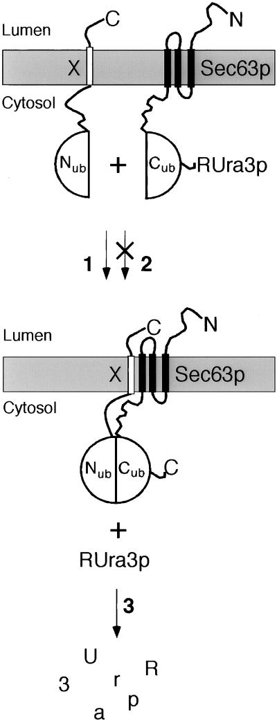
Sec63p was extended at its C terminus with Cub that was linked to an N-terminally modified version of the enzyme Ura3p (RUra3p) to create Sec63-Cub-RUra3p (Sec63CRUp) (Figures 1 and 2). Due to the topology of Sec63p, CRUp points into the cytosol of the cell (Feldheim et al., 1992). By coexpressing a set of Nub-fusion proteins (Nub-X in Figure 1), we first attempted to distinguish between Sec63p-interacting and -noninteracting proteins. Pathway 1: X is a protein that strongly interacts with Sec63p. Nub and Cub reassemble to the quasi-native Ub, and RUra3p is cleaved by the UBPs. Since the N-terminal residue of the released RUra3p is an arginine, rapid degradation of RUra3p by the enzymes of the N-end rule ensures that the cells stop dividing on plates lacking uracil (Ura−). 5-FOA is converted by Ura3p into 5-fluorouracil, which is toxic for the cell. Therefore the rapid degradation of RUra3p due to the interaction between protein X and Sec63p allows the cells to grow on plates containing 5-FOA (FOAR) (Ghislain et al., 1996; Johnsson and Varshavsky, 1997; Varshavsky, 1997). Pathway 2: X is a protein that does not interact with Sec63p. The linked Nub and Cub do not or only partially reassemble to the quasi-native Ub. The cells retain sufficient unclipped Sec63CRUp to stay Ura+ and 5-FOA-sensitive (FOAS). As an alternative to the RUra3p reporter, Sec63p-Cub was extended by the enzyme dihydrofolate reductase that carries an ha tag at its C terminus (Sec63-Cub-Dha). The cleaved Dha remains stable in the cytosol and can be detected together with the unclipped fusion protein by immunoblotting with antibodies directed against the ha epitope (Johnsson and Varshavsky, 1994).
Figure 1.
The split-Ubiquitin technique and its application to the analysis of membrane proteins using a metabolic marker. Cub-RUra3p was linked to the C terminus of Sec63p, and Nub was linked to the N terminus of the membrane protein X. Pathway 1: Nub is coupled to a protein that binds to Sec63p. The complex brings Nub and Cub into close proximity. Nub and Cub reconstitute the quasi-native Ub that is cleaved by the Ub-specific proteases to release RUra3p from Cub. The cleaved RUra3p is targeted for rapid destruction by the enzymes of the N-end rule (3) to yield cells that are uracil auxotrophs and 5-FOA resistant. Pathway 2: Nub is linked to a protein that does not bind to Sec63p. The two fusion proteins do not improve the reconstitution of Nub and Cub into the quasi-native Ub. Thus, RUra3p stays linked to Sec63-Cub, and the cells are uracil prototrophs and 5-FOA sensitive.
Figure 2.
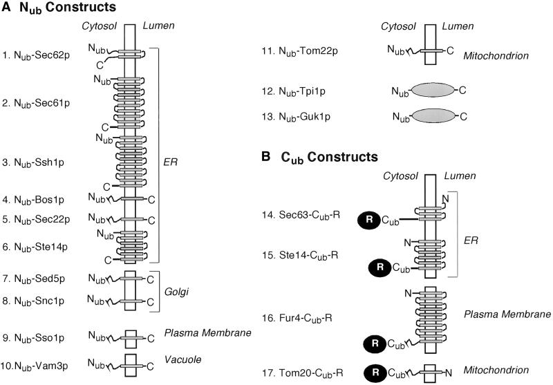
Nub and Cub fusions. (A) Nub (residues 1–36 of Ub) was fused to the N terminus of either a transmembrane protein (constructs 1–11) or a cytosolic protein (constructs 12–13). The N termini of all proteins are located in the cytosol. The orientation and the numbers of the membrane-spanning domains were obtained from published studies. The orientation of the N and the C terminus of Ste14p and its subcellular localization was a subject of this study. The Nub-attached proteins of constructs 1–5 are localized in the ER (Deshaies and Schekman, 1990; Shim et al., 1991; Finke et al., 1996; Wilkinson et al., 1996; Ballensiefen et al., 1998). The localization of the Nub-attached protein of construct 6 was a subject of this study. The Nub-attached protein of construct 7 resides in the early Golgi and of construct 8 in the late Golgi/plasma membrane (Protopopov et al., 1993; Banfield et al., 1994). The Nub-attached protein of construct 9 was shown to be in the plasma membrane (Aalto et al., 1993). The Nub-attached protein of construct 10 was found in the vacuole, and the Nub-attached protein of construct 11 was found in the outer membrane of the mitochondrion (Kiebler et al., 1993; Darsow et al., 1997; Wada et al., 1997; Srivastava and Jones, 1998). (B) Cub (residues 35–76 of Ub) was linked to the C terminus of a transmembrane protein and extended at its own C terminus by a reporter protein. The C termini of all proteins are localized in the cytosol. The information on the orientation of the N- and C-termini, the numbers of the membrane-spanning domains, and the localization of the unmodified proteins were obtained from published studies except for construct 15, where the number of membrane-spanning domains is still tentative. The Cub-attached protein of construct 14 is localized in the ER, that of construct 16 is found in the plasma membrane, and that of construct 17 is localized in the outer membrane of the mitochondrion (Jund et al., 1988; Feldheim et al., 1992; Moczko et al., 1997). The reporter (R) is RUra3p for the constructs 15–17 and RUra3p or DHFRha (Dha) for construct 14.
The Interaction between the Two Membrane Proteins, Sec62p and Sec63p, Can Be Monitored by the Split-Ub Assay In Vivo
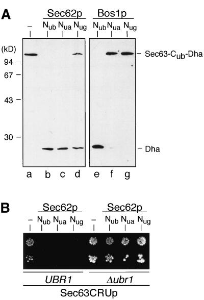
Sec63CRUp and Sec63-Cub-Dha were integrated into diploid cells via homologous recombination to replace one native copy of Sec63p. Tetrad analysis of the sporulated diploids validated that both Sec63-Cub-fusion proteins are functional (our unpublished observation). Since the two spores containing the modified versions of Sec63p grew slightly slower, the interaction assay was performed in diploid cells. To test the interaction between Sec62p and Sec63p, the Nub-moiety was linked to the cytosolic N-terminus of Sec62p (Figure 2). Nub-Sec62p is functional (Dünnwald et al., 1999). Immunoblot analysis of protein extracts from cells expressing Sec63-Cub-Dha together with Nub- or Nua-Sec62p showed that Sec63-Cub-Dha is completely converted into Sec63-Cub and Dha. Nug-Sec62p still induces more than 60% cleavage (Figure 3A). The ratio of cleaved to uncleaved Cub-Dha matches the ratio seen for the interaction between two correspondingly labeled Nub- and Cub-zipper proteins, reinforcing the interpretation of a tight interaction between Sec62p and Sec63p (Johnsson and Varshavsky, 1994). Bos1p, a membrane protein of the ER that does not interact with Sec63p, induces significant cleavage of Sec63-Cub-Dha when labeled with Nub, but hardly induces any cleavage when labeled with Nua or Nug (Figures 2 and 3A).
Figure 3.
Split-Ub monitors the interaction between Sec63p and Sec62p in vivo. (A) Immunoblot analysis of cells expressing Sec63-Cub-Dha together with an empty plasmid (lane a) or together with Nub-, Nua-, or Nug-Sec62p (lanes b, c, and d, respectively) or Nub-, Nua-, or Nug-Bos1p (lanes e, f, and g, respectively). The nitrocellulose membrane was probed with the anti-ha antibody that recognizes the uncleaved Cub fusion and the cleaved Dha. (B) Growth assay of the interaction between Sec63p and Sec62p based on split-Ub and a short-lived Ura3p (RUra3p) as a reporter. Sec63CRUp-containing cells bearing either the UBR1 gene or a UBR1 deletion were transformed with an empty plasmid or Nub-, Nua-, or Nug-Sec62p. Cells were pregrown in selective media containing uracil. Cells (103 or 102) were spotted on selective plates lacking uracil and also lacking leucine and tryptophan to select for the presence of the Cub- and Nub-constructs.
Cells harboring Sec63CRUp grow on medium lacking uracil. The same cells coexpressing Nub-, Nua- or Nug-Sec62p grow on medium containing uracil but fail to grow on medium lacking uracil (Figure 3B). To test whether this new phenotype of the Sec63CRUp containing cells is due to the ability of Nub-Sec62p to induce cleavage and the rapid degradation of RUra3p, we expressed the same Nub/Cub combination in congenic yeast cells harboring a deletion of UBR1 (Figure 3B). UBR1 encodes the recognition component of the N-end rule pathway, and proteins bearing destabilizing N-terminal residues that are rapidly degraded in wild-type cells are stabilized in Δubr1 cells (Bartel et al., 1990). Since Δubr1 cells carrying Nub-Sec62p and Sec63CRUp are still Ura+, we conclude that in wild-type cells bearing Sec63CRUp, Nub-Sec62p causes the cleavage and degradation of RUra3p.
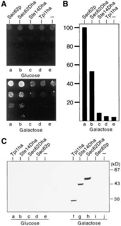
The measured proximity between Nub-Sec62p and Sec63CRUp is a strong indicator, albeit not proof, that Sec63p and Sec62p are components of one protein complex. If the efficient reassociation of Nug-Sec62p and Sec63CRUp is a consequence of a direct protein interaction, overexpression of the unlabeled Sec62p should displace its Nub-labeled counterpart in the complex. As a consequence, the local concentration between Nub-Sec62p and Sec63CRUp will decrease, less RUra3p will be cleaved, and the cells will start to grow on plates lacking uracil. We expressed the unmodified Sec62p and a Sec62p derivative that carries the Dha extension at its C terminus (Sec62-Dha) from the inducible PGAL1-promoter in the presence of Nug-Sec62p and Sec63CRUp. The triply transformed cells were spotted on plates lacking uracil that either contained glucose to repress or contained galactose to induce the expression of Sec62p or Sec62-Dha. The growth of the cells on plates that lacked uracil but contained galactose confirmed the displacement of Nug-Sec62p by Sec62p or Sec62-Dha (Figure 4A). To verify the specificity of this experiment, the competition was repeated with the membrane protein Ste14p and the cytosolic Triose phosphate isomerase (Tpi1p) that were expressed from the PGAL1-promoter and C-terminally extended by the Dha module (Ste14-Dha) or the ha-epitope (Tpi1-ha). Dha and ha served in these constructs as a tag to allow the immunodetection of the correspondingly labeled proteins. In contrast to the expression of Sec62p or Sec62-Dha, the overexpression of Ste14-Dha and Tpi1-ha had no effect on the growth of the cells harboring Sec63CRUp and Nug-Sec62p (Figure 4A). Immunoblots confirmed the expression of all ha-bearing proteins (Figure 4C), and a Sec62p-specific antibody confirmed the expression of the PGAL1-driven Sec62p (our unpublished observation). Using the Sec62p-specific antibody, we could also demonstrate that the expression of Nug-Sec62p was not influenced by galactose (our unpublished observation). To semiquantitatively measure the influence of Sec62p overexpression on the interaction between Nug-Sec62p and Sec63CRUp, roughly 10,000 cells were plated on galactose-containing medium without uracil, and the yeast colonies were counted after 4 d (Figure 4B). Approximately 800 colonies were recovered upon overexpression of Sec62p, and 400 colonies were recovered upon overexpression of Sec62-Dha, suggesting that the extension at the C terminus of Sec62p might already interfere with the ability of the molecule to interact with Sec63p. Around 30 colonies were recovered from yeast cells carrying the empty PGAL1-promoter, and an average of 60 and 40 colonies were recovered upon coexpression of Ste14-Dha and Tpi1-Dha. The competition of Nug-Sec62p by Sec62p shows that the split-Ub measured proximity between Sec62p and Sec63p is a consequence of both proteins being components of one protein complex.
Figure 4.
The measured proximity between Sec62p and Sec63p is due to both proteins being in one complex. (A) Cells bearing Sec63CRUp and Nug-Sec62p were transformed with a plasmid containing either Sec62p, Sec62Dha, Ste14Dha, Tpi1ha, or an empty plasmid, all under the control of the PGAL1-promoter (lanes a–e). Approximately 105, 104, 103, and 102 cells were spotted on selective media lacking uracil and containing either glucose to repress or galactose to induce the PGAL1 promoter. (B) S. cerevisiae cells (104) were plated as described in panel A on selective media containing galactose and lacking uracil, and colonies were counted after 4 d. The average of seven independent experiments is shown. Approximately 800 colonies were recovered upon overexpression of Sec62p. This number was arbitrarily set as 100. (C) Overexpression of the ha epitope-bearing proteins was confirmed by immunoblot analysis of extracts of S. cerevisiae cells coexpressing Sec63CRUp, Nug-Sec62p, and the following constructs: Tpi1ha (lanes a and f), Ste14Dha (lanes b and g), Sec62Dha (lanes c and h), Sec62p (lanes d and i), and empty vector (lanes e and j). Cells were grown in glucose (lanes a–e) to repress and grown in galactose (lanes f–j) to induce the expression of the proteins.
The Response in the Split-Ub Assay Correlates with the Distance of the Unlabeled Protein to Sec63p
Every protein displays a characteristic spectrum of local concentrations toward the other proteins inside the cell. Split-Ub allows comparison of the local concentrations that exist between different Nub-labeled proteins and a common Cub-fusion. The proteins of high local concentration will need a Nub with a lower affinity to Cub to achieve Nub-Cub reassembly than the proteins of low local concentration. The RUra3p reporter will translate these differences into the growth rate of the yeasts. Cells harboring a Nub-labeled protein that is close to a CRUp-fusion do not grow or grow slower than cells carrying a Nub-labeled protein that is more distant. We started to map the spectrum of local concentrations of Sec63p by comparing the interactions of Sec63CRUp with 13 different Nub-, Nua-, and Nug fusions. The proteins were chosen to cover a wide range of local concentrations by predominantly selecting membrane proteins, whose distances to Sec63p are adjusted by their distinct distribution in the cell. Sec61p as a member of the heptameric Sec complex should be very close, whereas Tom22p as a membrane protein of the outer mitochondrial membrane should be very distant to Sec63p. The topology of all Nub-modified proteins and the cellular localization of the unmodified proteins are shown in Figure 2. Since the local concentration of two proteins is influenced by their amount and their cellular distribution, we tried to minimize the differences in total amount by expressing all Nub-fusions from the noninduced PCUP1-promotor.
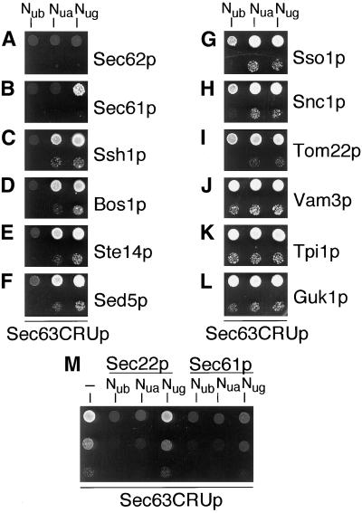
The different growth of the transformed cells on SD-ura allows us to clearly separate the Nub constructs of the two known Sec63p-interacting proteins, Sec62p and Sec61p, from all the other Nub constructs (Figure 5 and Table 1). The Nub and Nua constructs of both proteins completely inhibit the growth of the Sec63CRUp-bearing cells. The Nug construct inhibits growth in the case of Sec62p and strongly impairs growth in the case of Sec61p. Sec63CRUp-containing cells transformed with any other Nug construct show unimpaired growth on media lacking uracil. Furthermore, the assay allows us to distinguish between the Nub constructs of those proteins that do not bind to Sec63p (Figure 5 and Table 1). According to the growth of the transformed yeasts, we could arrange the Nub constructs into five groups of decreasing proximity to Sec63p. The classification approximately reflects the localization of the unlabeled proteins (see Figure 1 and Table 1). Groups 1 and 2 comprise the Sec63p-binding proteins Sec62p and Sec61p.
Figure 5.
Split Ub measures the proximity between Sec63p and membrane-associated proteins in vivo. Sec63CRUp containing cells expressing Nub, Nua, and Nug constructs of Sec62p (A), Sec61p (B), Ssh1p (C), Bos1p (D), Ste14p (E), Sed5p (F), Sso1p (G), Snc1p (H), Tom22p(I), Vam3p (J), Tpi1p (K), and Guk1p (L) were spotted (105 and 103 cells) on selective media lacking uracil (A–M) and leucine and histidine (A and D) or leucine and tryptophan (B, C, and E–M) to select for the presence of the Cub and Nub constructs. (M) Sec63CRUp-containing cells bearing either the empty plasmid, Nub-, Nua-, -Nug-Sec22p or Nub-, Nua-, Nug-Sec61p were spotted (105, 104, 103 cells) on plates lacking uracil. Cells were grown for 4 d.
Table 1.
Growth of cells containing Sec63CRUp and different Nub constructs
| Protein | Nub | Nua | Nug | FOA | Group |
|---|---|---|---|---|---|
| Sec62p | − | − | − | R | 1 |
| Sec61p | − | − | + | R | 2 |
| Sec22p | − | (+) | +++ | R | 3 |
| Ssh1p | − | ++ | +++ | S | 3 |
| Bos1p | − | ++ | +++ | S | 3 |
| Ste14p | − | ++ | +++ | S | 3 |
| Sed5p | (+) | ++ | +++ | S | 3 |
| Sso1p | + | +++ | +++ | S | 4 |
| Snc1p | + | +++ | +++ | S | 4 |
| Tom22p | + | ++ | +++ | ND | 4 |
| Vam3p | +++ | +++ | +++ | S | 5 |
| Tpi1p | +++ | +++ | +++ | S | 5 |
| Guk1p | +++ | +++ | +++ | S | 5 |
Growth was scored on plates lacking uracil. The number of pluses denotes the robustness of the growth of the colonies. The column FOA indicates the behavior of the corresponding Nua construct-bearing cells on plates containing 5-FOA. R, the cells are 5-FOA resistant and grow; S, the cells are 5-FOA sensitive.
Group 3 includes the proteins whose Nub constructs abolish the growth of Sec63CRUp cells, whose Nua constructs inhibit their growth to varying degrees but whose Nug constructs allow full growth on media lacking uracil (Figure 5 and Table 1). Group 3 includes the proteins Ssh1p, Bos1p, Ste14p, Sec22p, and Sed5p (Figure 5 and Table 1). Sec22p, Bos1p, and Ssh1p localize in the ER, whereas Sed5p resides in the early Golgi, the compartment that is functionally adjacent to the ER (Shim et al., 1991; Hardwick and Pelham, 1992; Banfield et al., 1994; Finke et al., 1996; Ballensiefen et al., 1998).
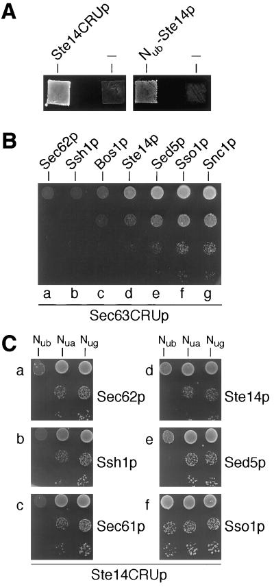
In contrast to all the other analyzed proteins, the localization and topology of Ste14p were unknown when we started its analysis. STE14 encodes an enzyme that methylates the C terminus of the CAAX box motif-containing proteins such as the small GTPases, Ras1p, Cdc42p, or Rho1p (Sapperstein et al., 1994; Zhang and Casey, 1996). The corresponding activity in mammalian cells was shown to be associated with a microsomal membrane fraction (Stephenson and Clarke, 1990). Functionality of Nub-Ste14p was confirmed by complementing the mating defect of a STE14 deletion strain (Figure 6A). Nub-Ste14p induces the cleavage of Cubs that are localized in the cytosol, implying that the N terminus of the protein is in the cytosol of the cell (Figure 5; Dünnwald et al., 1999). Since the interaction between Nub-Ste14p and Sec63CRUp is comparable to the interactions of the correspondingly labeled Bos1p, Ssh1p, and Sed5p, Ste14p might be localized in the ER, the Golgi, or in both compartments. To better resolve the localization of Ste14p, we had to search for a Nub mutant whose affinity to Cub falls between the affinities of wild-type Nub and Nua. This was accomplished by exchanging isoleucine 3 of Nub against a valine (Nvi) (Eckert, Raquet, and Johnsson, unpublished observation). Figure 6B shows the growth of the Sec63CRUp- containing cells transformed with Nvi-Sec62p, -Ssh1p, -Bos1p, -Ste14p, -Sed5p, -Sso1p, and -Snc1p. Nvi increases the resolution among the proteins of group 3. Specifically we can clearly separate Sed5p from the known membrane proteins of the ER. According to the growth of the Nvi-transformed Sec63CRUp-containing cells, Sec63p is closer to Ssh1p and Bos1p than to Sed5p and still closer to Sed5p than to Sso1p or Snc1p. We conclude that Sed5p is situated between the ER proteins, Ssh1p and Bos1p, and the proteins of the late Golgi/plasma membrane, Snc1p and Sso1p (Aalto et al., 1993; Protopopov et al., 1993). Our analysis places Ste14p between Bos1p and Sed5p.
Figure 6.
(A) Nub and Cub constructs of Ste14p are functional. Nub-Ste14p and Ste14CRUp were expressed in cells containing a STE14 deletion and mated with an appropriate tester strain of the opposite mating type. The mated cells were patched on media selecting for the formation of diploids. (B) Ste14p is located between Bos1p and Sed5p. Sec63CRUp containing cells expressing Nvi-Sec62p (a),-Ssh1p (b),-Bos1p (c),-Ste14p (d),-Sed5p (e),-Sso1p (f), and -Snc1p (g) were spotted (105, 104, 103, and 102 cells) on SD-ura plates that also lacked leucine and tryptophan to select for the presence of the Cub and Nvi constructs. Cells were grown for 3 d. (C) Sec62p, Ssh1p, and Sec61p are equidistant to Ste14p. Ste14CRUp-containing cells expressing Nub, Nua, and Nug constructs of Sec62p (a), Ssh1p (b), Sec61p (c), Ste14p (d), Sed5p (e), and Sso1p (f) were spotted (105, 103, and 102 cells) on selective media lacking uracil, leucine, and tryptophan and containing 500 μM methionine to reduce the expression of Ste14CRUp. Cells were grown for 3 d.
The faint growth of the Nvi-Bos1p–containing cells in the second dilution of Figure 6B may indicate a slightly closer proximity between Sec63p and Ssh1p than between Sec63p and Bos1p. Ssh1p is a homologue of Sec61p (Figure 2). Ssh1p was found in a heterotrimeric complex that is very similar to the trimeric Sec61 complex. However, unlike Sec61p, Ssh1p did not copurify with the Sec62/63p complex and was not coimmunoprecipitated with antibodies to members of the Sec62/63p complex (Finke et al., 1996). Does the inability to demonstrate interaction by these techniques reflect the situation in living cells or an inherent instability of this complex that causes its disruption during purification? By comparing the growth of the Sec63CRUp cells expressing Nua-Sec61p and Nua-Ssh1p, we conclude that Sec63p is closer to Sec61p than to Ssh1p in vivo (Figure 5 and Table 1). To confirm that the measured difference is specific and not caused by a general higher cellular activity of the Nua-Sec61p, we compared the two different Nub constructs toward a Cub landmark that is known not to interact with Sec61p or Ssh1p. We constructed a Ste14p derivative that bears the Cub-RUra3p module at its C terminus (Figure 2, Ste14CRUp). Ste14CRUp is functional (Figure 6A). The unimpaired growth of the Ste14CRUp-containing cells on media lacking uracil demonstrates that the Cub-RUra3p moiety most likely points into the cytosol of the cell (our unpublished observation). The nearly identical growth characteristics of the cells bearing Ste14CRUp and the Nubs of Sec62p, Sec61p, and Ssh1p document a comparable activity of the Nub fusion proteins (Figure 6C), i.e., no growth of Ste14CRUp cells bearing the Nub, reduced but significant growth of the cells bearing the Nua, and unimpaired growth of the cells bearing the Nug constructs. We conclude that the differences in the interaction between Nua-Sec62p, -Sec61p, -Ssh1p, and Sec63CRUp are real and reflect the differences in the interaction between the unlabeled molecules. Therefore, Ssh1p is a membrane protein of the ER but does not interact with Sec63p in vivo.
Figure 6C also shows that Ste14CRUp is closer to the Nub fusions of the ER than to the Nub fusions of any other compartment. Again, the difference between Nub-Ste14p and Nub-Sed5p is very subtle. However, we can discriminate between Sed5p and Ste14p more clearly by using the corresponding Nvis. Nvi-Ste14p is closer to Ste14CRUp than is Nvi-Sed5p (our unpublished observation). Nub-Sso1p and -Snc1p differ from the known Nub-labeled proteins of the ER and Nub-Sed5p by permitting unimpaired growth of the Ste14CRUp-containing cells (Figure 6C and our unpublished observation).
Characterizing Proteins That Are Very Distant to Sec63p
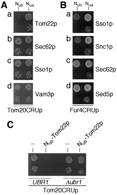
Group 4 includes the proteins whose Nub constructs impair, but do not abolish, the growth of the Sec63CRUp-containing cells. This group is very heterogeneous and thereby documents the increasing difficulty to assign a correct localization as the distance between the Cub landmark and the Nub protein gets larger (Figure 5 and Table 1). Tom22p is localized at the outer mitochondrial membrane, while Sso1p and Snc1p, a t- and v-SNARE, are localized at the plasma membrane and the late Golgi, respectively (Figure 2) (Aalto et al., 1993; Kiebler et al., 1993; Protopopov et al., 1993). We assumed that the assay could establish the correct localization of Nub-Tom22p, Nub-Snc1p, and Nub-Sso1p by selecting the appropriate Cub landmarks. To localize Tom22p, the Cub-RUra3p module was attached to the C terminus of Tom20p (Figure 2, Tom20CRUp). Tom20p and Tom22p are both subunits of the translocation complex of the outer mitochondrial membrane (Schatz, 1997). Tom20p has an N-terminal membrane anchor and a C-terminal domain pointing into the cytosol of the cell (Moczko et al., 1997). Nub-Tom22p strongly impairs the growth of Tom20CRUp-containing cells on medium lacking uracil, whereas all other Nub constructs have no influence (Figure 7A and our unpublished observation). This effect depends on a functional N-end rule pathway (Figure 7C). We conclude that Tom22p colocalizes with Tom20p at the outer mitochondrial membrane.
Figure 7.
Tom22p is close to Tom20p; Sso1p and Snc1p are close to Fur4p. (A) Tom20CRUp-containing S. cerevisiae cells expressing the Nub and Nua constructs of Tom22p (a), Sec62p (b), Sso1p (c), and Vam3p (d) were spotted (103 and 102 cells) on selective media lacking uracil. Cells were grown for 3 d. (B) Fur4CRUp containing S. cerevisiae cells expressing the Nub and Nua constructs of Sso1p (a), Snc1p (b), Sec62p (c), and Sed5p (d) were spotted (105 and 103 cells) on selective media lacking uracil. Cells were grown for 3 d. (C) Tom20CRUp-containing cells bearing the UBR1 gene or a UBR1 deletion were transformed with a plasmid harboring Nub-Tom22p or the empty vector pRS314. Cells (103 and 102) were spotted on selective media lacking uracil. Plates were incubated for 3 d.
To address the localization of Sso1p and Snc1p, we constructed Fur4CRUp (Figure 2). Fur4p belongs to the superfamily of membrane transporters, is localized in the plasma membrane, and transports uracil or 5-FOA across the membrane (Jund et al., 1988; Silve et al., 1991). The C terminus of the protein is very probably localized in the cytosol of the cell and is not important for the activity of the molecule (Jund et al., 1988). Yeast cells containing Fur4CRUp instead of the native Fur4p are still FOA sensitive, thereby demonstrating the functionality and indirectly the correct localization of the fusion protein (our unpublished observation). A subset of Nub and Nua constructs was transformed into the Fur4CRUp-expressing cells, and their growth on plates lacking uracil was scored. We observe a change in the order of proximity that was obtained for Sso1p, Snc1p, Sed5p, and Sec62p toward the Cub landmarks, Sec63p and Ste14p, of the ER. According to the growth of the Fur4CRUp-containing cells harboring the corresponding Nub constructs, Fur4p is closer to Sso1p and Snc1p than to Sed5p and Sec62p (Figure 7B). Nub-Sec62p inhibits the growth of the Fur4CRUp-containing cells slightly more than Nub-Sed5p (Figure 7B). Taken together, the activity of Nub-Sso1p and -Snc1p toward the landmarks, Fur4-, Sec63-, and Tom20-CRUp, is compatible with their localization at or close to the plasma membrane.
Group 5 includes the proteins Vam3p, Tpi1p, and Guk1p. Even the Nub constructs of these proteins do not significantly impair the growth of the Sec63CRUp-bearing cells (Figure 5 and Table 1). The Nub constructs of all three proteins were also tested against Tom20CRUp (Figure 7A for Vam3p), Fur4CRUp, and Ste14CRUp (our unpublished observation). The proteins of this group display no significant proximity to any of the three Cub landmarks. Tpi1p and Guk1p very probably have a homogenous distribution in the cytosol and therefore are equally distant from the tested landmarks. Vam3p, as a protein of the vacuole, is in a compartment that seems to be the least accessible to all three Cub fusions (Darsow et al., 1997; Wada et al., 1997; Srivastava and Jones, 1998).
In this modified form, split-Ub correlates close proximity between two proteins with poor growth of the transformed yeasts. The feature of Ura3p to transform the nontoxic 5-FOA to the toxic 5-fluorouracil makes it possible to reverse this correlation. The cells bearing Sec63CRUp and the Nub constructs that inhibit growth on plates lacking uracil should survive in the presence of the drug. We spotted the cells carrying Sec63CRUp and the different Nua-constructs onto 5-FOA containing plates. A summary of the growth assay is given in Table 1. As expected, the cells that do not grow or grow very poorly on medium lacking uracil display 5-FOA resistance, whereas the cells that survive on SD-ura are 5-FOA sensitive.
DISCUSSION
In this paper, we describe the molecular environment of a membrane protein in vivo. We attached Cub to the membrane protein Sec63p and measured the reassociation of the Cub moiety with different Nub-fusion proteins. The extent of cleavage at the C terminus of Cub reflects the local concentrations between the tested Nub constructs and the Cub landmark. By attaching RUra3p behind Cub we were able to translate this microscopic parameter into the growth rate of yeast cells bearing different Nub constructs.
Monitoring the Interactions between Members of the Sec Complex In Vivo
By using Nub constructs of proteins that are known not to interact with Sec63p as a reference, we were able to monitor the residence of Sec63p within the heptameric and the tetrameric Sec complex for the first time in vivo (Figures 3–5 and Table 1). Nug, the Nub mutant with the weakest affinity to Cub, revealed a lower reassociation efficiency of Sec63CRUp with Nug-Sec61p than with Nug-Sec62p. We propose that this difference reflects the higher stability of the tetrameric Sec62/63p complex compared with the heptameric Sec complex in vivo (Deshaies et al., 1991; Brodsky and Schekman, 1993; Panzner et al., 1995).
Ssh1p is by sequence closely related to Sec61p and shares some of the biochemical features of Sec61p (Finke et al., 1996). The direct comparison between the activities of Nua-Sec61p and Nua-Ssh1p toward Sec63CRUp showed that Ssh1p is a membrane protein of the ER, but does not bind to Sec63p in vivo (Figure 5 and Table 1). It is assumed that the presence of the tetrameric Sec62/63p complex enables the heptameric Sec complex to translocate proteins whose signal sequences guide them into the posttranslational pathway of translocation (Panzner et al., 1995; Ng et al., 1996). If Ssh1p constitutes a translocation pore, it should translocate a subset of those proteins that cross the membrane independently of Sec62/63p.
A Gradient of Local Concentrations of v- and t-SNARES Is Visualized by Split-Ub In Vivo
The Nub and the Nvi constructs of different t- and v-SNARES of the secretion pathway (reviewed by Rothman, 1994), revealed a gradient of local concentrations of the labeled proteins toward Sec63p that is compatible with the localization of the unlabeled molecules. Ordered by their decreasing local concentration, Sec22p is followed by Bos1p, Sed5p, Sso1p, and Snc1p, and finally Vam3p (Figures 5 and 6B and Table 1). Since proximity in this assay stems from the frequency of the encounters between the labeled molecules, the differences between Sed5p and Snc1p or Sso1p toward Sec63p are not trivial. To account for the higher frequency of encounters, Sed5p, Sec63p, or both molecules have to shuttle between the ER and the Golgi. The assay cannot distinguish which of the two molecules actually move, but a recent study showed that Sed5p indeed cycles through the ER (Wooding and Pelham, 1998). We therefore propose that the short-lived stay of Sed5p during its cycling through the ER accounts for its increased proximity toward Sec63p.
A Network of Cub Landmarks to Map Membrane Proteins
The relative distance to a Cub landmark can reveal the localization of a given Nub-fusion protein. The localization of the membrane protein Ste14p provided a first test. Ste14p is situated between Bos1p and Sed5p on our Sec63CRUp-derived linear distance map (Figure 6B). Bos1p and Sed5p are localized in the ER and the Golgi, respectively. The intermediate localization of Ste14p might be explained by its dynamic distribution between these two compartments. However, by additionally showing that Ste14CRUp behaves like a membrane protein of the ER, we propose that the main residence of the protein is the ER. During our study we became aware of a report that localized Ste14p in the ER and showed Ste14p to change its cellular distribution toward the Golgi once the N or the C terminus are extended artificially by an epitope tag (Romano et al., 1998). Our findings of an intermediate position of Ste14p can be therefore explained by a partial redistribution of Ste14p upon extending the N terminus with Nub or the C terminus with Cub. The extensions might mask a signal or a binding site and thereby interfere with the proper sorting of the molecule. However, both Ub modifications leave the protein functional. We conclude from the efficient reassociation of Nub-Ste14p and Ste14-Cub with different cytosolic Nub- and Cub-fusion proteins that both N and C termini of Ste14p are on the cytosolic side of the membrane. The fact that one of the modification enzymes of the isoprenylated proteins is localized in the ER should stimulate a closer look into the trafficking of these proteins.
The limit of using one Cub landmark to localize proteins became evident by our difficulty in distinguishing between Sso1p, Snc1p, and Tom22p on our Sec63p-derived distance map. We had to introduce two further Cub landmarks to resolve the localization of these proteins. Our assay confirmed that Nub-Sso1p and -Snc1p are closer to the plasma membrane protein Fur4CRUp than are the Nub constructs of Sec62p, Sed5p, Tom22p, Tpi1p, and Guk1p or the vacuolar Vam3p (Figure 7B and our unpublished results).
The growth of the cells containing Sec63CRUp and Nub-Sso1p or Nub-Snc1p on media lacking uracil is impaired (Figure 5 and Table 1). This can be explained by both Nub proteins being first integrated into the ER membrane before being transported to their final destination. Why does Nub-Tom22p show any interaction with Sec63CRUp? We suggest that the measured proximity between Sec63CRUp and Nub-Tom22p stems from a fraction of mislocalized Nub-Tom22p. Mislocalization of Tom22p into the ER might occur since its hydrophobic C-terminal tail is quite similar to the C-terminal membrane anchors of proteins that reside in the ER or other compartments of the secretion pathway. A more speculative interpretation invokes a specific proximity between the outer membrane of the mitochondrion and the membrane of the ER. The two membranes are sometimes seen adjacent to each other in electron microscopic pictures of cells. A functional proximity is postulated by certain models of lipid transfer between the two organelles (Paltauf et al., 1992; Ardail et al., 1993).
A Genetic Selection for Binding Partners of Cub-RUra3p–Labeled Proteins
A modification of the split-Ub assay based on the release of a transcription factor was recently introduced to monitor the interaction between the two proteins of the oligosaccharyl transferase complex, Wbp1p and Ost1p. The assembly of the Nub- and Cub-labeled proteins releases a Cub-linked transcription factor to enter the nucleus and to initiate the transcription of lacZ (Stagljar et al., 1998). The features of RUra3p as the reporter of the split-Ub assay make it possible to select for binding partners of Sec63CRUp or any other CRUp-labeled protein. The Nua constructs of Sec63p-binding proteins were identified by enabling cells to grow on medium containing 5-FOA (Table 1). Since the critical parameter in the split-Ub technique is the local concentration between the two Nub- and Cub-labeled molecules, the assay senses not only a protein’s direct interaction partners but also its near neighbors. Therefore, this parameter can be the source of false positives and negatives. False negatives are Nub constructs that show no specific proximity to any Cub landmark, although the unlabeled proteins form either a complex or are localized in the same compartment. Here inaccessibility of the coupled Nub, instability of the fusion protein, or mislocalization upon Nub labeling are some of the more obvious possibilities. A false positive suggests a proximity between a pair of Nub- and Cub-labeled proteins that does not exist for the unlabeled molecules. Nub-Sec22p is closer to Sec63p than any other Nub-labeled membrane protein of the ER that does not interact with Sec63p (Figure 5 and Table 1). This becomes most obvious when Nub-Sec22p is directly compared with Nub-Bos1p. Sec22p and Bos1p are both v-SNAREs involved in the vesicular transport between the ER and the Golgi (Figure 2). Both proteins have a very similar topology and were both expressed from the heterologous PCUP1 promotor yet show a different interaction with Sec63CRUP (Table 1). To test whether the tighter interaction of Sec22p is specific for Sec63p, we measured both proteins against Ste14CRUP. Since Nub-Sec22p displays also a closer proximity toward Ste14CRUP, we conclude that its proximity toward Sec63p is due to a higher nonspecific activity of the Sec22p-coupled Nub (our unpublished observation). The 5-FOA resistance of the Sec63CRUP cells harboring Nua-Sec22p emphasizes the need for additional assays to confirm a Nub-labeled protein as a true binding partner of a Cub-fusion protein. The established competition assay will serve as a control that still operates in the frame of the split-Ub technique (Figure 4). Coprecipitation and similar techniques that are used as independent tests for the two-hybrid system should also be applied for proteins that are identified by the split-Ub technique. In addition, as more independent Cub landmarks become available, it will become easier to discriminate true interactions from false positives and negatives.
The selection for conditions, compounds, or proteins that disrupt a specific protein interaction is an interesting feature of the RUra3p reporter system. Starting with a pair of Nub- and Cub-labeled proteins that inhibit growth on media lacking uracil, any compound interfering with this interaction can be identified by its capacity to induce colony-forming cells. We confirmed the feasibility of this approach by overexpressing unmodified Sec62p in the presence of Nug-Sec62p and Sec63CRUp.
A related scheme to search for molecules interfering with a given protein interaction was devised on the basis of the two-hybrid system (Vidal et al., 1996; Huang and Schreiber, 1997). However, this assay is limited to interactions that can be reconstituted in the nucleus. The split-Ub system makes it possible to extend this approach to the analysis of membrane proteins or other proteins whose interactions cannot be reconstituted in the nucleus.
Table 2.
Yeast strains
| Strain | Relevant genotype | Source/comment |
|---|---|---|
| JD53 | MATα his3-Δ200 leu2-3,112 lys2-801 trp1-Δ63 ura3-52 | Dohmen et al., 1995 |
| NJY73-I | MATα his3-Δ200 leu2-3,112 lys2-801 trp1-Δ63 ura3-52 NUB-BOS1::pRS303 | Derivative of JD53 |
| NJY73-A | MATα his3-Δ200 leu2-3,112 lys2-801 trp1-Δ63 ura3-52 NUA-BOS1::pRS303 | Derivative of JD53 |
| NJY73-G | MATα his3-Δ200 leu2-3,112 lys2-801 trp1-Δ63 ura3-52 NUG-BOS1::pRS303 | Derivative of JD53 |
| NJY73-VI | MATα his3-Δ200 leu2-3,112 lys2-801 trp1-Δ63 ura3-52 NUVI-BOS1::pRS304 | Derivative of JD53 |
| NJY61-I | MATα his3-Δ200 leu2-3, 112 lys2-801 trp1-Δ63 ura3-52 NUB-SEC61::pRS304 | Derivative of JD53 |
| NJY61-A | MATα his3-Δ200 leu2-3,112 lys2-801 trp1-Δ63 ura3-52 NUA-SEC61::pRS304 | Derivative of JD53 |
| NJY61-G | MATα his3-Δ200 leu2-3,112 lys2-801 trp1-Δ63 ura3-52 NUG-SEC61::pRS304 | Derivative of JD53 |
| NJY78-I | MATα his3-Δ200 leu2-3,112 lys2-801 trp1-Δ63 ura3-52 NUB-SSH1::pRS304 | Derivative of JD53 |
| NJY78-A | MATα his3-Δ200 leu2-3,112 lys2-801 trp1-Δ63 ura3-52 NUA-SSH1::pRS304 | Derivative of JD53 |
| NJY78-G | MATα his3-Δ200 leu2-3,112 lys2-801 trp1-Δ63 ura3-52 NUG-SSH1::pRS304 | Derivative of JD53 |
| NJY78-VI | MATα his3-Δ200 leu2-3,112 lys2-801 trp1-Δ63 ura3-52 NUVI-SSH1::pRS304 | Derivative of JD53 |
| NJY79RU | MATa/α his3-Δ200/his3-Δ200 leu2-3,112/leu2-3,112 lys2-801/lys2-801 trp1-Δ63/trp1-Δ63 ura3-52/ura3-52 SEC63/SEC63-CUB-RURA3::pRS305 | Derivative of JD51 |
| NJY79DH | MATa/α his3-Δ200/his3-Δ200 leu2-3,112/leu2-3,112 lys2-801/lys2-801 trp1-Δ63/trp1-Δ63 ura3-52/ura3-52 SEC63/SEC63-CUB-DHA::pRS305 | Derivative of JD51 |
| NJY80RU | MATα his3-Δ200 leu2-3,112 lys2-801 trp1-Δ63 ura3-52 SEC63-CUB-RURA3:::pRS305 | Derivative of JD53 |
| NJY80DH | MATα his3-Δ200 leu2-3,112 lys2-801 trp1-Δ63 ura3-52 SEC63-CUB-DHA::pRS305 | Derivative of JD53 |
| NJY81RU | MATα his3-Δ200 leu2-3,112 lys2-801 trp1-Δ63 ura3-52 SEC63-CUB-RURA3::pRS305 UBR1::HIS3 | Derivative of JD55 |
| Ghislain et al., 1996 | ||
| NYJ82 | MATα his3-Δ200 leu2-3,112 lys2-801 trp1-Δ63 ura3-52 FUR4-CUB-RURA3::pRS303 | Derivative of JD53 |
| NJY83 | MATα ade2-1 his3-11.3-15 trp1-1 ura3-1 can1-100 STE14::kanr | Derivative of W303 |
ACKNOWLEDGMENTS
We thank David Banfield, Jürgen Dohmen, Walther Mothes, Tom Rapoport, Hans Ronne, and Randy Schekman for the gifts of plasmids, yeast strains, and antisera; Sonja Kind for cloning the TOM22 and TOM20 constructs; Jörg H. Eckert and Xavier Raquet for constructing and testing the Nvi-mutant; and G. Dues, J.H. Eckert, V. Gerke, X. Raquet, and D. Schmitt for critically reading the manuscript. We thank D. Schmitt for providing the plasmids harboring Nub-Sec22p. This work was supported by grant 0311107 to N.J. from the Bundesministerium für Bildung, Wissenschaft, Forschung und Technologie. N.L. was supported by a stipend from the Max-Planck-Society.
REFERENCES
- Aalto MK, Ronne H, Keranen S. Yeast syntaxins Sso1p and Sso2p belong to a family of related membrane proteins that function in vesicular transport. EMBO J. 1993;12:4095–4104. doi: 10.1002/j.1460-2075.1993.tb06093.x. [DOI] [PMC free article] [PubMed] [Google Scholar]
- Ardail D, Gasnier F, Lerme F, Simonot C, Louisot P, Gateau-Roesch O. Involvement of mitochondrial contact sites in the subcellular compartmentalization of phospholipid biosynthetic enzymes. J Biol Chem. 1993;268:25985–25992. [PubMed] [Google Scholar]
- Banfield DK, Lewis MJ, Rabouille C, Warren G, Pelham HR. Localization of Sed5, a putative vesicle targeting molecule, to the cis-Golgi network involves both its transmembrane and cytoplasmic domains. J Cell Biol. 1994;127:357–371. doi: 10.1083/jcb.127.2.357. [DOI] [PMC free article] [PubMed] [Google Scholar]
- Ballensiefen W, Ossipov D, Schmitt D. Recycling of the yeast v-SNARE Sec22p involves COPI-proteins and the ER transmembrane proteins Ufe1p and Sec20p. J Cell Sci. 1998;111:1507–1520. doi: 10.1242/jcs.111.11.1507. [DOI] [PubMed] [Google Scholar]
- Bartel B, Wünning I, Varshavsky A. The recognition component of the N-end rule pathway. EMBO J. 1990;9:3179–3189. doi: 10.1002/j.1460-2075.1990.tb07516.x. [DOI] [PMC free article] [PubMed] [Google Scholar]
- Beckmann R, Bubeck D, Grassucci R, Penczek P, Verschoor A, Blobel G, Frank J. Alignment of conduits for the nascent polypeptide chain in the ribosome-Sec61 complex. Science. 1997;278:2123–2126. doi: 10.1126/science.278.5346.2123. [DOI] [PubMed] [Google Scholar]
- Brodsky JL, Schekman R. A Sec63p-BiP complex from yeast is required for protein translocation in a reconstituted proteoliposome. J Cell Biol. 1993;123:1355–1363. doi: 10.1083/jcb.123.6.1355. [DOI] [PMC free article] [PubMed] [Google Scholar]
- Darsow T, Rieder SE, Emr SD. A multispecificity syntaxin homologue, Vam3p, essential for autophagic and biosynthetic protein transport to the vacuole. J Cell Biol. 1997;138:517–529. doi: 10.1083/jcb.138.3.517. [DOI] [PMC free article] [PubMed] [Google Scholar]
- Deshaies RJ, Sanders SL, Feldheim DA, Schekman R. Assembly of yeast Sec proteins involved in translocation into the endoplasmic reticulum into a membrane-bound multisubunit complex. Nature. 1991;349:806–808. doi: 10.1038/349806a0. [DOI] [PubMed] [Google Scholar]
- Deshaies RJ, Schekman R. Structural and functional dissection of Sec62p, a membrane-bound component of the yeast endoplasmic reticulum protein import machinery. Mol Cell Biol. 1990;10:6024–6035. doi: 10.1128/mcb.10.11.6024. [DOI] [PMC free article] [PubMed] [Google Scholar]
- Dohmen RJ, Stappen R, McGrath JP, Forrova H, Kolarov J, Goffeau A, Varshavsky A. An essential yeast gene encoding a homolog of ubiquitin-activating enzyme. J Biol Chem. 1995;270:18099–18109. doi: 10.1074/jbc.270.30.18099. [DOI] [PubMed] [Google Scholar]
- Dünnwald M, Varshavsky A, Johnsson N. Detection of transient interactions between substrate and transporter during protein translocation into the endoplasmic reticulum. Mol Biol Cell. 1999;10:329–344. doi: 10.1091/mbc.10.2.329. [DOI] [PMC free article] [PubMed] [Google Scholar]
- Feldheim D, Rothblatt J, Schekman R. Topology and functional domains of Sec63p, an endoplasmic reticulum membrane protein required for secretory protein translocation. Mol Cell Biol. 1992;12:3288–3296. doi: 10.1128/mcb.12.7.3288. [DOI] [PMC free article] [PubMed] [Google Scholar]
- Finke K, Plath K, Panzner S, Prehn S, Rapoport TA, Hartmann E, Sommer T. A second trimeric complex containing homologs of the Sec61p complex functions in protein transport across the ER membrane of S. cerevisiae. EMBO J. 1996;15:1482–1494. [PMC free article] [PubMed] [Google Scholar]
- Ghislain M, Dohmen RJ, Levy F, Varshavsky A. Cdc48p interacts with Ufd3p, a WD repeat protein required for ubiquitin-mediated proteolysis in Saccharomyces cerevisiae. EMBO J. 1996;15:4884–4899. [PMC free article] [PubMed] [Google Scholar]
- Görlich D, Prehn S, Hartmann E, Kalies KU, Rapoport TA. A mammalian homolog of SEC61p and SECYp is associated with ribosomes and nascent polypeptides during translocation. Cell. 1992;71:489–503. doi: 10.1016/0092-8674(92)90517-g. [DOI] [PubMed] [Google Scholar]
- Güldener U, Heck S, Fielder T, Beinhauer J, Hegemann JH. A new efficient gene disruption cassette for repeated use in budding yeast. Nucleic Acids Res. 1996;24:2519–2524. doi: 10.1093/nar/24.13.2519. [DOI] [PMC free article] [PubMed] [Google Scholar]
- Hardwick KG, Pelham HR. SED5 encodes a 39-kDa integral membrane protein required for vesicular transport between the ER and the Golgi complex. J Cell Biol. 1992;119:513–521. doi: 10.1083/jcb.119.3.513. [DOI] [PMC free article] [PubMed] [Google Scholar]
- Huang J, Schreiber SL. A yeast genetic system for selecting small molecule inhibitors of protein-protein interactions in nanodroplets. Proc Natl Acad Sci USA. 1997;94:13396–13401. doi: 10.1073/pnas.94.25.13396. [DOI] [PMC free article] [PubMed] [Google Scholar]
- Johnsson N, Varshavsky A. Split ubiquitin as a sensor of protein interactions in vivo. Proc Natl Acad Sci USA. 1994;91:10340–10344. doi: 10.1073/pnas.91.22.10340. [DOI] [PMC free article] [PubMed] [Google Scholar]
- Johnsson N, Varshavsky A. Split Ubiquitin: A sensor of protein interactions in vivo. In: Bartel PL, Fields S, editors. The Yeast Two Hybrid System. Oxford, UK: Oxford University Press; 1997. pp. 316–332. [Google Scholar]
- Jund R, Weber E, Chevallier MR. Primary structure of the uracil transport protein of Saccharomyces cerevisiae. Eur J Biochem. 1988;171:417–424. doi: 10.1111/j.1432-1033.1988.tb13806.x. [DOI] [PubMed] [Google Scholar]
- Kiebler M, Keil P, Schneider H, van der Klei IJ, Pfanner N, Neupert W. The mitochondrial receptor complex: a central role of MOM22 in mediating preprotein transfer from receptors to the general insertion pore. Cell. 1993;74:483–492. doi: 10.1016/0092-8674(93)80050-o. [DOI] [PubMed] [Google Scholar]
- Michaelis S, Herskowitz I. The a-factor pheromone of Saccharomyces cerevisiae is essential for mating. Mol Cell Biol. 1988;8:1309–1318. doi: 10.1128/mcb.8.3.1309. [DOI] [PMC free article] [PubMed] [Google Scholar]
- Moczko M, Bömer U, Kübrich M, Zufall N, Hönlinger A, Pfanner N. The intermembrane space domain of mitochondrial Tom22 functions as a trans binding site for preproteins with N-terminal targeting sequences. Mol Cell Biol. 1997;17:6574–6584. doi: 10.1128/mcb.17.11.6574. [DOI] [PMC free article] [PubMed] [Google Scholar]
- Ng DT, Brown JD, Walter P. Signal sequences specify the targeting route to the endoplasmic reticulum membrane. J Cell Biol. 1996;134:269–278. doi: 10.1083/jcb.134.2.269. [DOI] [PMC free article] [PubMed] [Google Scholar]
- Ng DT, Walter P. ER membrane protein complex required for nuclear fusion. J Cell Biol. 1996;132:499–509. doi: 10.1083/jcb.132.4.499. [DOI] [PMC free article] [PubMed] [Google Scholar]
- Paltauf F, Kohlwein S, Henry S. Regulation and compartmentalization of lipid synthesis in yeast. In: Jones EJ, Pringle J, Broach J, editors. The Molecular and Cellular Biology of the Yeast Saccharomyces: Gene Expression. Cold Spring Harbor, NY: Cold Spring Harbor Laboratory Press; 1992. pp. 415–500. [Google Scholar]
- Panzner S, Dreier L, Hartmann E, Kostka S, Rapoport TA. Posttranslational protein transport in yeast reconstituted with a purified complex of Sec proteins and Kar2p. Cell. 1995;81:561–570. doi: 10.1016/0092-8674(95)90077-2. [DOI] [PubMed] [Google Scholar]
- Plemper RK, Böhmler S, Bordallo J, Sommer T, Wolf DH. Mutant analysis links the translocon and BiP to retrograde protein transport for ER degradation. Nature. 1997;388:891–895. doi: 10.1038/42276. [DOI] [PubMed] [Google Scholar]
- Protopopov V, Govindan B, Novick P, Gerst JE. Homologs of the synaptobrevin/VAMP family of synaptic vesicle proteins function on the late secretory pathway in S. cerevisiae. Cell. 1993;74:855–861. doi: 10.1016/0092-8674(93)90465-3. [DOI] [PubMed] [Google Scholar]
- Rapoport TA, Jungnickel B, Kutay U. Protein transport across the eukaryotic endoplasmic reticulum and bacterial inner membranes. Annu Rev Biochem. 1996;65:271–303. doi: 10.1146/annurev.bi.65.070196.001415. [DOI] [PubMed] [Google Scholar]
- Romano JD, Schmidt WK, Michaelis S. The Saccharomyces cerevisiae prenylcysteine carboxyl methyltransferase Ste14p is in the endoplasmic reticulum membrane. Mol Biol Cell. 1998;9:2231–2247. doi: 10.1091/mbc.9.8.2231. [DOI] [PMC free article] [PubMed] [Google Scholar]
- Rothblatt JA, Deshaies RJ, Sanders SL, Daum G, Schekman R. Multiple genes are required for proper insertion of secretory proteins into the endoplasmic reticulum in yeast. J Cell Biol. 1989;109:2641–2652. doi: 10.1083/jcb.109.6.2641. [DOI] [PMC free article] [PubMed] [Google Scholar]
- Rothman JE. Mechanisms of intracellular protein transport. Nature. 1994;372:55–63. doi: 10.1038/372055a0. [DOI] [PubMed] [Google Scholar]
- Sapperstein S, Berkower C, Michaelis S. Nucleotide sequence of the yeast STE14 gene, which encodes farnesylcysteine carboxyl methyltransferase, and demonstration of its essential role in a-factor export. Mol Cell Biol. 1994;14:1438–1449. doi: 10.1128/mcb.14.2.1438. [DOI] [PMC free article] [PubMed] [Google Scholar]
- Schatz G. Just follow the acid chain. Nature. 1997;388:121–122. doi: 10.1038/40510. [DOI] [PubMed] [Google Scholar]
- Shim J, Newman AP, Ferro-Novick S. The BOS1 gene encodes an essential 27-kDa putative membrane protein that is required for vesicular transport from the ER to the Golgi complex in yeast. J Cell Biol. 1991;113:55–64. doi: 10.1083/jcb.113.1.55. [DOI] [PMC free article] [PubMed] [Google Scholar]
- Sikorski RS, Hieter P. A system of shuttle vectors and yeast host strains designed for efficient manipulation of DNA in Saccharomyces cerevisiae. Genetics. 1989;122:19–27. doi: 10.1093/genetics/122.1.19. [DOI] [PMC free article] [PubMed] [Google Scholar]
- Silve S, Volland C, Garnier C, Jund R, Chevallier MR, Haguenauer-Tsapis R. Membrane insertion of uracil permease, a polytopic yeast plasma membrane protein. Mol Cell Biol. 1991;11:1114–1124. doi: 10.1128/mcb.11.2.1114. [DOI] [PMC free article] [PubMed] [Google Scholar]
- Srivastava A, Jones EW. Pth1/Vam3p is the syntaxin homolog at the vacuolar membrane of Saccharomyces cerevisiae required for the delivery of vacuolar hydrolases. Genetics. 1998;148:85–98. doi: 10.1093/genetics/148.1.85. [DOI] [PMC free article] [PubMed] [Google Scholar]
- Stagljar I, Korostensky C, Johnsson N, te Heesen S. A genetic system based on split-ubiquitin for the analysis of interactions between membrane proteins in vivo. Proc Natl Acad Sci USA. 1998;95:5187–5192. doi: 10.1073/pnas.95.9.5187. [DOI] [PMC free article] [PubMed] [Google Scholar]
- Stephenson RC, Clarke S. Identification of a C-terminal protein carboxyl methyltransferase in rat liver membranes utilizing a synthetic farnesyl cysteine-containing peptide substrate. J Biol Chem. 1990;265:16248–16254. [PubMed] [Google Scholar]
- Varshavsky A. The N-end rule pathway of protein degradation. Genes Cells. 1997;2:13–28. doi: 10.1046/j.1365-2443.1997.1020301.x. [DOI] [PubMed] [Google Scholar]
- Vidal M, Brachmann RK, Fattaey A, Harlow E, Boeke JD. Reverse two-hybrid and one-hybrid systems to detect dissociation of protein-protein and DNA-protein interactions. Proc Natl Acad Sci USA. 1996;93:10315–10320. doi: 10.1073/pnas.93.19.10315. [DOI] [PMC free article] [PubMed] [Google Scholar]
- Wada Y, Nakamura N, Ohsumi Y, Hirata A. Vam3p, a new member of syntaxin related protein, is required for vacuolar assembly in the yeast Saccharomyces cerevisiae. J Cell Sci. 1997;110:1299–1306. doi: 10.1242/jcs.110.11.1299. [DOI] [PubMed] [Google Scholar]
- Walter P, Johnson AE. Signal sequence recognition and protein targeting to the endoplasmic reticulum membrane. Annu Rev Cell Biol. 1994;10:87–119. doi: 10.1146/annurev.cb.10.110194.000511. [DOI] [PubMed] [Google Scholar]
- Wilkinson BM, Critchley AJ, Stirling CJ. Determination of the transmembrane topology of yeast Sec61p, an essential component of the endoplasmic reticulum translocation complex. J Biol Chem. 1996;271:25590–25597. doi: 10.1074/jbc.271.41.25590. [DOI] [PubMed] [Google Scholar]
- Wooding S, Pelham HRB. The dynamics of Golgi protein traffic visualized in living yeast cells. Mol Biol Cell. 1998;9:2667–2680. doi: 10.1091/mbc.9.9.2667. [DOI] [PMC free article] [PubMed] [Google Scholar]
- Zhang FL, Casey PJ. Protein prenylation: molecular mechanisms and functional consequences. Annu Rev Biochem. 1996;65:241–269. doi: 10.1146/annurev.bi.65.070196.001325. [DOI] [PubMed] [Google Scholar]


