

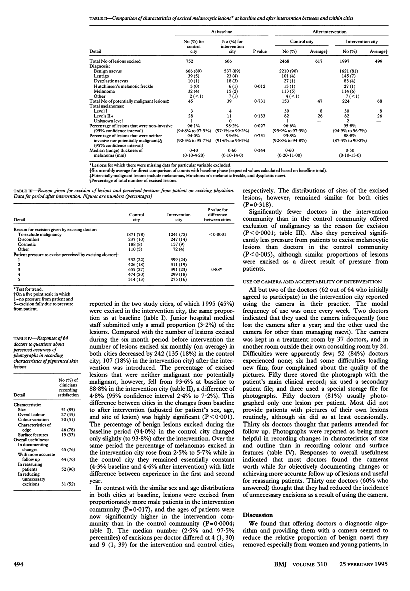
Abstract
OBJECTIVE--To evaluate an intervention designed to reduce the number of benign melanocytic lesions excised from the skin. DESIGN--A randomised controlled field trial based in the medical practices of two cities. Examination of histopathological reports of 5823 melanocytic skin lesions excised over the intervention period and in the preceding six months. INTERVENTION--Medical practitioners were offered an algorithm and use of an instant developing camera. SETTING AND SUBJECTS--Over 50 medical practitioners, mostly in general practice, in each of two cities in tropical Queensland, Australia. MAIN OUTCOME MEASURES--Percentages of benign (neither malignant nor potentially malignant) melanocytic lesions excised during the two year intervention period. RESULTS--There were no significant differences in the percentages of benign lesions reported in the intervention and control cities before the intervention started (93.6% and 94.0%, respectively), but there was a significant difference afterwards (88.8% and 93.8%, P < 0.001). There was no difference in the percentage of invasive melanomas excised per month in the intervention city (3.4%) compared with control city (3.4%). CONCLUSION--Clinical diagnostic accuracy may be enhanced by offering to clinicians managing suspicious melanocytic skin lesions a simple algorithm and a camera with which to record the appearance of lesions objectively.
Full text
PDF


Selected References
These references are in PubMed. This may not be the complete list of references from this article.
- Cooke K., McNoe B., Spears G. General practice consultations involving pigmented naevi presented for assessment of malignancy. N Z Med J. 1993 Nov 24;106(968):493–495. [PubMed] [Google Scholar]
- Curley R. K., Cook M. G., Fallowfield M. E., Marsden R. A. Accuracy in clinically evaluating pigmented lesions. BMJ. 1989 Jul 1;299(6690):16–18. doi: 10.1136/bmj.299.6690.16. [DOI] [PMC free article] [PubMed] [Google Scholar]
- DeCoste S. D., Stern R. S. Diagnosis and treatment of nevomelanocytic lesions of the skin. A community-based study. Arch Dermatol. 1993 Jan;129(1):57–62. [PubMed] [Google Scholar]
- Del Mar C., Green A., Cooney T., Cutbush K., Lawrie S., Adkins G. Melanocytic lesions excised from the skin: what percentage are malignant? Aust J Public Health. 1994 Jun;18(2):221–223. doi: 10.1111/j.1753-6405.1994.tb00232.x. [DOI] [PubMed] [Google Scholar]
- Green A., Martin N., McKenzie G., Pfitzner J., Quintarelli F., Thomas B. W., O'Rourke M., Knight N. Computer image analysis of pigmented skin lesions. Melanoma Res. 1991 Nov-Dec;1(4):231–236. doi: 10.1097/00008390-199111000-00002. [DOI] [PubMed] [Google Scholar]
- Kenet R. O., Kang S., Kenet B. J., Fitzpatrick T. B., Sober A. J., Barnhill R. L. Clinical diagnosis of pigmented lesions using digital epiluminescence microscopy. Grading protocol and atlas. Arch Dermatol. 1993 Feb;129(2):157–174. [PubMed] [Google Scholar]
- MacKie R. M. Clinical recognition of early invasive malignant melanoma. BMJ. 1990 Nov 3;301(6759):1005–1006. doi: 10.1136/bmj.301.6759.1005. [DOI] [PMC free article] [PubMed] [Google Scholar]