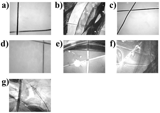
Abstract
This study compares the efficacy of six disaccharides and glucose for the preservation of solid supported lipid bilayers (SLBs) upon exposure to air. Disaccharide molecules containing an α,α-(1→1) linkage, such as α,α-trehalose and α,α-galacto-trehalose, were found to be effective at retaining bilayer structure in the absence of water. These sugars are known to crystallize in a clam shell conformation. Other saccharides, which are found to crystallize in more open structures, did not preserve the SLB structure during the drying process. These included the non-reducing sugar, sucrose, as well as maltose, lactose and the monosaccharide, glucose. In fact, even close analogs to α,α-trehalose, such as α,β-trehalose, which connects its glucopyranose rings via a (1→1) linkage in an axial, equatorial fashion, permitted nearly complete delamination and destruction of supported bilayers upon exposure to air. Lipids with covalently attached sugar molecules such as ganglioside GM1, lactosyl phosphatidylethanolamine, and glucosylcerebroside were also ineffective at preserving bilayer structure. The liquid crystalline-to-gel phase transition temperature of supported phospholipid bilayers was tested in the presence of sugars in a final set of experiments. Only α,α-trehalose and α,α-galacto-trehalose depressed the phase transition temperature, while the introduction of other sugar molecules into the bulk solution caused the phase transition temperature of the bilayer to increase. These results point to the importance of the axial-axial linkage of disaccharides for preserving supported lipid bilayer structure.
Introduction
While water is a biological necessity, anhydrobiotic organisms can persist in a dehydrated state for long periods of time.1 Ultimate survival depends on their ability to maintain cell integrity upon drying. A central concern is that the cell’s lipid membrane can be damaged by the drying process.2 To overcome this problem, many organisms employ specialized proteins or synthesize large amounts of di- and tri-saccharides, which upon synthesis, are exported to the extracellular side of the biomembrane.3 Perhaps the most common of these sugars are α,α-trehalose4 and sucrose,5 although nature sometimes employs glucose and larger fructans as lipopreservatives.6 It has been shown in a number of experiments that α,α-trehalose, a disaccharide consisting of two α-glucopyranose rings connected via a (1→1) glycosidic linkage, is particularly effective at preventing membrane damage upon drying in comparison to other sugar molecules.7 The question arises, therefore, as to whether the specific structure of this molecule leads to a distinct mechanism of anhydrobiotic preservation compared with other saccharides. This is especially relevant from a chemical point of view because no other naturally occurring disaccharide contains an α,α-(1→1) glycosidic linkage.
In vivo and in vitro8 experimentation provide key information about the combination of mechanisms by which disaccharides preserve membranes.9 These mechanisms include (a) the replacement of water by sugar molecules, (b) the formation of an amorphous sugar glass, and (c) the retention of water at the membrane interface. According to the first mechanism, sugar molecules such as trehalose should be able to replace water at the membrane surface during the drying process. In fact, several studies have shown that sugar molecules directly interact with the lipid headgroup causing, for example, a frequency shift in the infrared bands arising from the phosphate moiety.10–13 This is critical because the gel-to-liquid crystalline phase transition temperature, Tm, of the lipid membrane increases sharply upon drying. In fact, a hydrated liquid crystalline phase bilayer usually goes through its phase transition into the gel phase when dried at room temperature. This causes contraction, cracking, and fissure. The introduction of α,α-trehalose depresses the Tm value of the bilayer, thus allowing the system to remain in the liquid crystalline state upon drying at room temperature.14 This is believed to occur by the direct interaction (hydrogen bonding) of the disaccharide with the headgroups, which prevents the contraction of the surface area per lipid headgroup.15, 16
A second critical influence of the sugar involves the formation of a vitrified coating at the membrane surface upon drying.11, 17 It has been argued that such an amorphous sugar glass can suppress the Tm of the dried bilayer.18, 19 This should occur because the solid sugar coating is rigid and, hence, hinders volumetric changes (i.e. expansion and contractions) that the lipid membrane must undergo at the phase transition. Significantly, α,α-trehalose has a higher glass transition temperature, Tg, than other disaccharides.19 This is true both for pure α,α-trehalose as well as α,α-trehalose/water mixtures. Finally, it has been hypothesized that the sugar layer can help prevent water evaporation in a dehydrating environment.13
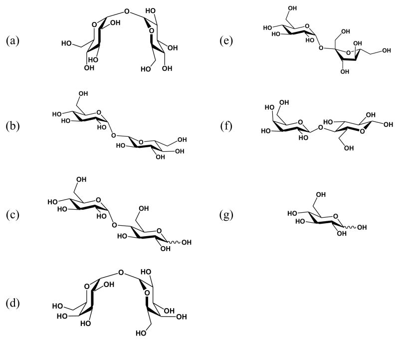
Despite physical evidence supporting the idea that a combination of mechanisms is probably involved in membrane preservation upon drying, an underlying chemical level understanding of anhydrobiosis has yet to be laid out. This is significant because the structures of various disaccharides are substantially different. As noted above, α,α-trehalose is unique among naturally occurring disaccharides in that its two glucopyranose rings are connected by their anomeric carbons in an axial, axial fashion. This type of bonding leads to the stability of a clam shell conformation relative to a more open conformation (Figure 1a). In fact, X-ray measurements have shown that α,α-trehalose crystallizes in the clam shell configuration.20 Also, the (1→1) connection makes trehalose a non-reducing sugar (i.e. the rings are not in equilibrium with their corresponding linear structures).
Figure 1.
Structure of the mono- and disaccharides employed in this study: (a) α,α-trehalose, (b) α,β-trehalose, (c) maltose, (d) α,α-galacto-trehalose, (e) sucrose, (f) lactose, and (g) glucose.
If two glucose moieties are linked by a (1→1) glycosidic linkage in an α,β (axial, equatorial) fashion, then the more open conformation will be highly favored on steric grounds.21 Such a molecule, called α,β-trehalose, is also a non-reducing sugar (Figure 1b). Maltose, which consists of two glucopyranose rings fused via a (1→4) linkage in an axial, equatorial fashion, should also adopt a more extended conformation (Figure 1c). This expectation is born out by X-ray crystal structure data.22 On the other hand, a predominantly clam shell configuration is obtained when two α-galactose molecules are attached via their anomeric carbons (Figure 1d).23 This molecule, α,α-galacto-trehalose, has not been found in nature and to the best of our knowledge has never been previously tested as a lipopreservative. Herein, this non-reducing sugar has been used to test the importance of the axial, axial linkage in anhydrobiosis.
Two additional molecules of interest in this study are sucrose and lactose. Sucrose consists of an α-D-glucopyranose ring linked to β-D-fructofuranose via a (1→2) glycosidic linkage (Figure 1e). This molecule is a non-reducing sugar and its rings are linked in an axial, equatorial fashion. As expected, the crystal structure of this molecule shows it adopts an open structure.24 Lactose consists of a galactose ring linked to a glucose ring in a (1→4) fashion. The rings are linked in an equatorial, equatorial manner and X-ray diffraction confirms that it crystallizes in an open conformation as shown in Figure 1f.25 Finally, glucose is a simple monosaccharide (Figure 1g).
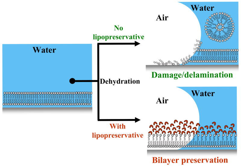
To date, the majority of sugar-stabilizing studies have either involved in vivo experimentation on cells4 or in vitro investigations of liposomes.14, 26 An alternative system for studying anhydrobiosis involves the use of supported phospholipid bilayers.27 SLBs are a useful model system that mimic the cell membrane and have been employed to investigate cellular processes such as multivalent ligand-receptor binding and cell signaling events.28–31 A very attractive property of these systems is that they exhibit the same two-dimensional fluidity found in native biomembranes.32–35 SLBs are unstable upon exposure to air since the lipids spontaneously rearrange themselves. This results in the delamination of the thin film from the solid support.36–45 It was therefore reasoned that the effectiveness of a lipopreservative against supported bilayer destruction could provide insight into the mechanism of anhydrobiosis (Figure 2).
Figure 2.
The dehydration of supported phospholipid membranes. Upper-Right Panel: In the absence of a lipopreservative the thin lipid film spontaneously reorganizes and delaminates from the solid support. Lower-Right Panel: The presence of a lipoprotectant suppresses damage and delamination.
Herein, a supported lipid bilayer platform was employed to study the protection of SLBs upon drying in the presence of the sugar molecules shown in Figure 1. These systematic studies of water removal in the presence of mono- and disaccharides demonstrated that trehalose was far superior to other common sugars for preserving the integrity of the supported bilayer upon drying. Most significantly, the data indicated that the α,α-(1→1) linkage of the trehalose molecule was the key to its efficacy. In fact, α,β-trehalose afforded almost no protection. On the other hand, replacing the two glucopyranose rings with galactopyranose rings, while maintaining the same α,α-(1→1) linkage, worked nearly as well as α,α-trehalose in preserving the long-range bilayer structure in the dry state. Furthermore, maltose, which has a high glass transition temperature, Tg,46 was capable of preventing delamination of the supported bilayer from the interface upon drying. However, the lipid material at the interface was completely immobile upon rehydration, which indicated that the bilayer structure was disrupted upon air exposure. Additional studies showed that α,α-trehalose depressed the liquid crystalline-to-gel phase transition temperature, Tm, of supported bilayers immersed in bulk water. α,α-Galacto-trehalose could also do this, although the magnitude of the depression was more modest. All other disaccharides, which lacked the axial-axial disaccharide linkage, caused the Tm value to increase. A final set of control experiments revealed that the presence of glycolipids in the membrane did not afford protection against the destruction of the bilayer upon drying.
Experimental
Materials
1-Palmitoyl-2-oleoyl-sn-glycero-3-phosphocholine (POPC), 1,2-dimyristoyl-sn-glycero-3-phosphocholine (DMPC), ganglioside GM1, 1,2-dioleoyl-sn-glycero-3-phosphoethanolamine-N-lactosyl (lactosyl PE), and glucosylcerebroside were purchased from Avanti Polar Lipids (Alabaster, AL). N-(Texas Red sulfonyl)-1,2-dihexadecanoyl-sn-glycero-3-phophoethanolamine (Texas Red DHPE) was obtained from Molecular Probes (Eugene, OR). D(+)-Glucose, D(+)-maltose monohydrate (90%), and D(+)-sucrose (99%) were purchased from Acros Organics (Morris Plains, NJ). α,α-Trehalose (99.5%) was purchased from Fluka BioChemika (Buchs, CH). β-Lactose (99%) and α,β-trehalose (98%) were purchased from Sigma-Aldrich (St. Louis, MO). α,α-Galacto-trehalose (98.5%) was synthesized and characterized by Sussex Research (Ottawa, Canada). Purified water with a minimum resistivity of 18.2 MΩ·cm was obtained from a NANOpure Ultrapure water system (Barnstead, Dubuque, IA) and was used in the preparation of all buffer solutions. Phosphate-buffered saline (PBS) solution was prepared with 10 mM sodium phosphate and 150 mM NaCl (Sigma-Aldrich). The pH of the buffer was adjusted to 7.4 by adding NaOH (EM Science). Poly(dimethylsiloxane) (PDMS) was used to prepare PDMS wells.47 The polymer and crosslinker were purchased from Dow Corning (Sylgard Silicone Elastomer-184, Krayden Inc.). Glass microscope slides (VWR International) were cleaned and annealed by standard procedures.47
Preparation of Unilamellar Vesicles and Bilayer Formation
Small unilamellar vesicles (SUVs)48 were prepared from either POPC or DMPC. In both cases 0.1 mol % Texas Red DHPE was incorporated as a fluorescent probe. For all studies, solutions with the appropriate concentrations of each lipid component were mixed in chloroform. Vesicles for the glycolipid studies were prepared with an appropriate mol % of either GM1, lactosyl PE, or glucosylcerebroside in addition to POPC and the dye-conjugated lipid. A stream of dry nitrogen was used to evaporate the solvent and the remaining dried lipids were desiccated under vacuum for 4 hours. The lipids were then rehydrated in PBS to a final concentration of 2.5 mg/ml and were subjected to 10 freeze-thaw cycles. The resulting vesicle solutions were extruded through polycarbonate filters (VWR International) with an average pore size of 50 nm. Dynamic light scattering by a 90Plus particle size analyzer (Brookhaven Instruments Corp.) showed that the vesicles had diameters of ~90 nm. Supported lipid bilayers were formed via vesicle fusion inside PDMS wells adhered to clean glass coverslips. After an incubation period of 5 minutes, the wells were rinsed thoroughly with deionized water. A nascently formed supported bilayer sample was then observed with a 10x objective on an inverted epifluorescence Nikon Eclipse TE2000-U microscope. Images were obtained with a MicroMax 1024b CCD camera (Princeton Instruments) and subsequent data analysis was performed with MetaMorph software (Universal Imaging).
Fluorescence Recovery after Photobleaching (FRAP)
FRAP49 curves were obtained by exposing a supported bilayer sample to laser irradiation from a 2.5 W mixed gas Ar+/Kr+ laser (Stabilite 2018, Spectra Physics). The SLBs were irradiated at 568.2 nm with 100 mW of power for times not exceeding 1 sec. A 17.0 μm full-width at half-max bleach spot was made by focusing the light onto the bilayer through a 10x objective. The recovery of the photobleached spot was monitored by time-lapse imaging. The fluorescence intensity of the bleached spot was then determined as a function of time after background subtraction and intensity normalization. All fluorescence recovery curves were fit to a single exponential to obtain both the mobile fraction of dye-labeled lipids and the half-time of recovery, t1/2, following standard procedures.49 The diffusion constant, D, could be obtained from the τ1/2 value by employing the following equation:
| (1) |
Where w is the full-width-at-half-max of the Gaussian profile of the focused beam and γD is a correction factor that depends on the bleach time and geometry of the laser beam.49 Herein, w = 17.0 μm and the value of γD was 1.2.
Determination of the Phase Transition Temperature
The Tm values of DMPC bilayers supported on borosilicate were determined using a temperature controlled system. The device, which was adapted for our earlier temperature gradient designs,50, 51 consisted of two square brass tubes (1/8″×1/8″, K&S Engineering, IL) spaced ~1 mm apart and were bonded to a metal ring. The brass tubes were connected to rubber hoses to allow liquid to flow through them. The liquid, an ethylene glycol/water mixture, was subject to temperature control via a circulation bath (VWR Scientific). The close spacing between the brass tubes allowed very precise temperature control as has been described previously.52
The phase transition temperature of an SLB was determined by placing the backside of the glass substrate directly onto the brass tubes. The aqueous solution above the bilayers was held in place by a small PDMS well. The temperature of the PDMS/glass system could be directly monitored by a thermocouple (Omega Engineering, CT). FRAP measurements were performed as a function of temperature from 11 to 35 °C. The relative diffusion constant, y, was plotted as a function of temperature (°C), T, and the data were fit to the following equation in order to obtain Tm:
| (2) |
The constant, y0, represents the value of the relative diffusion constant at 35 °C while a and b are fitting parameters. The value of y0 is normalized to 1; therefore, the relative diffusion constant as a function of temperature takes on a value between 0 and 1 as the system it cooled. It should be noted that this method led to high precision in the measurement of Tm. In fact, the error was found to be no greater than ±0.2 °C.
Dehydration and Rehydration of Lipid Bilayers
Solutions of the sugars shown in Figure 1 were prepared in PBS. Herein, the amount of sugar employed in each aqueous solution is given as a weight percent (w/w %) as is common for the literature in this field. It should be noted, for example, that a 20 w/w % solution of α,α-trehalose corresponds to a concentration of 0.6 M. Freshly formed bilayers were initially examined by fluorescence microscopy and analyzed by FRAP before the introduction of the sugar. After FRAP measurements were made, the buffer solution above the supported bilayer was exchanged for the desired sugar solution and allowed to incubate for 1 hour. FRAP measurements were performed again. To dehydrate the bilayer, the bulk sugar solution was removed from the PDMS well with a pipette and the platform was placed under vacuum in a desiccator for 1 hour followed by exposure to ambient air for another 20 hours. After desiccation, the dried bilayer was again observed under a fluorescence microscope to inspect for damage and delamination. Next, PBS solution was placed in the well and the system was allowed to rehydrate for 1 hour at which time a thorough rinsing was performed. Finally, epifluorescence images and FRAP data were taken of the rehydrated bilayer. The identical procedures were performed for the series of experiments involving glycolipids, except that no sugar solution was introduced above the bilayer.
In all experiments delaminated areas were quantitatively assayed with the aide of MetaMorph software (Universal Imaging, Downingtown, PA) by dividing the area of non-uniform fluorescence by the total area of the sample to obtain a percent damage value. Such values were averaged over twelve individual regions on three separate samples for each sugar solution. To measure background fluorescence, the bilayer-coated substrates were scratched with the tip of a pair of sharp tweezers to produce a non-fluorescent region.
Results
Protection of Supported Lipid Bilayers by Sugars
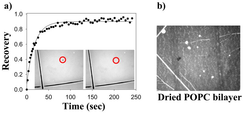
In a first set of experiments, solid supported POPC bilayers in PBS containing 0.1 mol% Texas Red DHPE were formed by the vesicle fusion method.30, 53–55 Fluorescence microscopy revealed that these bilayers were uniform down to the diffraction limit (Figure 3a, inset). FRAP measurements demonstrated that the membranes were two-dimensionally fluid with diffusion constants of 4.7 (± 0.4) × 10−8 cm2/sec and mobile fractions of 98 ± 1%. The aqueous solution was removed from above the bilayer followed by sample desiccation. Upon inspection by fluorescence microscopy, it was found that nearly 100% of the bilayer had been delaminated from the substrate surface (Figure 3b). This observation did not change upon rehydration.
Figure 3.
(a) A FRAP curve for a fully hydrated POPC bilayer. Inserts: Fluorescence micrographs of the same bilayer showing the laser bleach spot both before and after recovery (red circles). (b) A fluorescence micrograph of the POPC bilayer after exposure to air.
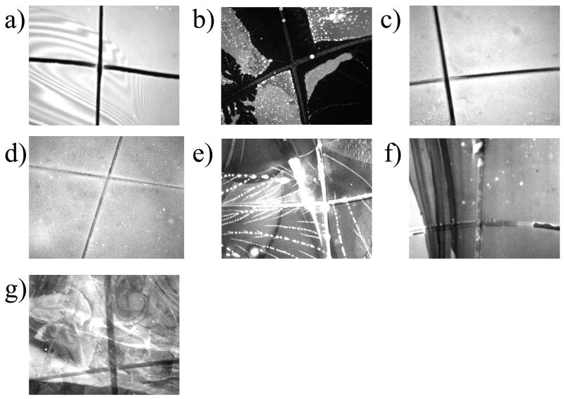
In a second set of experiments, 20 w/w % solutions of α,α-trehalose, α,β-trehalose, maltose, α,α-galacto-trehalose, sucrose, lactose, and glucose were each used as candidate bilayer protectants. Solid supported POPC bilayers were formed in PBS buffer and the aqueous solution was replaced by the appropriate sugar solution. Next, the samples were imaged by fluorescence microscopy. In all cases it was found that the bilayer remained uniform down to the diffraction limit. FRAP measurements showed that the lipids were still highly mobile with a 98 ± 1% mobile fraction for the dye-conjugated lipid. The bilayers were dried, desiccated for 20 hours, and imaged by fluorescence microscopy (Figure 4). It was found that a far larger fraction of the lipid molecules remained on the surface under most conditions in comparison to the control experiments performed without sugar. Only the bilayers protected with glucose and α,β-trehalose showed evidence for near complete delamination. The bilayer protected with α,α-trehalose displayed nearly uniform fluorescence and showed little evidence for delamination. Also, the bilayer protected by maltose had some areas of highly uniform fluorescence, although evidence for damage could be found in other regions. The bilayers protected with α,α-galacto-trehalose, sucrose, and lactose all retained substantial fluorescence. In these cases, however, there was evidence for varying amounts of damage depending upon the region of the membrane observed. Moreover, there was very substantial delamination for sucrose and lactose, which showed up as dark regions in the images shown in Figure 4.
Figure 4.
Images of supported POPC lipid membranes after drying from 20 w/w % solutions of (a) α,α-trehalose, (b) α,β-trehalose, (c) maltose, (d) α,α-galacto-trehalose, (e) sucrose, (f) lactose, and (g) glucose.
As with the control bilayers, each of the seven SLBs dried in the presence of mono- and disaccharides was rehydrated in PBS without any sugar. The supported bilayers were again examined by fluorescence microscopy (Figure 5). In most cases, there was little if any difference in the appearance of the bilayer in the dry and rehydrated states. Curiously, the fluorescence intensity from bilayers protected by α,α-galacto-trehalose consistently looked more uniform upon rehydration. FRAP measurements were again performed on the rehydrated bilayers. The supported membranes protected by α,α-trehalose showed nearly the same diffusion constant and mobile fraction of dye-conjugated lipids as before drying and rehydration (3.4 ± 0.5 × 10⁻⁸ cm2/sec and 93 ± 1%). The bilayers protected with α,α-galacto-trehalose also displayed some mobility. In this case the diffusion constant was reduced to 0.8 ± 0.5 × 10⁻⁸ cm2/sec and the mobile fraction of dye-conjugated lipids was 75 ± 5%. This indicated that although some damage was sustained, a substantial faction of the supported bilayer still remained intact at the interface. By contrast, the bilayers protected with maltose showed no fluorescence recovery despite the fact that they looked uniform by fluorescence. As expected, the glucose, lactose, sucrose, and α,β-trehalose-protected membranes showed no evidence for lipid mobility as determined by FRAP. These data are summarized in Table 1.
Figure 5.
Images of rehydrated POPC lipid membranes exposed to 20 w/w % solutions of (a) α,α-trehalose, (b) α,β-trehalose, (c) maltose, (d) α,α-galacto-trehalose, (e) sucrose, (f) lactose, and (g) glucose.
Table 1.
Diffusion constants and percent recovery values for supported POPC bilayers before and after drying in the presence of sugar solutions. The percent delamination values upon rehydration are also provided. SD = Severely Damaged, NM = Not Mobile. All experiments were performed at 22 ºC.
| Sugar Solution 20 w/w % | Hydrated 10−8 cm2/s | % Recovery | Dried- Rehydrated, 10−8 cm2/s | % Recovery | Approx. % Delamination |
|---|---|---|---|---|---|
| α,α-Trehalose | 3.2 ± 0.4 | 96 ± 1 | 3.4 ± 0.5 | 97 ± 2 | 5 |
| 20% α,β-Trehalose | 5.2 ± 1.6 | 97 ± 2 | SD | 0 | 100 |
| 20% Maltose | 3.5 ± 0.4 | 96 ± 2 | NM | 0 | 44 |
| 20% α,α-Galacto Trehalose | 5.6 ± 1.6 | 97 ± 1 | 0.8 ± 0.2 | 71 ± 5 | 30 |
| 20% Sucrose | 3.4 ± 0.9 | 96 ± 1 | SD | 0 | 61 |
| 20% Lactose | 4.6 ± 0.5 | 96 ± 3 | SD | 0 | 95 |
| 20% Glucose | 3.7 ± 0.8 | 96 ± 2 | SD | 0 | 100 |
| No Sugar | 4.7 ± 0.4 | 98 ± 1 | SD | 0 | 100 |
In the next set of experiments, we aimed to determine the concentration of α,α-trehalose needed to afford protection from damage and delamination. To this end, 20, 15, 10, and 5 w/w % α,α-trehalose solutions were employed. It was found that the area fraction of the membrane which was damaged and delaminated upon desiccation increased continuously as the solution concentration of the sugar was lowered. However, those portions of the surface which still displayed uniform fluorescence had diffusion constants and mobile fractions that were identical to bilayers that had not been dried. These data are summarized in Table 2.
Table 2.
Diffusion constants and percent recovery values for supported POPC bilayers before and after drying in the presence of various solution concentrations of α,α-trehalose. The percent delamination values upon rehydration are also provided.
| α,α-Trehalose Concentration | Hydrated 10−8 cm2/s | % Recovery | Dried-Rehydrated, 10−8 cm2/s | % Recovery | Approx. % Delamination |
|---|---|---|---|---|---|
| 20 w/w % | 3.2 ± 0.4 | 96 ± 1 | 3.4 ± 0.5 | 97 ± 2 | 5 |
| 15 w/w % | 3.6 ± 0.7 | 96 ± 1 | 3.0 ± 0.6 | 98 ± 2 | 11 |
| 10 w/w % | 4.8 ± 0.7 | 98 ± 1 | 4.9 ± 0.3 | 98 ± 2 | 40 |
| 5 w/w % | 5.1 ± 1.0 | 97 ± 1 | 4.3 ± 1.0 | 98 ± 3 | 45 |
Drying Phospholipid Bilayers in the Presence of Glycolipids
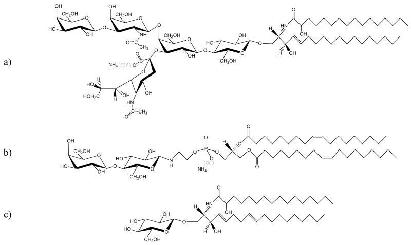
The incorporation of glycolipids into the membrane was tested to assess their ability to prevent membrane delamination upon air exposure. Three candidate glycolipids were chosen: ganglioside GM1, lactosyl PE, and glucosylcerebroside (Figure 6). Vesicles were prepared at concentrations of 10 mol % glycolipid in POPC with 0.1 mol % Texas Red DHPE. The motivation for choosing each glycolipid was straightforward. Lactosyl PE and glucosylcerebroside are lipid-conjugated analogs of lactose and glucose. Therefore, we wished to determine whether covalently attaching the sugar moieties to the membrane would afford air stability even though the free sugars did not. The last species, ganglioside GM1, was chosen because a particularly large glycolipid headgroup might be expected to protect the bilayer against delamination by increasing its bending elastic modulus.7
Figure 6.
Structures of three glycolipids: (a) GM1, (b) lactosyl-PE, and (c) glucosylcerebroside.
FRAP data were obtained before and after rehydration of the three glycolipid systems. As shown in Figure 7a, the bilayer with GM1 showed some protection against delamination upon drying, while the bilayers in the other two cases were completely destroyed (Figure 7b & 7c). FRAP measurements revealed that even the membrane containing GM1 was completely immobile after air exposure. These results are summarized in Table 3.
Figure 7.
Fluorescence micrographs of dehydrated POPC lipid membranes containing: (a) 10 mol % GM1, (b) 10 mol % lactosyl-PE, and (c) 10 mol % glucosylcerebroside.
Table 3.
Diffusion constants and percent recovery values for supported glycolipid/POPC bilayer systems before and after drying. The percent delamination values upon rehydration are also provided. SD = Severely Damaged
| Bilayer System | control, 10−8 cm2/s | % Recovery | Dried-Rehydrated 10−8 cm2/s | % Recovery | Approx. % Delamination |
|---|---|---|---|---|---|
| 10% GM1 | 2.9 ± 1.1 | 94 ± 2 | SD | 0 | 32 |
| 10% Lactosyl-PE | 3.3 ± 0.5 | 98 ± 1 | SD | 0 | 79 |
| 10% Glucosylcerebroside | 3.3 ± 0.7 | 97 ± 1 | SD | 0 | 62 |
Tm Values of Hydrated Bilayers in the Presence of Sugar Solutions
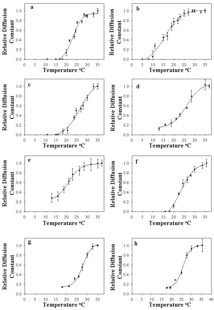
DMPC bilayers containing 0.1 mol% Texas Red DHPE were employed to determine the effect of sugar solutions upon cooling SLBs through the liquid crystalline-to-gel phase transition. DMPC bilayers (Tm = 23.9 ºC) were chosen instead of POPC (Tm = −1 ºC) because these membranes have a phase transition temperature nearer to room temperature,56, 57 thus allowing Tm to be more easily measured. In this set of experiments, a DMPC bilayer in PBS was formed by vesicle fusion to the glass support at 37 °C and rinsed with copious amounts of deionized water. The membranes were again characterized by fluorescence microscopy and appeared to be uniform down to the diffraction limit. The diffusion constants of the bilayers were obtained during the cooling process. Data for the relative diffusion constant vs. temperature for a control DMPC bilayer are shown in Figure 8a. The value of the phase transition temperature abstracted from these data, Tm = 23.1 °C, is in good agreement with literature values.56, 57 These experiments were repeated in the presence of 20 w/w % of each of the seven test sugars. These data are also plotted in Figure 8. The Tm values, abstracted using eqn. 2, are summarized in Table 4. As can be seen, the Tm value was increased modestly in most cases. This is consistent with the known kosmotropic properties of the sugar molecules.58 The exceptions to this finding are for α,α-trehalose and α,α-galacto-trehalose. In these cases, the Tm values were depressed by 7.4 °C and 2.2 °C, respectively, compared with the control DMPC bilayer. The transitions also appear to be somewhat broader. Such results are consisted with the notion that α,α-trehalose strongly interacts with the headgroup region and that α,α-galacto-trehalose does so as well, albeit to a lesser extent.
Figure 8.
Plots of the relative diffusion constant of DMPC bilayers vs. temperature in the presence of 20 w/w % sugar solutions: (a) control DMPC membrane in PBS buffer, (b) α,α-trehalose, (c) maltose, (d) α,β-trehalose, (e) α,α-galacto-trehalose, (f) sucrose, (g) lactose, and (h) glucose.
Table 4.
The effect 20 w/w % sugar solutions on the liquid crystalline-to-gel phase transition temperature of supported DMPC bilayers.
| Sugar Solution 20 w/w % | TmºC |
|---|---|
| DMPC (Control) | 23.1 ± 0.6 |
| α,α-Trehalose | 15.7 ± 0.6 |
| Maltose | 26.7 ± 0.8 |
| α,β-Trehalose | 25.2 ± 1.0 |
| α,α-Galacto-Trehalose | 20.9 ± 0.8 |
| Sucrose | 23.3 ± 0.5 |
| Lactose | 27.9 ± 0.7 |
| Glucose | 25.7 ± 0.7 |
Discussion
Importance of Disaccharide Structure
A well hydrated phospholipid membrane exhibits two-dimensional fluidity and has relatively disordered alkyl chain structure when the bilayer is above its phase transition temperature.59 As water is removed at constant temperature, the individual lipids are brought into closer contact as membrane contraction starts to occur.60 After loss of sufficient water, increased van der Waals interactions start to increase the order of the alkyl chains in the tail region. This decrease in entropy is balanced by the liberation of heat as the bilayer undergoes a phase transition into the gel state.61 This process is concomitant with the cracking and rupture of the drying bilayer. Agents that protect membranes upon drying should preserve the spacing of the lipid headgroups and prevent membrane cracking and rupture.
Our studies showed that α,α-trehalose was far more effective than other naturally occurring disaccharides in preserving the integrity of SLBs upon removal of water (Figures 4&5). The efficacy of α,α-trehalose for membrane preservation in comparison to other disaccharides appears to involve its α,α-(1→1) glycosidic linkage. As pointed out above, the axial, axial linkage allows this molecule to adopt a clam shell structure20 that probably facilitates interactions between the sugar and the headgroup region by creating the appropriate hydrogen bonding geometry to adjacent lipids. This hypothesis is consistent with previous FTIR data in which changes are observed in the local hydrogen bonding environment of the lipid’s phosphoryl group when α,α-trehalose is present.62 Others studies have reported a shift in the resonance of the fatty acid carbonyl group as well.63 In the present studies, α,α-trehalose suppressed the Tm value of DMPC bilayers, while the other naturally occurring sugars actually raised the phase transition temperature (Figure 8 and Table 4). The behavior of α,α-trehalose is somewhat reminiscent of chaotropes, which can interact strongly with amphiphilic interfaces.64 Our results for the other naturally occurring disaccharides correspond more closely with standard kosmotropic behavior that sugar molecules typically display.58 It should be pointed out that kosmotropes are assumed to be preferentially excluded from penetrating amphiphilic interfaces.64
Strong evidence for the importance of the axial, axial linkage came from the synthetic molecule, α,α-galacto-trehalose. This disaccharide suppressed the Tm value of a lipid bilayer, although not to the same extent as α,α-trehalose (Table 4). Such behavior is consistent with its more modest ability to preserve the integrity of fluid supported phospholipid membranes upon air exposure (Figures 4&5). As noted above, this molecule, like α,α-trehalose, crystallizes in a clam shell conformation.23 By contrast, the other disaccharides, including α,β-trehalose, adopt more open conformations.21, 22, 24, 25 Thus, the ability of a disaccharide to adopt the appropriate conformation appears to be crucial for replacing water at the interface with sugar molecules upon drying. On the other hand, it does not seem to be sufficient for a disaccharide to be a non-reducing sugar in order for it to protect an SLB. Indeed, both sucrose and α,β-trehalose are non-reducing sugars and they afforded little if any protection to supported membranes.
Glycolipids and Volumetric Effects
The mere presence of conjugated sugar molecules at the lipid headgroup/water interface does not appear to be sufficient to afford protection of an SLB upon drying. In fact, neither lactosyl-PE nor glucosylcerebroside provided any protection to the membrane upon dehydration (Figure 7). This is again consistent with the notion that the sugar needs to adopt the proper conformation in order to perform its role as a lipopreservative. It should be noted that certain glycolipids such as digalactosyldiacylglycerol (DGDG) have been reported to have an influence on the physical behavior of lipid membranes in the dry and hydrated states.65 However, whether these effects were due to sugar interactions with the PC headgroups or merely the influence of their highly unsaturated acyl chains, which strongly depressed the gel-to-liquid crystalline phase transition temperature of the membrane, remains unclear.66 DGDG might also have a volumetric effect.17
Volumetric effects may certainly be important for α,α-trehalose as well. As noted in the introduction, this sugar has the highest glass transition temperature of any known disaccharide.19 In fact, its Tg value may be raised even further by the presence of phosphate ions in the buffer.67 It is certainly possible that the axial, axial linkage and the resultant clam shell structure help provide lipid membrane protection upon drying because they lead to a high glass transition temperature. This idea would be consistent with a combined mechanism involving water replacement and volumetric effects.
Bilayer Damage, but not Delamination: Maltose and GM1
The data in Figure 4 showed that maltose prevented delamination of lipid material from the glass surface upon removal of water from the supported membrane. Unlike α,α-trehalose, however, these bilayers experienced a complete loss in lateral fluidity upon rehydration. A similar result was found for bilayers containing ganglioside GM1. In this case, a considerable amount of lipid material also remained at the interface upon removal of bulk water, but again was completely immobile after rehydration as judged by FRAP.
We hypothesize that maltose may give rise to a volumetric effect, but lacks the appropriate conformation to protect supported lipid membranes against lipid rearrangements after delamination is prevented. It should be noted that maltose has a rather high Tg value, although not quite as high as α,α-trehalose.19 Whether or not the ability to form a vitrified layer by itself affects the phase transition temperature of a lipid bilayer has been an issue of debate in the literature.11, 18, 68 Our results are consistent with the notion that volumetric effects may be sufficient to prevent lipid delamination from the solid substrate as bulk water is removed. Therefore, the ability to form a sugar glass layer may be a necessary, but not sufficient condition to preserve supported lipid bilayers upon drying.
Symmetry and Bivalency
Because of its clam shell conformation, α,α-trehalose has several structural characteristics that may also help explain its unusual anhydrobiotic properties. For example, unlike other naturally occurring disaccharides, α,α-trehalose possesses a formal C2 axis of symmetry in the clam shell conformation. Therefore, both of its glucopyranose rings can hydrogen bond in a very similar fashion with surround lipids molecules when the disaccharide is present in the headgroup region of the bilayer.15, 16 Indeed, it is also almost exactly the right size in the clam shell conformation to span the distance between the headgroups of two adjacent lipid molecules. Such bivalency may provide stability upon drying or even facilitate a more extensive hydrogen bonding network. Moreover, the specific arrangement of the hydroxyl groups in α,α-trehalose may optimize the hydrogen bonding arrangement for water replacement. Evidence for this last statement comes from the somewhat less protective behavior afforded by α,α-galacto-trehalose, which only differs from the structure of α,α-trehalose by the epimerization of the two 4-hydroxyl groups to the axial position from the equatorial position (Figure 1). Such a change may slightly weaken anhydrobiotic preservation by putting these OH groups in a less favorable position for simultaneous hydrogen bonding with two phosphatidylcholine headgroups.
Acknowledgments
The authors thank DARPA (FA9550-06-C-0006) and the NIH (R01 GM070622) for support. The authors also wish to thank Prof. Eric E. Simanek for useful discussions.
References
- 1.Crowe JHCJS. Anhydrobiosis. Dowden, Hutchinson & Ross; Stroudsburg, PA: 1973. [Google Scholar]
- 2.Crowe JH, Crowe LM, Chapman D. Science. 1984;223:701–703. doi: 10.1126/science.223.4637.701. [DOI] [PubMed] [Google Scholar]
- 3.Crowe JH, Crowe LM, Oliver AE, Tsvetkova N, Wolkers W, Tablin F. Cryobiology. 2001;43:89–105. doi: 10.1006/cryo.2001.2353. [DOI] [PubMed] [Google Scholar]
- 4.Sussman AS, Lingappa BT. Science. 1959;130:1343–1343. doi: 10.1126/science.130.3385.1343. [DOI] [PubMed] [Google Scholar]
- 5.Strauss G, Hauser H. Proc Natl Acad Sci USA. 1986;83:2422–2426. doi: 10.1073/pnas.83.8.2422. [DOI] [PMC free article] [PubMed] [Google Scholar]
- 6.Hincha DK, Hellwege EM, Heyer AG, Crowe JH. Eur J Biochem. 2000;267:535–540. doi: 10.1046/j.1432-1327.2000.01028.x. [DOI] [PubMed] [Google Scholar]
- 7.Crowe LM, Crowe JH. Biochim Biophys Acta. 1988;946:193–201. doi: 10.1016/0005-2736(88)90392-6. [DOI] [PubMed] [Google Scholar]
- 8.Crowe JH, Crowe LM, Carpenter JF, Wistrom CA. Biochem J. 1987;242:1–10. doi: 10.1042/bj2420001. [DOI] [PMC free article] [PubMed] [Google Scholar]
- 9.Crowe JH, Hoekstra FA, Crowe LM. Annu Rev Physiol. 1992;54:579–599. doi: 10.1146/annurev.ph.54.030192.003051. [DOI] [PubMed] [Google Scholar]
- 10.Tsvetkova NM, Phillips BL, Crowe LM, Crowe JH, Risbud SH. Biophys J. 1998;75:2947–2955. doi: 10.1016/S0006-3495(98)77736-7. [DOI] [PMC free article] [PubMed] [Google Scholar]
- 11.Crowe JH, Hoekstra FA, Nguyen KHN, Crowe LM. Biochim Biophys Acta. 1996;1280:187–196. doi: 10.1016/0005-2736(95)00287-1. [DOI] [PubMed] [Google Scholar]
- 12.Lee CWB, Waugh JS, Griffin RG. Biochemistry. 1986;25:3737–3742. doi: 10.1021/bi00361a001. [DOI] [PubMed] [Google Scholar]
- 13.Lee SL, Debenedetti PG, Errington JR. J Chem Phys. 2005;122(204511):1–10. doi: 10.1063/1.1917745. [DOI] [PubMed] [Google Scholar]
- 14.Ohtake S, Schebor C, Palecek SP, de Pablo JJ. Biochim Biophys Acta. 2005;1713:57–64. doi: 10.1016/j.bbamem.2005.05.001. [DOI] [PubMed] [Google Scholar]
- 15.Doxastakis M, Sum AK, de Pablo JJ. J Phys Chem B. 2005;109:24173–24181. doi: 10.1021/jp054843u. [DOI] [PubMed] [Google Scholar]
- 16.Sum AK, Faller R, de Pablo JJ. Biophys J. 2003;85:2830–2844. doi: 10.1016/s0006-3495(03)74706-7. [DOI] [PMC free article] [PubMed] [Google Scholar]
- 17.Crowe JH, Carpenter JF, Crowe LM. Annu Rev Phys. 1998;60:73–103. doi: 10.1146/annurev.physiol.60.1.73. [DOI] [PubMed] [Google Scholar]
- 18.Koster KL, Lei YP, Anderson M, Martin S, Bryant G. Biophys J. 2000;78:1932–1946. doi: 10.1016/S0006-3495(00)76741-5. [DOI] [PMC free article] [PubMed] [Google Scholar]
- 19.Green JL, Angell CA. J Phys Chem B. 1989;93:2880–2882. [Google Scholar]
- 20.Brown GM, Rohrer DC, Berking B, Beevers CA, Gould RO, Simpson R. Acta Cryst. 1972 Nov15;B 28:3145–3158. [Google Scholar]
- 21.French AD, Kelterer AM, Johnson GP, Dowd MK. J Mol Struct. 2000;556:303–313. [Google Scholar]
- 22.Quigley GJ, Sarko A, Marchess Rh. J Am Chem Soc. 1970;92:5834. [Google Scholar]
- 23.Linden A, Lee CK. Acta Crystallogr, Sect C: Cryst Struct Commun. 1995;51:1007–1012. [Google Scholar]
- 24.Brown GM, Levy HA. Science. 1963;141:921. doi: 10.1126/science.141.3584.921. [DOI] [PubMed] [Google Scholar]
- 25.Beevers CA, Hansen HN. Acta Crystallogr, Sect B: Struct Sci. 1971;B 27:1323. [Google Scholar]
- 26.Crowe JH, Crowe LM. Preservation of Liposomes During Freeze-Drying, Liposome Technology. Boca Raton, FL: 1993. [Google Scholar]
- 27.Chiantia S, Kahya N, Schwille P. Langmuir. 2005;21:6317–6323. doi: 10.1021/la050115m. [DOI] [PubMed] [Google Scholar]
- 28.Sackmann E. Science. 1996;271:43–48. doi: 10.1126/science.271.5245.43. [DOI] [PubMed] [Google Scholar]
- 29.Groves JT, Boxer SG. Acc Chem Res. 2002;35:149–157. doi: 10.1021/ar950039m. [DOI] [PubMed] [Google Scholar]
- 30.McConnell HM, Watts TH, Weis RM, Brian AA. Biochim Biophys Acta. 1986;864:95–106. doi: 10.1016/0304-4157(86)90016-x. [DOI] [PubMed] [Google Scholar]
- 31.Cremer PS, Yang TL. J Am Chem Soc. 1999;121:8130–8131. [Google Scholar]
- 32.Cremer PS, Groves JT, Kung LA, Boxer SG. Langmuir. 1999;15:3893–3896. [Google Scholar]
- 33.Groves JT, Boxer SG, McConnell HM. J Phys Chem B. 2000;104:11409–11415. [Google Scholar]
- 34.Lipowsky R. Colloids Surf, A. 1997;128:255–264. [Google Scholar]
- 35.Baumgart T, Offenhauser A. Biophys J. 2002;83:1489–1500. doi: 10.1016/S0006-3495(02)73919-2. [DOI] [PMC free article] [PubMed] [Google Scholar]
- 36.Cremer PS, Boxer SG. J Phys Chem B. 1999;103:2554–2559. [Google Scholar]
- 37.Morigaki K, Kiyosue K, Taguchi T. Langmuir. 2004;20:7729–7735. doi: 10.1021/la049340e. [DOI] [PubMed] [Google Scholar]
- 38.Ross EE, Bondurant B, Spratt T, Conboy JC, O’Brien DF, Saavedra SS. Langmuir. 2001;17:2305–2307. [Google Scholar]
- 39.Conboy JC, Liu SC, O’Brien DF, Saavedra SS. Biomacromolecules. 2003;4:841–849. doi: 10.1021/bm0256193. [DOI] [PubMed] [Google Scholar]
- 40.Morigaki K, Schonherr H, Frank CW, Knoll W. Langmuir. 2003;19:6994–7002. [Google Scholar]
- 41.Morigaki K, Baumgart T, Jonas U, Offenhausser A, Knoll W. Langmuir. 2002;18:4082–4089. [Google Scholar]
- 42.Petralli-Mallow T, Brigmann KA, Richter LJ, Stephenson JC, Plant AL. Proc SPIE-Int Soc Opt Eng. 1999;3858:25–31. [Google Scholar]
- 43.Phillips KS, Dong Y, Carter D, Cheng Q. Anal Chem. 2005;77:2960–2965. doi: 10.1021/ac0500481. [DOI] [PubMed] [Google Scholar]
- 44.Holden MA, Jung SY, Yang TL, Castellana ET, Cremer PS. J Am Chem Soc. 2004;126:6512–6513. doi: 10.1021/ja048504a. [DOI] [PubMed] [Google Scholar]
- 45.Albertorio F, Diaz AJ, Yang TL, Chapa VA, Kataoka S, Castellana ET, Cremer PS. Langmuir. 2005;21:7476–7482. doi: 10.1021/la050871s. [DOI] [PubMed] [Google Scholar]
- 46.Green JL, Angell CA. J Phys Chem. 1989;93:2880–2882. [Google Scholar]
- 47.Yang TL, Jung SY, Mao HB, Cremer PS. Anal Chem. 2001;73:165–169. doi: 10.1021/ac000997o. [DOI] [PubMed] [Google Scholar]
- 48.Barenholz Y, Gibbes D, Litman BJ, Goll J, Thompson TE, Carlson FD. Biochemistry. 1977;16:2806–2810. doi: 10.1021/bi00631a035. [DOI] [PubMed] [Google Scholar]
- 49.Axelrod D, Koppel DE, Schlessinger J, Elson E, Webb WW. Biophys J. 1976;16:1055–1069. doi: 10.1016/S0006-3495(76)85755-4. [DOI] [PMC free article] [PubMed] [Google Scholar]
- 50.Mao HB, Yang TL, Cremer PS. J Am Chem Soc. 2002;124:4432–4435. doi: 10.1021/ja017625x. [DOI] [PubMed] [Google Scholar]
- 51.Mao HB, Yang TL, Cremer PS. Anal Chem. 2002;74:379–385. doi: 10.1021/ac010822u. [DOI] [PubMed] [Google Scholar]
- 52.Mao HB, Li CM, Zhang YJ, Bergbreiter DE, Cremer PS. J Am Chem Soc. 2003;125:2850–2851. doi: 10.1021/ja029691k. [DOI] [PubMed] [Google Scholar]
- 53.Brian AA, McConnell HM. Proc Nat Acad Sci USA. 1984;81:6159–6163. doi: 10.1073/pnas.81.19.6159. [DOI] [PMC free article] [PubMed] [Google Scholar]
- 54.Tamm LK, McConnell HM. Biophys J. 1985;47:105–113. doi: 10.1016/S0006-3495(85)83882-0. [DOI] [PMC free article] [PubMed] [Google Scholar]
- 55.Kalb E, Frey S, Tamm LK. Biochim Biophys Acta. 1992;1103:307–316. doi: 10.1016/0005-2736(92)90101-q. [DOI] [PubMed] [Google Scholar]
- 56.Charrier A, Thibaudau F. Biophys J. 2005;89:1094–1101. doi: 10.1529/biophysj.105.062463. [DOI] [PMC free article] [PubMed] [Google Scholar]
- 57.Yang J, Appleyard J. J Phys Chem B. 2000;104:8097–8100. [Google Scholar]
- 58.Koynova R, Brankov J, Tenchov B. Eur Biophys J. 1997;25:261–274. doi: 10.1007/s002490050038. [DOI] [PubMed] [Google Scholar]
- 59.Xie AF, Yamada R, Gewirth AA, Granick S. Phys Rev Lett. 2002;89 doi: 10.1103/PhysRevLett.89.246103. [DOI] [PubMed] [Google Scholar]
- 60.Wolfe J, Bryant G. Cryobiology. 1999;39:103–129. doi: 10.1006/cryo.1999.2195. [DOI] [PubMed] [Google Scholar]
- 61.Kirchner S, Cevc G. Europhys Lett. 1994;28:31–36. [Google Scholar]
- 62.Wolkers WF, Oliver AE, Tablin F, Crowe JH. Carbohydr Res. 2004;339:1077–1085. doi: 10.1016/j.carres.2004.01.016. [DOI] [PubMed] [Google Scholar]
- 63.Luzardo MC, Amalfa F, Nunes AM, Diaz S, Biodi de Lopez AC, Disalvo AE. Biophys J. 2000;78:2452–2458. doi: 10.1016/s0006-3495(00)76789-0. [DOI] [PMC free article] [PubMed] [Google Scholar]
- 64.Gurau MC, Lim SM, Castellana ET, Albertorio F, Kataoka S, Cremer PS. J Am Chem Soc. 2004;126:10522–10523. doi: 10.1021/ja047715c. [DOI] [PubMed] [Google Scholar]
- 65.Popova AV, Hincha DK. Glycobiology. 2005;15:1150–1155. doi: 10.1093/glycob/cwj001. [DOI] [PubMed] [Google Scholar]
- 66.Luzardo MC, Bernik DL, Pazos FI, Figueroa S, Lanio ME, Verz V, Disalvo AE. Arch Biochem Biophys. 1999;363:81–90. doi: 10.1006/abbi.1998.1017. [DOI] [PubMed] [Google Scholar]
- 67.Ohtake S, Schebor C, Palecek SP, de Pablo JJ. Pharm Res. 2004;21:1615–1621. doi: 10.1023/b:pham.0000041456.19377.87. [DOI] [PubMed] [Google Scholar]
- 68.Mobley CW, Schreier H. J Controlled Release. 1994;31:73–87. [Google Scholar]


