Abstract
A hybrid protein, tPA/GFP, consisting of rat tissue plasminogen activator (tPA) and green fluorescent protein (GFP) was expressed in PC12 cells and used to study the distribution, secretory behavior, and dynamics of secretory granules containing tPA in living cells with a neuronal phenotype. High-resolution images demonstrate that tPA/GFP has a growth cone-biased distribution in differentiated cells and that tPA/GFP is transported in granules of the regulated secretory pathway that colocalize with granules containing secretogranin II. Time-lapse images of secretion reveal that secretagogues induce substantial loss of cellular tPA/GFP fluorescence, most importantly from growth cones. Time-lapse images of the axonal transport of granules containing tPA/GFP reveal a surprising complexity to granule dynamics. Some granules undergo canonical fast axonal transport; others move somewhat more slowly, especially in highly fluorescent neurites. Most strikingly, granules traffic bidirectionally along neurites to an extent that depends on granule accumulation, and individual granules can reverse their direction of motion. The retrograde component of this bidirectional transport may help to maintain cellular homeostasis by transporting excess tPA/GFP back toward the cell body. The results presented here provide a novel view of the axonal transport of secretory granules. In addition, the results suggest that tPA is targeted for regulated secretion from growth cones of differentiated cells, strategically positioning tPA to degrade extracellular barriers or to activate other barrier-degrading proteases during axonal elongation.
INTRODUCTION
Neuronal cells must efficiently transport a broad spectrum of proteins over large distances, from sites of protein synthesis in the cell body out to the tips of axons that can be many centimeters long. Moreover, like other cells, neuronal cells must accurately sort and target proteins before they are transported to their final destination (for review see Craig and Banker, 1994). Although considerable progress has been made in understanding the distribution and dynamics of proteins in neuronal cells, many of the underlying molecular events have yet to be elucidated.
It is now well established that protein transport along axons in neuronal cells proceeds via fast and slow transport systems (for reviews see Grafstein and Forman, 1980; Sheetz and Martenson, 1991; Hirokawa, 1993). Fast axonal transport is the better characterized of the two transport systems, involving the anterograde and retrograde motors, kinesin and dynein, respectively, which move some cellular constituents along microtubules using energy derived from ATP hydrolysis (Brady, 1985; Vale et al., 1985a; Paschal and Vallee, 1987; Shpetner et al., 1988; Vallee et al., 1988; Schnapp and Reese, 1989; Schroer et al., 1989; for reviews see Schroer, 1992; Hirokawa, 1993; Vallee and Sheetz, 1996). Proteins associated with small vesicles move along axons via fast (∼1–5 μm/s) transport, whereas cytoskeletal constituents and soluble enzymes move via slow (∼10−3 to 3 × 10⁻² μm/s) transport (for reviews see Grafstein and Forman, 1980; Vale et al., 1992a; Hirokawa, 1993). Organelles like mitochondria move at intermediate rates (∼0.5 μm/s), slightly slower than those characteristic of the fast transport system (Allen et al., 1982; Vale et al., 1985c; Morris and Hollenbeck, 1993).
Recent advances in fluorescence microscopy promise to enhance further our understanding of protein dynamics and axonal transport in living cells. Key among these advances is the discovery that functional, fluorescent derivatives of many proteins can be expressed in living cells as protein fusions involving wt green fluorescent protein (GFP) (Chalfie et al., 1994) or a fluorescent GFP mutant (Heim et al., 1995; Cormack et al., 1996). This advance has made it possible to monitor the distribution and dynamics of a single type of protein within living cells while taking advantage of the sensitivity of fluorescence (see, e.g., Kaether and Gerdes, 1995; Cole et al., 1996; Rizzuto et al., 1996; Shelby et al., 1996; Haseloff et al., 1997; Lang et al., 1997). In contrast, optical microscopy techniques (such as differential interference contrast and phase contrast) that have traditionally been used to study axonal transport are nonspecific, revealing the motion of many cellular constituents simultaneously.
Here we have used fluorescence microscopy and the new GFP technology to study the distribution and axonal transport of an important secretory protein in living cells with a neuronal phenotype. We have studied tissue plasminogen activator (tPA), a serine protease best known for its role in degrading fibrin-containing thrombi (for a recent review see Hajjar, 1995) that has also been implicated in pivotal degradative processes occurring at the growth cone during axonal elongation and in mediating neuronal degeneration (Tsirka et al., 1995). In particular, tPA has been postulated to be involved in regulating adherence to the substratum and penetration of the extracellular matrix (ECM) by the growth cone during axonal elongation (Pittman et al., 1989; Verrall and Seeds, 1989; McGuire and Seeds, 1990; Leprince et al., 1991; Garcia-Rocha et al., 1994; Pittman and DiBenedetto, 1995; Seeds et al., 1995). We have expressed a hybrid protein consisting of rat tPA and GFP, tPA/GFP, in living PC12 cells using transient transfection techniques. We have also expressed two additional hybrid proteins, tPA/GFP(SIG−) and GFP(SIG+). The first consists of tPA/GFP without the rat tPA signal sequence; the second consists of only GFP and the rat tPA signal sequence. Using fluorescence microscopy, we have monitored the distribution of all three fusion proteins, and the axonal transport of tPA/GFP, in living PC12 cells. These data reveal novel features of the dynamics, distribution, and secretory behavior of secretory granules containing tPA/GFP, including bidirectional transport of granules, direction reversal of individual granules, extensive accumulation of granules in growth cones, and regulated secretion of granules from growth cones. These data also support tPA’s postulated role in regulating adherence to the substratum and ECM degradation during axonal elongation.
MATERIALS AND METHODS
Cell Culture
The cell line studied here, PC12, is a common model neuronal tissue culture cell line originally derived from a rat pheochromocytoma (Greene and Tischler, 1976). Stock cells were grown on Primaria Plates (Becton Dickinson, Lincoln, NJ) at 37°C in a 5% CO2/95% air incubator in DMEM (Life Technologies, Gaithersburg, MD) supplemented with 5% FCS and 5% horse serum. When grown in a standard tissue culture medium, PC12 cells have a chromaffin-like appearance; however, after transfer into serum-free medium containing nerve growth factor (NGF), the cells extend neurites that are similar to axons. The PC12 cell line used here (subclone GR-5, developed by Dr. Rae Nishi, Oregon Health Sciences University) is particularly responsive to induction by NGF.
In preparation for high-resolution fluorescence microscopy, PC12 cells were plated onto 25-mm diameter round glass no. 1 coverslips (Fisher Scientific, Pittsburgh, PA) or 22-mm square no. 1.5 Gold Seal coverslips (Becton Dickinson) that had been acid washed and coated with a thin layer of dilute Matrigel (Collaborative Research, Waltham, MA). Cells were allowed to settle onto the coverslips and were then transferred into serum-free N2 medium (Bottenstein and Sato, 1979) to induce the production of NGF receptors. Twenty four hours after transfer into N2, the cells were induced to differentiate with 50 ng/ml NGF (Life Technologies). For some experiments, cells were left undifferentiated.
For live imaging, cells were mounted in Sykes-Moore Chambers (Bellco Glass, Vineland, NJ) in imaging medium consisting of Dulbecco’s glucose-supplemented PBS (Life Technologies) that was further supplemented with 2.5% FCS and 2.5% horse serum. The sample chamber was maintained at 31–37°C, using a custom-heated holder for the Sykes-Moore Chambers (constructed by electronics technician, Alan Younis).
In some cases, cells were fixed in PBS containing 4% paraformaldehyde for 30 min and then washed three times in PBS. Cells were fixed to prepare them for immunostaining or to avoid motion-induced blurring in three-dimensional images of transfected samples. Fixed cells were imaged after being mounted in 90% buffered glycerol containing the photobleaching inhibitor n-propyl gallate (2% wt/vol) (Sigma Chemical, St. Louis, MO). The coverslip bearing the cells was sealed to the microscope slide using clear nail polish (Wet ‘n’ Wild, Pavion, Nyack, NY).
Construction of Hybrid DNA and Transient Transfection
The gene for rat tPA was kindly provided by Dr. Randall N. Pittman (University of Pennsylvania School of Medicine, Philadelphia, PA). PCR was used to introduce SalI and SacII sites onto the N-terminal and C-terminal coding regions of the gene, respectively. The PCR-amplified fragment was subcloned into pCR-2.1 (Invitrogen, San Diego, CA), digested with SalI and SacII, and ligated into pEGFP N-1, an N-terminal enhanced GFP fusion vector that contains the immediate early promoter of human cytomegalovirus (Clontech Laboratories, Palo Alto, CA). The enhanced GFP mutant used here (GFPmut1) produces a fluorescence signal that is ∼35-fold stronger than the wt GFP fluorescence signal when expressed in mammalian cells; the mutant also has a red-shifted excitation maximum (Cormack et al., 1996). The vector for tPA/GFP(SIG−) was prepared in an analogous manner except that the SalI restriction site was introduced immediately upstream of the sequence specified by the second exon of tPA. The tPA/GFP(SIG−) vector thus coded for a fusion protein that lacked the coding information for the first 21 amino acids of tPA, which include the signal sequence.
The vector for GFP(SIG+) was prepared from the tPA/GFP vector by first engineering a unique BspE1 restriction site at the end of the sequence specified by the first exon of tPA. The BspE1 site was introduced using the Quick Change Site Directed Mutagenesis Kit (Stratagene, La Jolla, CA). Digestion of the engineered vector with BspE1 and NotI released the tPA/GFP(SIG−) coding information. The GFP gene was then subcloned into the BspE1, NotI digested vector. DNA sequence analysis confirmed that the resulting construct contained the coding information for the first 21 amino acids of the tPA gene product followed directly by the coding information for GFP.
PC12 cells were transiently transfected following a standard cationic lipid-based DNA delivery protocol. In brief, 15 μl lipofectamine were combined with 85 μl OPTI-MEM (both from Life Technologies), and 1–4 μg DNA were diluted into 100 μl OPTI-MEM. These two solutions were combined, mixed gently, and allowed to sit for ∼45 min at 23°C. The lipofectamine-DNA complex was then combined with 0.8 ml of DMEM (37°C), and plated over cells in a 35-mm culture dish. Five hours later, the transfection mixture was diluted with 1 ml N2 and, if the cells were differentiated, 2 μl NGF (50 μg/ml). The cells were typically imaged ∼24 h post-transfection, after being washed three times with PBS. For a few experiments, cells were imaged ∼10 h post-transfection while maintaining sterile conditions, returned to the incubator, and then reimaged ∼24 h post-transfection. In this manner, build-up of tPA/GFP fluorescence could be followed as a function of time.
Secretion Assays
Regulated secretion of tPA/GFP induced by the Ca2+ ionophore A23187 (Sigma) or the cholinergic agonist carbachol (Sigma) was assayed by first imaging readily identified, transfected, PC12 cells in imaging medium that did not contain A23187 or carbachol. The imaging medium in the Sykes-Moore Chamber was then replaced with imaging medium containing 30 μM A23187 or 10 mM carbachol, and specific cells that had been imaged before addition of the ionophore or carbachol were quickly relocated. Images of these cells were then taken at defined time intervals after addition.
Loss of cellular fluorescence induced by the ionophore or carbachol was determined in two different ways. First, loss of fluorescence from entire cells was determined by calculating and comparing fluorescence signals emitted by entire cells before and after addition. Second, loss of fluorescence from growth cones was determined by calculating and comparing fluorescence signals emitted from growth cones before and after addition. Fluorescence signals emitted by entire cells or growth cones were calculated by outlining the appropriate area and summing intensities inside the outlined area, using Priism image visualization software (Applied Precision, Issaquah, WA).
Antibody Staining for Endogenous tPA and Secretogranin II
To compare the distribution of tPA/GFP and endogenous tPA, PC12 cells were immunostained as follows. Forty-eight to 72 h after the induction of differentiation, PC12 cells were fixed for 30 min in PBS containing 4% paraformaldehyde, permeabilized for 5 min in 0.1% Triton X-100 (Bio-Rad, Hercules, CA), and blocked for 20 min in 10% rabbit serum (Vector Laboratories, Burlingame, CA). Cells were then incubated for 2 h with a goat anti-human melanoma tPA antibody (American Diagnostica, Greenwich, CT) that has been shown to cross-react with rat tPA (Sumi et al., 1992), followed by a 1-h incubation with a TRITC-conjugated rabbit anti-goat secondary antibody (Sigma). In preparation for fluorescence microscopy, cells were mounted in glycerol/PBS containing 2% n-propyl gallate. Specificity of tPA staining was verified with controls that involved leaving out the primary antibody or preabsorbing the primary antibody with a 5-fold M excess of tPA (American Diagnostica).
To test for colocalization of tPA/GFP with a known marker for the regulated secretory pathway, PC12 cells expressing tPA/GFP were stained with a rabbit primary antibody against secretogranin II (SgII) and then with a Texas Red-conjugated goat anti-rabbit secondary antibody (Jackson ImmunoResearch Laboratories, West Grove, PA), following a protocol similar to that used to stain for endogenous tPA. The antibody against SgII was a generous gift of Dr. Sharon Tooze (Imperial Cancer Research Institute) and has been shown previously to label regulated secretory granules in PC12 cells (Tooze et al., 1994).
Imaging
Time-lapse and three-dimensional images of the distribution and dynamics of fluorescent proteins in PC12 cells were collected on a DeltaVision wide-field optical-sectioning microscope system (Applied Precision) using a 60× (NA = 1.4) plan apochromatic Olympus objective (Olympus, Lake Success, NY). This system consists of an Olympus inverted microscope whose focus is under the control of a Nanomover microstepper motor (Melles Griot, Irvine, CA), a cooled 12-bit CCD camera (Photometrics, Tucson, AZ), a mercury arc lamp, various shutters and filters, and a Silicon Graphics workstation (Silicon Graphics, Mountain View, CA) that is used to visualize and deblur images. All aspects of data collection are under the automated control of the SGI and a PC, which makes it possible rapidly to collect time-lapse, multi-wavelength, three-dimensional images.
Time-lapse images of granule transport in living PC12 cells were generated by taking pictures of the same focal plane every few seconds and were not deblurred. Three-dimensional images of fixed PC12 cells were deblurred using a constrained iterative deconvolution algorithm, as described previously (Scalettar et al., 1996).
Data Analysis
Transfected PC12 cells varied considerably in their level of expression of tPA/GFP. To facilitate data analysis, transfected cells were divided into categories based on the linear density of granules containing tPA/GFP along neurites. This linear density was determined by counting fluorescent granules in one focal plane and dividing the resulting number by the length of the neurite in microns. For comparative purposes, the linear density of granules containing endogenous tPA was also assayed using untransfected cells that were immunostained for endogenous tPA.
Individual granules containing tPA/GFP were tracked visually by playing the time-lapse images as movies on a SGI workstation running Priism image visualization software (Hiraoka et al., 1991). Specifically, granules were tracked as long as their positions could be unambiguously determined, and speeds were computed by calculating distances from initial and final granule coordinates and dividing by the elapsed time. The dominant direction of motion was determined by drawing lines perpendicular to the neurite and counting the numbers of granules that crossed the lines moving in the anterograde and retrograde directions. In this way, directionality, rate, and net flux of granule transport along neurites were deduced from detailed tracking of 291 granules in movies generated from 10 representative cells that were ∼24 h post-transfection; granule dynamics in the cell body was also deduced using the movies.
RESULTS
Distribution
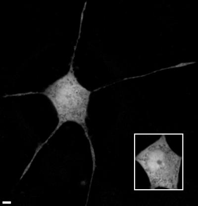
Figure 1 shows a computationally deblurred image of the distribution of soluble GFP in a fixed PC12 cell. Two different “optical sections” corresponding to two different depths through the cell are shown. In one optical section, the neurites are clearly in focus; in the other optical section, the nucleus is clearly in focus (see inset). The images show that GFP is uniformly distributed throughout the cell body and nucleus of PC12 cells but is largely excluded from the nucleoli. GFP’s presence in the nucleus reflects its relatively small size (∼27 kDa), which permits the protein to pass through nuclear pores via diffusion (Alberts et al., 1994). Similar uniform distribution patterns for GFP have been previously observed in other cell types (Ogawa et al., 1995; Haseloff et al., 1997).
Figure 1.
Deblurred images of a fixed GFP-expressing PC12 cell. The two images (including the inset) were obtained by optically sectioning the sample and correspond to two different depths through the cell. Bar, 4 μm.
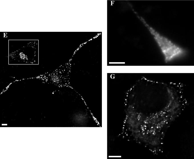
Figure 2 shows images of the distribution of the tPA/GFP hybrid in PC12 cells. Panels A, B, C, and D compare phase and fluorescence images of fixed cells; panels E, F, and G compare distribution in differentiated and undifferentiated cells. In differentiated and undifferentiated cells, the distribution of tPA/GFP is highly nonuniform and punctate, in marked contrast to the uniform distribution of GFP. Moreover, tPA/GFP is completely excluded from the nucleus, again in marked contrast to GFP. The punctate tPA/GFP distribution suggests that tPA/GFP is packaged in vesicles in PC12 cells.
Figure 2.
Images of tPA/GFP-expressing PC12 cells. The fluorescence images in E and G were deblurred, whereas the fluorescence images in B, D, and F were not. Phase (A and C) and fluorescence (B and D) images of a fixed, differentiated cell and a neurite taken with a 40× objective. Images of a fixed, differentiated cell (E), a bright growth cone from a living differentiated cell (F), and a fixed, undifferentiated cell (G). Note that the Matrigel is a source of background in the phase images. Bar, 4 μm.
In addition to being punctate, the fluorescence from the tPA/GFP hybrid is also enhanced in several regions of PC12 cells. First, the fluorescence is enhanced in the region of the cell body typically occupied by the Golgi, further suggesting that tPA/GFP traffics through the secretory pathway (see inset to Figure 2E). Second, in undifferentiated PC12 cells there is an enhancement of fluorescence near the plasma membrane, whereas in differentiated PC12 cells there is an enhancement of fluorescence in growth cones of neurites (see Figure 2). Similarly, in AtT20 cells (our unpublished data) there is an enhancement of fluorescence in tips of processes. Interestingly, we often observe enhanced fluorescence from growth cones even when the “neurites” are short. This growth cone-biased distribution pattern suggests that tPA/GFP traffics through the regulated secretory pathway, as does endogenous tPA (Possenti et al., 1989; Rivas and Moore, 1989; Harrison et al., 1996; Parmer et al., 1997). A similar distribution pattern has been observed for chromogranin B/GFP and preproatrial natriuretic factor/GFP hybrid proteins expressed in PC12 cells (Burke et al., 1997; Lang et al., 1997).
A potential artifact in interpretation could arise if expression of tPA/GFP perturbs distribution and dynamics. To assay for potential perturbation, the distribution of endogenous tPA in fixed PC12 cells was compared with the distribution of tPA/GFP in fixed and living PC12 cells. Figure 3 shows a deblurred image of the distribution of endogenous tPA in fixed PC12 cells obtained using immunofluorescence techniques. Endogenous tPA produces a punctate distribution pattern in the cell body and neurites, and enhanced staining in the growth cones of differentiated cells, that is similar to the distribution pattern produced by tPA/GFP (Figure 2). This similarity in distribution patterns suggests that expression of tPA/GFP is not leading to distribution artifacts in our system.
Figure 3.
Deblurred image of the distribution of endogenous tPA in a fixed, immunostained PC12 cell showing the similarity in distribution between endogenous tPA and tPA/GFP. Some surface staining is visible in the immunofluorescence image, which may reflect endogenous tPA that is bound to its membrane receptor. Bar, 4 μm.
Regulated Secretion
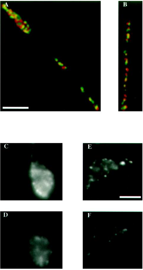
To show that tPA/GFP traffics through the regulated secretory pathway, we demonstrated that tPA/GFP colocalizes extensively with SgII, a known marker for the regulated secretory pathway in PC12 cells (Tooze et al., 1994). Figure 4, A and B, shows two representative high-magnification double-label images of regions of neurites containing a moderate number of secretory granules. Significantly, regions devoid of granules containing tPA/GFP are also basically devoid of granules containing SgII. Moreover, in Figure 4, A and B, respectively, 70% and 83% of the granules containing tPA/GFP overlap with a granule containing SgII, demonstrating extensive colocalization of the two types of protein. Overall, 220 granules containing tPA/GFP and SgII were counted in four representative double-labeled cells containing a low to moderate number of secretory granules, and the extent of overlap was 79 (± 7)%, where quoted errors are standard deviations. Such extensive, but not perfect, granule overlap is comparable to that obtained in previous attempts to colocalize another GFP hybrid protein with a marker for the regulated secretory pathway (Lang et al., 1997). We assayed granule overlap in cells that were not too congested to avoid fortuitous overlap resulting from high granule density. In determining the extent of granule overlap, we neglected slight (<0.2 μm) lack of registration in the center positions of overlapping granules, because wavelength-dependent image shifts are known to cause such slight registration problems in multicolor images (Hiraoka et al., 1991; Scalettar et al., 1996).
Figure 4.
Images demonstrating that tPA/GFP traffics through the regulated secretory pathway (A and B) and is secreted from growth cones after stimulation with carbachol (C–F). The images in panels A and B were deblurred, whereas the images in C–F were not. Representative two-color images (A and B) of regions of neurites from two different cells that demonstrate extensive colocalization of tPA/GFP and SgII. The distributions of tPA/GFP and SgII are shown in green and red, respectively. Areas of extensive overlap appear yellow in the images. Most green granules overlap with a red granule, although in some cases the sizes of the green and red spots differ; also, the centers of some spots are shifted slightly. Lack of perfect overlap could reflect antibody accessibility, wavelength-dependent image shifts, or differences in protein concentration in individual granules, especially in granules produced before transfection. Images (C–F) of two growth cones from living cells before (C and E) and 20 min after (D and F) the addition of 10 mM carbachol. Panels A and B and C–F share a scale bar. Bar, 4 μm.
We also assayed for regulated secretion of tPA/GFP induced by the calcium ionophore A23187 or the cholinergic agonist carbachol, as described under MATERIALS AND METHODS. A23187 and carbachol elevate intracellular calcium levels and are frequently used to assay for regulated secretion (Possenti et al., 1989; Harrison et al., 1996). After addition of either of these agents to each of six cells, we found that the total cellular fluorescence signal at each measured time point was smaller than at previously measured time points; average loss of total cellular fluorescence was 54 (± 9)% and 35 (± 11)% ∼20 min after addition of A23187 and carbachol, respectively. Similarly, in six of seven cells (∼86%), we observed a significant regional loss of fluorescence from growth cones induced by carbachol; specifically, average loss of fluorescence from growth cones ∼20 min after addition of carbachol was 39 (± 12)%. In the seventh cell, the fluorescence signal from the growth cone remained essentially unchanged. Figure 4, C–F, shows two growth cones for which the regional fluorescence was monitored at a series of time points; at each measured time point after the addition of 10 mM carbachol, the fluorescence signal was smaller than at previously measured time points. Moreover, for both growth cones, loss of fluorescence was ∼40% about 20 min after the addition of carbachol; such a dramatic regional or global loss of fluorescence was never observed in cells that were not exposed to carbachol or A23187. Potential reasons why loss of fluorescence from growth cones was observed in most, but not all, cells are supplied in the DISCUSSION.
Mutant Hybrid Proteins
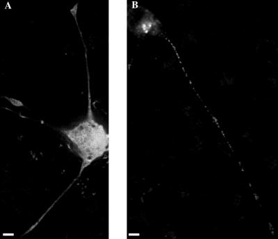
Hybrid proteins could be useful in studies of the putative signals that target proteins to specific regions or pathways within neuronal cells, if mutant hybrids exhibit readily identified changes in distribution and secretory behavior. To test this possibility, and to begin to dissect the role that tPA targeting signals may play in determining distribution and secretory behavior, we expressed two mutant hybrid proteins in PC12 cells. The first, tPA/GFP(SIG−), consists of tPA/GFP without the rat tPA signal sequence, whereas the second, GFP(SIG+), consists of GFP with the rat tPA signal sequence attached at the amino terminus. Significantly, the distributions and secretory behaviors of both mutants differ from tPA/GFP. Figure 5A shows a deblurred image of the distribution of tPA/GFP(SIG−) in a PC12 cell. Deletion of the signal sequence leads to a distribution pattern that is not punctate; rather, the pattern is similar to that produced by soluble GFP (see Figure 1), suggesting that tPA/GFP(SIG−) is a cytosolic protein, unlike tPA/GFP. Moreover, there is no measurable loss of fluorescence when cells expressing tPA/GFP(SIG−) are exposed to carbachol. Figure 5B shows a deblurred image of the distribution of GFP(SIG+) in a PC12 cell. Addition of the signal sequence to GFP leads to a distribution pattern that is nonuniform, punctate, and enhanced in regions of the cell body occupied by the Golgi, suggesting that GFP(SIG+) enters the endoplasmic reticulum and Golgi and is packaged in vesicles. In this respect, GFP(SIG+) and tPA/GFP behave similarly. Significantly, however, GFP(SIG+)-containing vesicles do not accumulate extensively in growth cones, and there is no measurable loss of fluorescence when cells expressing GFP(SIG+) are exposed to carbachol; in these respects, GFP(SIG+) and tPA/GFP behave differently.
Figure 5.
Deblurred images of fixed (A) tPA/GFP(SIG−) and (B) GFP(SIG+)-expressing PC12 cells. One significant difference in distribution between tPA/GFP and GFP(SIG+) is that the staining in growth cones and neurites is often so faint in GFP(SIG+)-expressing cells that these structures are almost invisible when viewed in fluorescence mode. The GFP(SIG+) labeling in the neurite and growth cone shown here is somewhat more intense than is typical, permitting these structures to be seen in the image. Differences in growth cone brightness exhibited by tPA/GFP and GFP(SIG+) are not due to a lower level of expression in GFP(SIG+)-expressing cells. In fact, GFP(SIG+)-expressing cells on average are brighter than tPA/GFP-expressing cells. Bar, 4 μm.
Dynamics
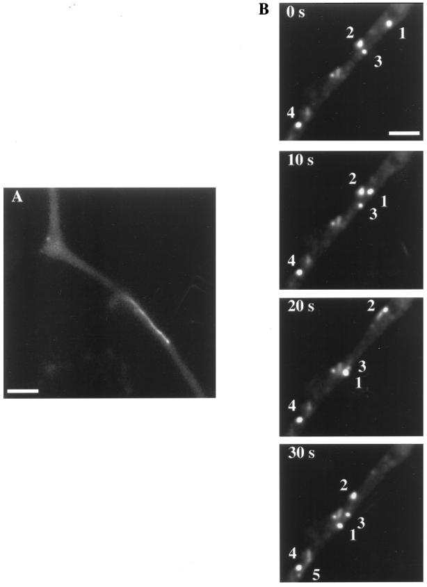
To monitor the dynamics of tPA/GFP in PC12 cells, time-lapse imaging was conducted. Figure 6 shows sequential time-lapse images of tPA/GFP-expressing PC12 cells. In the cell body, granules generally undergo saltatory motion in all directions (our unpublished data) (Wacker et al., 1997). However, some granules remain closely apposed to the plasma membrane, especially in undifferentiated PC12 cells (see Figure 2G). Plasma membrane-apposed distribution in undifferentiated cells presumably reflects accumulation of granules near potential sites of secretion.
Figure 6.
Time-lapse images of the axonal transport of secretory granules in PC12 cells. The granule in panel A is streaking in the anterograde direction along the neurite of a PC12 cell at a speed of ∼1.6 μm/s. This granule covered the entire length of the neurite in one long, unidirectional excursion. The sequential images in panel B exhibit a spectrum of generally less rapid granule motions. Granule 1 moves in the anterograde direction at a speed of ∼0.4 μm/s, slowing somewhat during the last frame. Granule 2 is motionless at first; it then starts moving in the retrograde direction at a speed of ∼0.4 μm/s, leaving a streak as it achieves an instantaneous speed of at least 0.9 μm/s. Granule 2 then reverses direction and finally moves in the anterograde direction at a speed of ∼0.7 μm/s. Granule 3 at first moves slowly in the anterograde direction; it then moves slightly out of the focal plane, reappearing more clearly in the last image. Granule 4 is essentially immobile during the entire observation period. Finally, granule 5 appears in the last frame near granule 4, streaking along the neurite at ∼1.3 μm/s. Bar, 4 μm.
In neurites, the dynamics is complex, as exemplified by the quantitative axonal transport data presented in Table 1. Some granules are observed to undergo long stretches of unidirectional anterograde motion at characteristic fast axonal transport speeds of 1–2 μm/s. These fast moving granules often appear as streaks in the images (see Figure 6). The remaining granules exhibit a spectrum of dynamic behaviors, including retrograde motion and direction reversal (see Figure 6). In addition, granules often saltate, and some granules remain relatively motionless during the entire observation period. Granule speeds ranged from ∼0 μm/s (stalled granules) to ∼1.6 μm/s (streaking granules). Sample movies of axonal transport of granules containing tPA/GFP can be viewed at http://www.lclark.edu/∼bethe/.
Table 1.
Average axonal transport data obtained by studying the dynamics of granules containing tPA/GFP in movies generated from 10 representative PC12 cells, as described in MATERIALS AND METHODS
| Linear density of granules containing tPA/GFP | v (μm/s) | % Anterograde | % Reversal |
|---|---|---|---|
| ∼0.1 fluorescent granules/μm | 0.92 ± 0.16 | 93 ± 10 | 14 ± 10 |
| ∼1.0 fluorescent granules/μm | 0.46 ± 0.23 | 63 ± 3 | 15 ± 7 |
| ∼3.0 fluorescent granules/μm | 0.28 ± 0.12 | 44 ± 10 | 31 ± 15 |
The data show that average granule speed and average percent anterograde flux depend on the linear density of granules along neurites. Average granule speed and average percent anterograde flux both steadily decrease as the linear density of granules containing tPA/GFP rises from ∼0.1 fluorescent granules/μm to ∼3 fluorescent granules/μm.
The percent of granules in the neurite exhibiting each type of motion varies considerably from cell to cell; this is especially true for the relative percents exhibiting anterograde and retrograde motion, as discussed below. However, over the time scale of most of our movies (<5 min), typically ∼20% of the granules remain relatively motionless and at least 10% of the granules exhibit one or more direction reversals. Over longer time scales, the percentages might be different.
The linear density of granules containing tPA/GFP is an important determinant of granule dynamics along neurites (see Table 1). To begin to identify the origin of this density effect, we compared densities of granules containing tPA/GFP in transfected cells with densities of granules containing endogenous tPA in immunostained, untransfected PC12 cells. Endogenous tPA densities lie in the range ∼0.5–1.0 granules/μm, whereas tPA/GFP densities lie in the range ∼0.1–3.0 granules/μm (see Table 1). Thus, we have studied granule dynamics in transfected cells over a range of granule densities representative of endogenous levels. In the DISCUSSION, we interpret these data, and our axonal transport results in the context of a regulatory model of secretory granule dynamics.
The dependence of axonal transport behavior on the density of granules containing tPA/GFP is illustrated in Table 1. In the least congested (fluorescent) neurites (∼0.1 fluorescent granules/μm), net granule fluxes are ∼93% anterograde. Moreover, in such neurites, many mobile granules undergo long stretches of unidirectional fast anterograde motion; specifically, average granule speed is 0.92 μm/s. Direction reversal also is observed occasionally, although its frequency (∼14%) is somewhat difficult to quantify because granule number is low in uncongested cells.
In more congested neurites, long stretches of fast unidirectional anterograde motion are less common, whereas retrograde motion and direction reversal are more common. In cells of intermediate congestion (∼1.0 fluorescent granules/μm), net granule fluxes are ∼63% anterograde, average percent reversal is 15%, and average speed is 0.46 μm/s. In the most congested cells (∼3.0 fluorescent granules/μm), net granule fluxes are ∼44% anterograde, average percent reversal is 31%, and average speed is 0.28 μm/s. These results suggest that motion slows and that retrograde and anterograde movements balance most completely in cells exhibiting the highest degree of congestion ∼24 h after transfection. Moreover, the results suggest that the attributes of transport can evolve with time. In particular, the net granule flux in congested cells must have been >50% anterograde at some point before observation; otherwise, granules would not have accumulated in growth cones (see Figure 2). Indeed, such an evolution in transport behavior is consistent with our observations that increased congestion reduces the percent anterograde flux and that PC12 cells become more congested on average as they advance from 10 to 24 h post-transfection.
DISCUSSION
The experiments described here were directed 1) at characterizing the distribution, dynamics, and secretory behavior of an important secretory protein, tPA, in living neuronal cells; and 2) at relating the results to current ideas about tPA’s role in axonal elongation during neuronal differentiation.
Implications of Distribution and Secretion
We have shown that both tPA/GFP and endogenous tPA display punctate distribution patterns that are enhanced in growth cones of PC12 cells. Moreover, we have obtained strong evidence suggesting that tPA/GFP is secreted from growth cones in response to secretagogues. We have also shown, in contrast, that GFP(SIG+) displays a punctate distribution pattern that is not enhanced in growth cones and that secretion of GFP(SIG+) is not influenced by secretagogues. Clearly, trafficking through the endoplasmic reticulum and Golgi is not sufficient to target tPA for accumulation in growth cones or into the regulated pathway but rather requires signals on tPA itself. Most significantly, regulated secretion of tPA/GFP from growth cones has important implications for tPA’s postulated function during neuronal differentiation.
During axonal elongation, movement of the growth cone requires clearing of proteinaceous barriers imposed by the ECM, as well as regulation of detachment of the growth cone from the ECM. For this reason, proteases that can degrade components of the ECM have frequently been hypothesized to play a pivotal role in axonal elongation. Members of the serine protease family, the plasminogen activators (PAs), have been intensely studied in this context, and a considerable body of data suggests a direct role for PAs and plasmin in degradative processes occurring at the growth cone during axonal elongation (Krystosek and Seeds, 1981, 1984; for review see Monard, 1988; Pittman et al., 1989; McGuire and Seeds, 1990; Garcia-Rocha et al., 1994; Pittman and DiBenedetto, 1995; Hayden and Seeds, 1996). PAs may also play an indirect role in degradative processes occurring at the growth cone. For example, plasmin, the major cleavage product of PAs, can cleave prostromelysin-1 and procollagenase, producing the active ECM-degrading proteases stromelysin-1 and collagenase (for reviews see Matrisian and Hogan, 1990; Matrisian, 1992). A proteolytic activation cascade involving PAs, plasmin, stromelysin-1, and collagenase may therefore be relevant to ECM degradation during neuronal differentiation.
Although two PAs have been hypothesized to play a role in axonal elongation, we have focused on tPA because it is the dominant PA in PC12 cells and the brain (Leprince et al., 1991; Seeds et al., 1995). Significantly, we have found that tPA/GFP makes a transition from a plasma membrane-apposed distribution to a growth cone-biased distribution during differentiation. In addition, we have shown that this biased distribution arises during differentiation because tPA/GFP is targeted into regulated granules that undergo predominantly anterograde axonal transport when the concentration of tPA/GFP in the growth cone is not too high. Most importantly, we have found that the secretagogue carbachol induces significant loss of tPA/GFP fluorescence from growth cones of differentiated PC12 cells, suggesting that growth cones are sites of regulated secretion of tPA/GFP. Given the similarity in distribution and secretory behavior exhibited by tPA/GFP and tPA, our results strongly suggest that growth cones are also sites of regulated secretion of endogenous tPA. Indeed, it is tempting to speculate, in the light of past work and the results presented here, that PC12 cells store secretory granules containing tPA at growth cones so that tPA can be readily and strategically released during neuronal differentiation upon receipt of an appropriate regulatory stimulus (Harrison et al., 1996).
We have observed a substantial reduction in tPA/GFP fluorescence from growth cones in most, but not all, cells exposed to carbachol. One possible explanation for this result is that tPA/GFP fluorescence from growth cones is influenced by two competing factors in the presence of carbachol. First, efflux of tPA/GFP induced by carbachol should cause a loss of fluorescence signal. Second, net anterograde axonal transport of tPA/GFP into growth cones, which occurs in most cells, should cause a gain in fluorescence signal. In the presence of these two competing factors, the fluorescence from a growth cone could in principle rise, fall, or remain relatively unchanged depending on the relative rates of efflux and influx. In contrast, total cellular fluorescence should fall in the presence of carbachol, assuming that new protein synthesis cannot keep pace with regulated secretion.
Implications of Dynamics
Granules containing tPA/GFP exhibit a spectrum of dynamic behaviors. Some granules undergo canonical fast axonal transport, whereas others move more slowly, saltate, or even reverse their direction of motion. This complex, bidirectional dynamics provides insight into processes that may regulate and drive the axonal transport of secretory granules.
An important and unique feature of the axonal transport described here is that it reflects the movement of secretory granules alone, which have been singled out for visualization using fluorescence microscopy by labeling tPA with GFP. In contrast, the axonal transport described in the past has generally reflected movement of a more heterogeneous population of axoplasmic organelles, because visualization was based on nonspecific optical microscopy techniques (such as differential interference contrast and phase contrast) (see, e.g., Allen et al., 1982). This difference is worthy of note because it can significantly alter the interpretation attached to the observed features of transport.
We have found that granules containing tPA/GFP undergo bidirectional axonal transport that is biased in the anterograde direction, with the degree of bias dependent on the degree of granule accumulation. Although bidirectional motion has frequently been observed in previous studies of axonal transport in heterogeneous systems, its origin in these systems is readily attributed to the diversity of organelle types observed (see, e.g., Allen et al., 1982; Vale et al., 1985c). For example, bidirectional transport could represent a combination of retrograde movement of endosomes and anterograde movement of secretory vesicles. In contrast, bidirectional motion in our system is more difficult to explain, because we are observing a homogeneous population of granules presumably all targeted for secretion.
Retrograde axonal transport of granules containing tPA/GFP may have its origin in regulatory mechanisms designed to maintain cellular homeostasis. Regulatory mechanisms have previously been invoked to explain increased retrograde transport of autophagic vacuoles when axonal elongation is blocked and macromolecules thus begin to accumulate at the growth cone (Hollenbeck and Bray, 1987; Hollenbeck, 1993). As in the case of blocked axonal elongation, in the absence of secretagogues, granules containing tPA/GFP appear to accumulate in growth cones. Moreover, retrograde transport of granules appears to increase as the number of fluorescent granules in the neurite increases. Retrograde transport of tPA/GFP may thus also reflect a control mechanism that is designed to maintain homeostasis by transporting excess tPA/GFP back toward the cell body. Alternatively, since secreted tPA is known to bind to the cell surface (Pittman et al., 1989; Verrall and Seeds, 1989; McGuire and Seeds, 1990), retrograde transport could represent movement of endocytosed tPA/GFP back toward the cell body. This is likely to be a small effect in our system, because secretion is low in the absence of secretagogues (Harrison et al., 1996).
We have also found that individual granules containing tPA/GFP can reverse their direction of transport along the neurite. In this respect, granules in PC12 cells behave like autophagic vacuoles, mitochondria, endosomes, and tubulovesicular organelles containing plasma membrane proteins in cultured neurons, as well as larger particles in squid giant axon (Allen et al., 1982; Hollenbeck, 1993; Morris and Hollenbeck, 1993; Overly et al., 1996; Nakata et al., 1998), and not like the large spectrum of axoplasmic organelles that are rarely observed to reverse direction (Allen et al., 1982; Schnapp et al., 1985; Kreis et al., 1989; Parton et al., 1992). Clearly, different classes of axoplasmic organelles exhibit somewhat different dynamic behaviors, highlighting the complexity and variability of axonal transport.
The microtubules in PC12 cell neurites have a uniform orientation, with their plus ends toward the distal tip (Okabe and Hirokawa, 1988); thus, direction reversal by granules presumably reflects, in large part, underlying properties of the motors directing transport, and not nonuniform microtubule polarity. Current evidence suggests that some organelles simultaneously bind kinesin and dynein (Hirokawa et al., 1990; Yu et al., 1992; Muresan et al., 1996; Nilsson et al., 1996), and the direction reversal observed here suggests that motors of opposite polarity are also simultaneously bound to secretory granules in PC12 cells. However, it is frequently assumed that if two opposite polarity motors are simultaneously bound to axoplasmic organelles, one is bound in an inactive form, leading to organelle movement in one direction only (see, e.g., Hirokawa et al., 1990; for review see Hirokawa, 1993). Although this seems reasonable for organelles that never reverse direction, it seems less reasonable for organelles like granules that sometimes reverse direction.
Direction reversal by granules could have its origin in mechanisms that have been postulated to cause direction reversal by microtubules bound to glass-adsorbed kinesin and dynein in vitro (Vale et al., 1992b). When bound at low motor densities to two opposite-polarity motors in vitro, some microtubules remain stationary, while others undergo several-micron-long unidirectional excursions punctuated by direction reversal. In contrast, when bound at high motor densities, microtubules undergo unidirectional motion, albeit relatively slowly. This complex, multifaceted dynamics shares many features in common with axonal transport of secretory granules in PC12 cells, suggesting that the underlying mechanistic origins may be similar. In the case of microtubules in vitro, the attributes of motion are apparently determined by the relative attachment times of the two opposite-polarity motors and their relative numbers. It is tempting to speculate, by analogy, that the complex dynamics we have observed for granules in PC12 cells is dictated by similar factors.
Comparison with Previous Studies of Axonal Transport
The squid giant axon and cultured neuronal cells are two prominent and pertinent systems that have commonly been used in optical microscopy studies of axonal transport (see, e.g., Allen et al., 1982, 1985; Brady et al., 1982; Schnapp et al., 1985; Vale et al., 1985b,c; Hollenbeck and Bray, 1987; Parton et al., 1992; Hollenbeck, 1993; Morris and Hollenbeck, 1995). In the squid giant axon studies, bidirectional motion of a spectrum of particles, ranging from small synaptic vesicles to relatively large mitochondria, has typically been observed. Small particles (<0.2 μm in diameter) exhibit the most rapid, least intermittent, and most unidirectional motion. Medium (0.2–0.6 μm) and large (0.8–5.0 μm) particles move more slowly and erratically, sometimes even reversing their direction of motion (Allen et al., 1982; Vale et al., 1985c). The movement of granules containing tPA/GFP described here corresponds most closely to the movement of medium and large particles along the squid giant axon.
In particularly pertinent work involving cultured cells, axonal transport of acridine orange-labeled secretory granules and endosomes was directly observed in AtT20 cells using video-enhanced fluorescence microscopy (Kreis et al., 1989). Because acridine orange labels both secretory granules and endosomes (i.e., acidic organelles), the motion of secretory granules alone in AtT20 cells had to be inferred indirectly using both fixed and living cells. Based on such indirect analysis, secretory granules in AtT20 cells were determined to move bidirectionally, although movement was biased in the anterograde direction. Endosomes were also found to move bidirectionally, although movement was biased in the retrograde direction. Thus, in AtT20 and PC12 cells, there are some common attributes to axonal transport of secretory granules, such as bidirectionality. However, in PC12 cells, the degree of transport bias appears to depend on the degree of granule accumulation in growth cones; no such effect was reported for AtT20 cells. Moreover, individual granules in AtT20 cells do not reverse direction, as they sometimes do in PC12 cells.
Conclusions
The distribution of tPA in living neuronal cells and the axonal transport of a homogeneous population of regulated secretory granules can be visualized at high resolution using fluorescence microscopy and a fluorescent tPA fusion protein involving GFP. Application of this methodology shows that 1) tPA/GFP is transported in PC12 cells in secretory granules that accumulate in growth cones in the absence of secretagogues and that are secreted from growth cones in the presence of secretagogues, and 2) axonal transport of granules containing tPA/GFP proceeds bidirectionally between the cell body and the growth cone with net granule flux in the anterograde direction in less congested neurites. Retrograde transport of granules containing tPA/GFP may represent a regulatory mechanism designed to maintain homeostasis, and the pool of tPA/GFP in growth cones may represent a store that can be strategically released during neuronal differentiation upon receipt of an appropriate regulatory stimulus.
ACKNOWLEDGMENTS
We thank Dr. Randall N. Pittman of the University of Pennsylvania School of Medicine for the gene for rat tPA and Dr. Sharon Tooze of the Imperial Cancer Research Institute for the antibody against SgII. We also thank Drs. Gary Thomas and Sheree Rybak of Oregon Health Sciences University and Dr. James Abney of Kolisch, Hartwell, Dickinson, McCormack and Heuser for a critical reading of the manuscript. This work was supported by National Science Foundation grant BIR-9510226 (to J.E.L. and B.A.S.).
REFERENCES
- Alberts B, Bray D, Lewis J, Raff M, Roberts K, Watson JD. Molecular Biology of the Cell. 3rd ed. New York: Garland; 1994. [Google Scholar]
- Allen RD, Metuzals J, Tasaki I, Brady ST, Gilbert SP. Fast axonal transport in squid giant axon. Science. 1982;218:1127–1129. doi: 10.1126/science.6183744. [DOI] [PubMed] [Google Scholar]
- Allen RD, Weiss DG, Hayden JH, Brown DT, Fujiwake H, Simpson M. Gliding movement of and bidirectional transport along single native microtubules from squid axoplasm: evidence for an active role of microtubules in cytoplasmic transport. J Cell Biol. 1985;100:1736–1752. doi: 10.1083/jcb.100.5.1736. [DOI] [PMC free article] [PubMed] [Google Scholar]
- Bottenstein JE, Sato GH. Growth of a rat neuroblastoma cell line in serum-free supplemented medium. Proc Natl Acad Sci USA. 1979;76:514–517. doi: 10.1073/pnas.76.1.514. [DOI] [PMC free article] [PubMed] [Google Scholar]
- Brady ST. A novel brain ATPase with properties expected for the fast axonal transport motor. Nature. 1985;317:73–75. doi: 10.1038/317073a0. [DOI] [PubMed] [Google Scholar]
- Brady ST, Lasek RJ, Allen RD. Fast axonal transport in extruded axoplasm from squid giant axon. Science. 1982;218:1129–1131. doi: 10.1126/science.6183745. [DOI] [PubMed] [Google Scholar]
- Burke NV, Han W, Li D, Takimoto K, Watkins SC, Levitan ES. Neuronal peptide release is limited by secretory granule mobility. Neuron. 1997;19:1095–1102. doi: 10.1016/s0896-6273(00)80400-6. [DOI] [PubMed] [Google Scholar]
- Chalfie M, Tu Y, Euskirchen G, Ward WW, Prasher DC. Green fluorescent protein as a marker for gene expression. Science. 1994;263:802–805. doi: 10.1126/science.8303295. [DOI] [PubMed] [Google Scholar]
- Cole NB, Smith CL, Sciaky N, Terasaki M, Edidin M, Lippincott-Schwartz J. Diffusional mobility of Golgi proteins in membranes of living cells. Science. 1996;273:797–801. doi: 10.1126/science.273.5276.797. [DOI] [PubMed] [Google Scholar]
- Cormack BP, Valdivia RH, Falkow S. FACS-optimized mutants of the green fluorescent protein (GFP) Gene. 1996;173:33–38. doi: 10.1016/0378-1119(95)00685-0. [DOI] [PubMed] [Google Scholar]
- Craig AM, Banker G. Neuronal polarity. Annu Rev Neurosci. 1994;17:267–310. doi: 10.1146/annurev.ne.17.030194.001411. [DOI] [PubMed] [Google Scholar]
- Garcia-Rocha M, Avila J, Armas-Portela R. Tissue-type plasminogen activator (tPA) is the main plasminogen activator associated with isolated rat nerve growth cones. Neurosci Lett. 1994;180:123–126. doi: 10.1016/0304-3940(94)90502-9. [DOI] [PubMed] [Google Scholar]
- Grafstein B, Forman DS. Intracellular transport in neurons. Physiol Rev. 1980;60:1167–1283. doi: 10.1152/physrev.1980.60.4.1167. [DOI] [PubMed] [Google Scholar]
- Greene LA, Tischler AS. Establishment of a noradrenergic clonal line of rat adrenal pheochromocytoma cells which respond to nerve growth factor. Proc Natl Acad Sci USA. 1976;73:2424–2428. doi: 10.1073/pnas.73.7.2424. [DOI] [PMC free article] [PubMed] [Google Scholar]
- Hajjar KA. Cellular receptors in the regulation of plasmin generation. Thromb Haemostasis. 1995;74:294–301. [PubMed] [Google Scholar]
- Harrison TM, Chidgey MAJ, Uff S. Novel markers for constitutive secretion used to show that tissue plasminogen activator is sorted to the regulated pathway in transfected PC12 cells. Cell Biol Int. 1996;20:293–300. doi: 10.1006/cbir.1996.0033. [DOI] [PubMed] [Google Scholar]
- Haseloff J, Siemering KR, Prasher DC, Hodge S. Removal of a cryptic intron and subcellular localization of green fluorescent protein are required to mark transgenic Arabidopsis plants brightly. Proc Natl Acad Sci USA. 1997;94:2122–2127. doi: 10.1073/pnas.94.6.2122. [DOI] [PMC free article] [PubMed] [Google Scholar]
- Hayden SM, Seeds NW. Modulated expression of plasminogen activator system components in cultured cells from dissociated mouse dorsal root ganglia. J Neurosci. 1996;16:2307–2317. doi: 10.1523/JNEUROSCI.16-07-02307.1996. [DOI] [PMC free article] [PubMed] [Google Scholar]
- Heim R, Cubitt AB, Tsien RY. Improved green fluorescence. Nature. 1995;373:663–664. doi: 10.1038/373663b0. [DOI] [PubMed] [Google Scholar]
- Hiraoka Y, Swedlow JR, Paddy MR, Agard DA, Sedat JW. Three-dimensional multiple-wavelength fluorescence microscopy for the structural analysis of biological phenomena. Semin Cell Biol. 1991;2:153–165. [PubMed] [Google Scholar]
- Hirokawa N. Axonal transport and the cytoskeleton. Curr Opin Neurobiol. 1993;3:724–731. doi: 10.1016/0959-4388(93)90144-n. [DOI] [PubMed] [Google Scholar]
- Hirokawa N, Sato-Yoshitake R, Yoshida T, Kawashima T. Brain dynein (MAP1C) localizes on both anterogradely and retrogradely transported membranous organelles in vivo. J Cell Biol. 1990;111:1027–1037. doi: 10.1083/jcb.111.3.1027. [DOI] [PMC free article] [PubMed] [Google Scholar]
- Hollenbeck PJ. Products of endocytosis and autophagy are retrieved from axons by regulated retrograde organelle transport. J Cell Biol. 1993;121:305–315. doi: 10.1083/jcb.121.2.305. [DOI] [PMC free article] [PubMed] [Google Scholar]
- Hollenbeck PJ, Bray D. Rapidly transported organelles containing membrane and cytoskeletal components: their relation to axonal growth. J Cell Biol. 1987;105:2827–2835. doi: 10.1083/jcb.105.6.2827. [DOI] [PMC free article] [PubMed] [Google Scholar]
- Kaether C, Gerdes H-H. Visualization of protein transport along the secretory pathway using green fluorescent protein. FEBS Lett. 1995;369:267–271. doi: 10.1016/0014-5793(95)00765-2. [DOI] [PubMed] [Google Scholar]
- Kreis TE, Matteoni R, Hollinshead M, Tooze J. Secretory granules and endosomes show saltatory movement biased to the anterograde and retrograde directions, respectively, along microtubules in AtT20 cells. Eur J Cell Biol. 1989;49:128–139. [PubMed] [Google Scholar]
- Krystosek A, Seeds NW. Plasminogen activator release at the neuronal growth cone. Science. 1981;213:1532–1534. doi: 10.1126/science.7197054. [DOI] [PubMed] [Google Scholar]
- Krystosek A, Seeds NW. Peripheral neurons and Schwann cells secrete plasminogen activator. J Cell Biol. 1984;98:773–776. doi: 10.1083/jcb.98.2.773. [DOI] [PMC free article] [PubMed] [Google Scholar]
- Lang T, Wacker I, Steyer J, Kaether C, Wunderlich I, Soldati T, Gerdes H-H, Almers W. Ca2+-triggered peptide secretion in single cells imaged with green fluorescent protein and evanescent-wave microscopy. Neuron. 1997;18:857–863. doi: 10.1016/s0896-6273(00)80325-6. [DOI] [PubMed] [Google Scholar]
- Leprince P, Rogister B, Delree P, Rigo JM, Andre B, Moonen G. Modulation of proteolytic activity during neuritogenesis in the PC12 nerve cell: differential control of plasminogen activator and plasminogen activator inhibitor activities by nerve growth factor and dibutyryl-cyclic AMP. J Neurochem. 1991;57:665–674. doi: 10.1111/j.1471-4159.1991.tb03798.x. [DOI] [PubMed] [Google Scholar]
- Matrisian LM. The matrix-degrading metalloproteinases. Bioessays. 1992;14:455–463. doi: 10.1002/bies.950140705. [DOI] [PubMed] [Google Scholar]
- Matrisian LM, Hogan BLM. Growth factor-related proteases and extracellular matrix remodeling during mammalian development. Curr Top Dev Biol. 1990;24:219–259. doi: 10.1016/s0070-2153(08)60089-7. [DOI] [PubMed] [Google Scholar]
- McGuire PG, Seeds NW. Degradation of underlying extracellular matrix by sensory neurons during neurite outgrowth. Neuron. 1990;4:633–642. doi: 10.1016/0896-6273(90)90121-u. [DOI] [PubMed] [Google Scholar]
- Monard D. Cell-derived proteases and protease inhibitors as regulators of neurite outgrowth. Trends Neurosci. 1988;11:541–544. doi: 10.1016/0166-2236(88)90182-8. [DOI] [PubMed] [Google Scholar]
- Morris RL, Hollenbeck PJ. The regulation of bidirectional mitochondrial transport is coordinated with axonal outgrowth. J Cell Sci. 1993;104:917–927. doi: 10.1242/jcs.104.3.917. [DOI] [PubMed] [Google Scholar]
- Morris RL, Hollenbeck PJ. Axonal transport of mitochondria along microtubules and F-actin in living vertebrate neurons. J Cell Biol. 1995;131:1315–1326. doi: 10.1083/jcb.131.5.1315. [DOI] [PMC free article] [PubMed] [Google Scholar]
- Muresan V, Godek CP, Reese TS, Schnapp BJ. Plus-end motors override minus-end motors during transport of squid axon vesicles on microtubules. J Cell Biol. 1996;135:383–397. doi: 10.1083/jcb.135.2.383. [DOI] [PMC free article] [PubMed] [Google Scholar]
- Nakata T, Terada S, Hirokawa N. Visualization of the dynamics of synaptic vesicle and plasma membrane proteins in living axons. J Cell Biol. 1998;140:659–674. doi: 10.1083/jcb.140.3.659. [DOI] [PMC free article] [PubMed] [Google Scholar]
- Nilsson H, Rutberg M, Wallin M. Localization of kinesin and cytoplasmic dynein in cultured melanophores from atlantic cod, Gadus morhua. Cell Motil Cytoskeleton. 1996;33:183–196. doi: 10.1002/(SICI)1097-0169(1996)33:3<183::AID-CM3>3.0.CO;2-C. [DOI] [PubMed] [Google Scholar]
- Ogawa H, Inouye S, Tsuji FI, Yasuda K, Umesono K. Localization, trafficking, and temperature-dependence of the Aequorea green fluorescent protein in cultured vertebrate cells. Proc Natl Acad Sci USA. 1995;92:11899–11903. doi: 10.1073/pnas.92.25.11899. [DOI] [PMC free article] [PubMed] [Google Scholar]
- Okabe S, Hirokawa N. Microtubule dynamics in nerve cells: analysis using microinjection of biotinylated tubulin into PC12 cells. J Cell Biol. 1988;107:651–664. doi: 10.1083/jcb.107.2.651. [DOI] [PMC free article] [PubMed] [Google Scholar]
- Overly CC, Rieff HI, Hollenbeck PJ. Organelle motility and metabolism in axons vs dendrites of cultured hippocampal neurons. J Cell Sci. 1996;109:971–980. doi: 10.1242/jcs.109.5.971. [DOI] [PubMed] [Google Scholar]
- Parmer RJ, Mahata M, Mahata S, Sebald MT, O’Connor DT, Miles LA. Tissue plasminogen activator (t-PA) is targeted to the regulated secretory pathway. Catecholamine storage vesicles as a reservoir for the rapid release of t-PA. J Biol Chem. 1997;272:1976–1982. doi: 10.1074/jbc.272.3.1976. [DOI] [PubMed] [Google Scholar]
- Parton RG, Simons K, Dotti CG. Axonal and dendritic endocytotic pathways in cultured neurons. J Cell Biol. 1992;119:123–137. doi: 10.1083/jcb.119.1.123. [DOI] [PMC free article] [PubMed] [Google Scholar]
- Paschal BM, Vallee RB. Retrograde transport by the microtubule-associated protein MAP 1C. Nature. 1987;330:181–183. doi: 10.1038/330181a0. [DOI] [PubMed] [Google Scholar]
- Pittman RN, DiBenedetto AJ. PC12 cells overexpressing tissue plasminogen activator regenerate neurites to a greater extent and migrate faster than control cells in complex extracellular matrix. J Neurochem. 1995;64:566–575. doi: 10.1046/j.1471-4159.1995.64020566.x. [DOI] [PubMed] [Google Scholar]
- Pittman RN, Ivins JK, Buettner HM. Neuronal plasminogen activators: cell surface binding sites and involvement in neurite outgrowth. J Neurosci. 1989;9:4269–4286. doi: 10.1523/JNEUROSCI.09-12-04269.1989. [DOI] [PMC free article] [PubMed] [Google Scholar]
- Possenti R, Eldridge JD, Paterson BM, Grasso A, Levi A. A protein induced by NGF in PC12 cells is stored in secretory vesicles and released through the regulated pathway. EMBO J. 1989;8:2217–2223. doi: 10.1002/j.1460-2075.1989.tb08345.x. [DOI] [PMC free article] [PubMed] [Google Scholar]
- Rivas RJ, Moore H-PH. Spatial segregation of the regulated and constitutive secretory pathways. J Cell Biol. 1989;109:51–60. doi: 10.1083/jcb.109.1.51. [DOI] [PMC free article] [PubMed] [Google Scholar]
- Rizzuto R, Brini M, De Giorgi F, Rossi R, Heim R, Tsien RY, Pozzan T. Double labelling of subcellular structures with organelle-targeted GFP mutants in vivo. Curr Biol. 1996;6:183–188. doi: 10.1016/s0960-9822(02)00451-7. [DOI] [PubMed] [Google Scholar]
- Scalettar BA, Swedlow JR, Sedat JW, Agard DA. Dispersion, aberration and deconvolution in multi-wavelength fluorescence images. J Microsc. 1996;182:50–60. doi: 10.1046/j.1365-2818.1996.122402.x. [DOI] [PubMed] [Google Scholar]
- Schnapp BJ, Reese TS. Dynein is the motor for retrograde axonal transport of organelles. Proc Natl Acad Sci USA. 1989;86:1548–1552. doi: 10.1073/pnas.86.5.1548. [DOI] [PMC free article] [PubMed] [Google Scholar]
- Schnapp BJ, Vale RD, Sheetz MP, Reese TS. Single microtubules from squid axoplasm support bidirectional movement of organelles. Cell. 1985;40:455–462. doi: 10.1016/0092-8674(85)90160-6. [DOI] [PubMed] [Google Scholar]
- Schroer TA. Motors for fast axonal transport. Curr Opin Neurobiol. 1992;2:618–621. doi: 10.1016/0959-4388(92)90028-j. [DOI] [PubMed] [Google Scholar]
- Schroer TA, Steuer ER, Sheetz MP. Cytoplasmic dynein is a minus end-directed motor for membranous organelles. Cell. 1989;56:937–946. doi: 10.1016/0092-8674(89)90627-2. [DOI] [PubMed] [Google Scholar]
- Seeds NW, Williams BL, Bickford PC. Tissue plasminogen activator induction in Purkinje neurons after cerebellar motor learning. Science. 1995;270:1992–1994. doi: 10.1126/science.270.5244.1992. [DOI] [PubMed] [Google Scholar]
- Sheetz MP, Martenson CH. Axonal transport: beyond kinesin and cytoplasmic dynein. Curr Opin Neurobiol. 1991;1:393–398. doi: 10.1016/0959-4388(91)90059-g. [DOI] [PubMed] [Google Scholar]
- Shelby RD, Hahn KM, Sullivan KF. Dynamic elastic behavior of α-satellite DNA domains visualized in situ in living human cells. J Cell Biol. 1996;135:545–557. doi: 10.1083/jcb.135.3.545. [DOI] [PMC free article] [PubMed] [Google Scholar]
- Shpetner HS, Paschal BM, Vallee RB. Characterization of the microtubule-activated ATPase of brain cytoplasmic dynein (MAP 1C) J Cell Biol. 1988;107:1001–1009. doi: 10.1083/jcb.107.3.1001. [DOI] [PMC free article] [PubMed] [Google Scholar]
- Sumi Y, Dent MAR, Owen DE, Seeley PJ, Morris RJ. The expression of tissue and urokinase-type plasminogen activators in neural development suggests different modes of proteolytic involvement in neuronal growth. Development. 1992;116:625–637. doi: 10.1242/dev.116.3.625. [DOI] [PubMed] [Google Scholar]
- Tooze SA, Hollinshead M, Dittie AS. Antibodies to secretogranin II reveal potential processing sites. Biochimie. 1994;76:271–276. doi: 10.1016/0300-9084(94)90157-0. [DOI] [PubMed] [Google Scholar]
- Tsirka S, Gualandris A, Amaral DG, Strickland S. Excitotoxin-induced neuronal degeneration and seizure are mediated by tissue plasminogen activator. Nature. 1995;377:340–344. doi: 10.1038/377340a0. [DOI] [PubMed] [Google Scholar]
- Vale RD, Banker G, Hall ZW. The neuronal cytoskeleton. In: Hall Z W, Sunderland, editors. An Introduction to Molecular Neurobiology. Sinauer Associates; 1992a. pp. 247–280. [Google Scholar]
- Vale RD, Malik F, Brown D. Directional instability of microtubule transport in the presence of kinesin and dynein, two opposite polarity motor proteins. J Cell Biol. 1992b;119:1589–1596. doi: 10.1083/jcb.119.6.1589. [DOI] [PMC free article] [PubMed] [Google Scholar]
- Vale RD, Reese TS, Sheetz MP. Identification of a novel force-generating protein, kinesin, involved in microtubule-based motility. Cell. 1985a;42:39–50. doi: 10.1016/s0092-8674(85)80099-4. [DOI] [PMC free article] [PubMed] [Google Scholar]
- Vale RD, Schnapp BJ, Mitchison T, Steuer E, Reese TS, Sheetz MP. Different axoplasmic proteins generate movement in opposite directions along microtubules in vitro. Cell. 1985b;43:623–632. doi: 10.1016/0092-8674(85)90234-x. [DOI] [PubMed] [Google Scholar]
- Vale RD, Schnapp BJ, Reese TS, Sheetz MP. Movement of organelles along filaments dissociated from the axoplasm of the squid giant axon. Cell. 1985c;40:449–454. doi: 10.1016/0092-8674(85)90159-x. [DOI] [PubMed] [Google Scholar]
- Vallee RB, Sheetz MP. Targeting of motor proteins. Science. 1996;271:1539–1544. doi: 10.1126/science.271.5255.1539. [DOI] [PubMed] [Google Scholar]
- Vallee RB, Wall JS, Paschal BM, Shpetner HS. Microtubule-associated protein 1C from brain is a two-headed cytosolic dynein. Nature. 1988;332:561–563. doi: 10.1038/332561a0. [DOI] [PubMed] [Google Scholar]
- Verrall S, Seeds NW. Characterization of 125I-tissue plasminogen activator binding to cerebellar granule neurons. J Cell Biol. 1989;109:265–271. doi: 10.1083/jcb.109.1.265. [DOI] [PMC free article] [PubMed] [Google Scholar]
- Wacker I, Kaether C, Kromer A, Migala A, Almers W, Gerdes H-H. Microtubule-dependent transport of secretory vesicles visualized in real time with a GFP-tagged secretory protein. J Cell Sci. 1997;110:1453–1463. doi: 10.1242/jcs.110.13.1453. [DOI] [PubMed] [Google Scholar]
- Yu H, Toyoshima I, Steuer ER, Sheetz MP. Kinesin and cytoplasmic dynein binding to brain microsomes. J Biol Chem. 1992;267:20457–20464. [PubMed] [Google Scholar]


