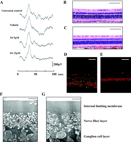
FIG. 8.
Histological and physiological examinations of eyes injected with simvastatin. Rabbits received intravitreal injections of 0.1 ml vehicle or vehicle with simvastatin (final concentration of 5 or 15 μmol/l simvastatin) on days 0, 1, 3, 5, and 7. A: Electroretinograms on day 28. A flash stimulus of intensity 1.30 log cd.s/m2 was superimposed on a background luminance of 1.15 log cd/m2. Light microscopy of the rabbit eye without any treatment (B) and that of the eye injected with 15 μmol/l simvastatin (C). Scale bar = 100 μm. Apoptotic and potentially necrotic cell death detected by TUNEL in the section from positive control (rabbit PVR model on day 7 after its onset) (D) and in the section from the 15 μmol/l simvastatin-treated eye (E). Scale bar = 50 μm. Transmission electron microscopy of the rabbit eye without treatment (F) and that of the eye injected with 15 μmol/l simvastatin (G). Scale bar = 10 μm. (Please see http://dx.doi.org/10.2337/db08-0302 for a high-quality digital representation of this image.)

