Abstract
Tarso-metatarsal injuries are rare but frequently missed. Due to the large variation in pathomorphic forms of these injuries, great precision is required when carrying out clinical and X-ray diagnostic procedures. The aim of the study was to describe the different forms of Lisfranc joint injuries and analyse the causes of delayed treatment. The treatment results of acute and chronic injuries were compared in 41 patients, with an average follow-up period of 16 years. Statistically significant poorer results were obtained in the group of chronic cases, based on two functional scores – the AOFAS evaluation questionnaire and the Lublin functional questionnaire. The main factor delaying the start of the proper treatment was diagnostic error during initial admission. The best results were achieved after closed reduction and percutaneous Kirschner wire fixation in acute cases.
Résumé
Les traumatismes tarso métatarsiens sont rares mais fréquemment négligés. Leurs différentes formes anatoma-pathologiques nécessitent des investigations précises sur le plan clinique et radio. Le but de cette étude est de présenter les différentes formes de traumatisme de l’articulation du Lisfranc et d’analyser les causes des diagnostics retardés. Par ailleurs, il est nécessaire de comparer le résultat des traitements des traumatismes aiguës et des lésions chroniques. L’étude consiste en 41 patients avec une période d’observation moyenne de 16 ans. Les différentes statistiques sont significatives. Les mauvais résultats sont plutôt obtenus dans le groupe des traumatismes avec lésions chroniques. Ces résultats ont été mesurés selon le score de l’AOFAS et selon notre propre score. La raison du diagnostic retardé est surtout le défaut de diagnostic au moment de l’accident. Les meilleurs résultats sont obtenus si l’on réalise une réduction fermée avec broches de Kirschner dans les lésions aiguëes.
Introduction
Tarso-metatarsal joints located between the surfaces of the cuneiform bones, the cuboid bone and the bases of the metatarsals are termed Lisfranc joints, after a French field doctor, Jacques Lisfranc, of Napoleon's army who introduced the amputation of the severely damaged forefoot at the level of these joints [5, 6].
The base of the II metatarsal bone, which reaches about 1 cm proximally relative to the base of the first metatarsal bone and about 0.5 cm proximally relative to the base of the third metatarsal bone plays a significant role in the stabilisation of the Lisfranc joint. This arrangement stabilises the transverse arch of the foot and allows minimal dorsal/plantar movement. Strong dorsal and plantar tarso-metatarsal ligaments and intermetatarsal ligaments as well as the insertions of the tibialis posterior tendon on the plantar side perfectly stabilise the Lisfranc joint [6]. The insertions of the tibialis anterior tendon strengthens the joint of the longitudinal arch on the plantar side. The Lisfranc ligament, which connects the lateral side of the medial cuneiform bone with the medial surface of the base of the second metatarsal bone, ensures stability between the first and the second foot radius [6]. This very stable structure ensures that injuries to the Lisfranc joint are relatively rare and that when they do occur, they are frequently the consequence of either severe indirect trauma through strenuous pressure to the foot set in an extreme plantar flexion or direct injuries when the foot has been crushed by a heavy weight [3, 9, 17].
Lisfranc joint injuries are not common. According to studies carried out by Aitken and Pauluson and English, they occur in one of 55,000 persons annually [1, 8]. Diagnostic delays and, consequently, delays in initiating the proper treatment have been assessed to occur in up to 20% of all cases [15].
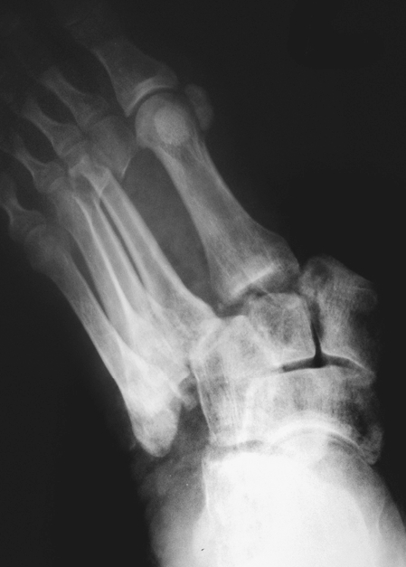
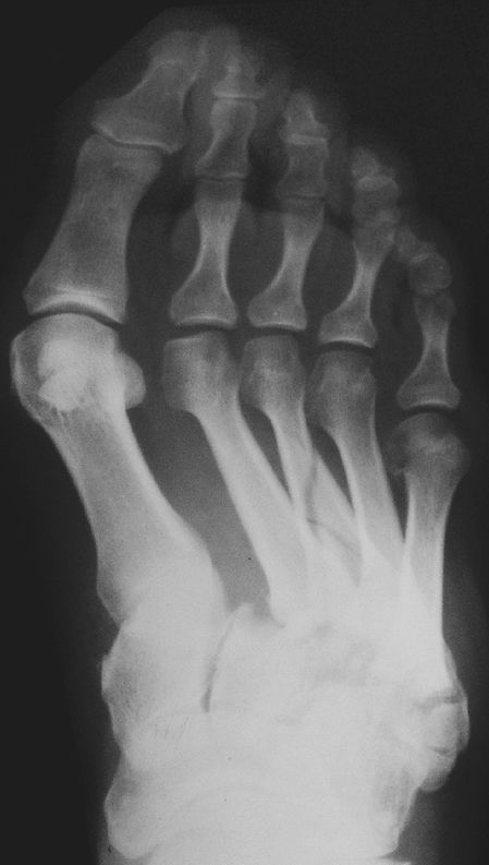
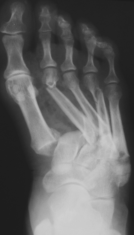
The classifications of Lisfranc joint injuries take into account the mechanics of the trauma, the type and direction of the dislocation as well as their pathomorphic forms [4, 7]. The classification suggested in 1909 by Quénu and Küss and modified by Hardcastle et al. in 1982 is undoubtedly the clearest [10, 16]. According to this classification, Lisfranc joint injuries are divided in three categories. The A form is characterised by the total dislocation of all tarso-metatarsal joints on one direction and corresponds to ‘homolateral’ dislocations in other classifications (Fig. 1). In the B form, the dislocations are partial and never involve all of the radii (the so-called ‘isolated’ dislocations in other classifications) (Fig. 2). In the C form, the dislocations are fully or partially divergent. As such, the C form contains injuries arising from cases in which the metatarsal bones have been displaced as a result of fractures or dislocations in various directions. The injury may include all or only some tarso-metatarsal joints. The ‘divergent’ dislocations of other classifications correspond to this group (Fig. 3).
Fig. 1.

Form A of Lisfranc joint injury
Fig. 2.

Form B of Lisfranc joint injury
Fig. 3.

Form C of Lisfranc joint injury
The aim of this study is to describe the pathomechanisms and pathomorphic forms of Lisfranc joint injuries based on the Hardcastle classification and to analyse the causes of diagnostic delays and mistakes based on the clinical experiences of the authors.
Material and methods
Material
Forty-one patients (13 women and 28 men), all treated between 1961 and 2004, comprised the study cohort. The causes of the Lisfranc joint injuries were traffic accidents (15 patients), the crushing of feet by significant weights (14 patients), sport injuries (six patients), falls from heights exceeding 1 m (five patients) and a fight (one patient).
Dislocations of tarso-metatarsal joints were found in 16 patients, dislocations with fractures of metatarsals or cuneiform bones and the cuboid were diagnosed in 23 patients and fractures of the metatarsal bone bases with dislocations of fragments were observed in the remaining two patients. The Lisfranc joint injuries were classified into 11 cases of the A form, 26 cases of the B form and four cases of the C form. The injuries in four feet were open.
As primary treatment, 11 patients received an open reduction with a stabilisation using Kirschner wires, 11 patients were immobilised in a below knee plaster for 4–6 weeks and ten patients underwent a closed reduction preceded by the application of plaster casts (five patients) or per cutaneous stabilisation with Kirschner wires (five patients). In the case of one patient with an open injury, immobilisation with a plaster cast was applied after the closure of the foot wound. Primary tarso-metatarsal arthrodesis was performed in only one patient, and three other patients initially treated for their injury in other institutions had cold compresses recommended. The injury resulting from the crushing of the foot with irreversible damage to the vascular system was an indication for the amputation of one foot soon after trauma. A delay in initiating treatment due to an erroneous diagnosis occurred in nine cases, with the delay varying from 6 months to 20 years. The observation/follow-up period varied from 2 to 37 years (average: 16 years).
Method
All of the patients who had been treated for the specified injuries were invited via mail for the follow-up examination. Nineteen patients responded. The examination was performed in accordance with the guidelines for the functional evaluation of the mid part of the foot set down by the American Foot and Ankle Society (AOFAS) in which pain intensity, function limitation, the necessity for special shoes, dependence on ground conditions for walking distance ability as well as the method of weight-bearing and its axis are taken into account. The score can range from 0 to 100 points (Table 1). The clinic's own (Lublin) foot functional scale was also used. This latter scale consists of an evaluation of swelling and foot skin changes as well as the ability to tiptoe and walk on the outer margins of the foot. This score can range from 0 to 80 points (Table 2). All patients also had a podoscopy examination as well as comparative radiograms of both feet in three standard projections. Questionnaires with both evaluation scales were sent to those people who were not able to attend the examination in person. Nine written responses were obtained by mail from these patients. Five patients were examined in their place of residence; these patients were not able to attend the follow-up examination due to significant medical problems. In total, roentgen and podoscopy examinations were performed in 19 patients, and the foot function of 14 people was evaluated according to the criteria of both of the applied scales. The final evaluation excluded eight patients who had either died or their current addresses were unknown. The scores of both evaluation tests were subjected to statistical analysis using the non-parametric Mann-Whitney U-test.
Table 1.
The American Foot and Ankle Society (AOFAS) midfoot scale
| Pain (40 points) | |
| None | 40 |
| Mild, occasional | 30 |
| Moderate, daily | 20 |
| Severe, almost always present | 0 |
| Function (45 points) | |
| Activity limitations, support | |
| No limitations, no support | 10 |
| No limitation of daily activities, limitation of recreational activities, no support | 7 |
| Limited daily and recreational activities, cane | 4 |
| Severe limitation of daily and recreational activities, walker, crutches, wheelchair | 0 |
| Footwear requirements | |
| Fashionable, conventional shoes, no insert required | 5 |
| Comfort footwear, shoe insert | 3 |
| Modified shoes or brace | 0 |
| Maximum walking distance, blocks | |
| Greater than 6 | 10 |
| 4–6 | 7 |
| 1–3 | 4 |
| Less than 1 | 0 |
| Walking surfaces | |
| No difficulty on any surface | 10 |
| Some difficulty on uneven terrain, stairs, inclines, ladders | 5 |
| Severe difficulty on uneven terrain, stairs, inclines, ladders | 0 |
| Gait abnormality | |
| None, slight | 10 |
| Obvious | 5 |
| Marked | 0 |
| Alignment (15 points) | |
| Good, plantigrade foot, midfoot well aligned | 15 |
| Fair, plantigrade foot, some degree foot midfoot malalignment observed, no symptoms | 8 |
| Poor, nonplantigrade foot, severe malalignment, symptoms | 0 |
| Total (100 points) | |
Table 2.
Lublin foot functional score
| Tiptoe walking (10 points) | |
| Without restrictions | 10 |
| Difficult but possible | 5 |
| Impossible | 0 |
| Jogging (10 points) | |
| Without restrictions | 10 |
| Difficult but possible | 5 |
| Impossible | 0 |
| Stair walking (10 points) | |
| Without restrictions | 10 |
| Difficult but possible | 5 |
| Impossible | 0 |
| Foot weight-bearing in supination (10 points) | |
| Without restrictions | 10 |
| Difficult but possible | 5 |
| Impossible | 0 |
| Skin corns (10 points) | |
| None | 10 |
| Present but small | 5 |
| Present diffused | 0 |
| Swelling (10 points) | |
| None | 10 |
| Present but small or temporary | 5 |
| Present persistent | 0 |
| Other complaints (10 points) | |
| None | 10 |
| Mild or temporary | 5 |
| Persistent | 0 |
| Superficial sensation abnormalities (10 points) | |
| None | 10 |
| Present, very local | 5 |
| Present, diffused | 0 |
| Total (80 points) | |
The podoscopy images of the examined feet were evaluated using the ElPodo ver. 2.10 computer programme, whereas the results were analysed statistically with the non-parametric Mann-Whitney U-test and the non-parametric Wilcoxon test. The programme examined the breadth of the anterior part of the foot and heel, the length of the whole foot and all toes, the angles of the hallux and the little toe, the Clark angle and the foot index.
The follow-up X-ray evaluation took into account the reconstruction of the correct anatomy of the correct tarso-metatarsal joints, the development of degenerative changes of the foot joints and the reconstruction of the bone structure following Lisfranc joint arthrodeses.
Results
The functional results on the AOFAS scale at the follow-up examination varied from 34 to 100 points (average: 75). The Lublin foot functional scale values ranged from 20 to 80 points, with an average of 53. In A form injuries, the values ranged from 44 to100 points (average: 76.5) on the AOFAS scale and from 20 to 80 points (average: 52) on the Lublin functional scale. In B form injuries, values ranged from 34 to 100 points (average: 71) on the AOFAS scale and from 20 to 80 (average: 52) on the Lublin functional scale. In C form injuries, values ranged from 68 to 100 points (average: 84) on the AOFAS scale and from 35 to 80 points (average: 58.8) on the Lublin functional scale. There was no significant difference between the scores of the two evaluation scores in terms of treatment results of the three forms of Lisfranc joint injuries according to the non-parametric Mann-Whitney U-test.
The comparison of treatment results revealed that those patients who had an open reduction with stabilisation using Kirschner wires scored on average of 76.5 points on the AOFAS scale and 55 on the Lublin functional scale. Those who had a closed reduction of dislocation preceded by a per cutaneous stabilisation with Kirschner wires or plaster immobilisation scored on average 92 points on the AOFAS scale and 74 points on the Lublin functional scale and those who had only plaster immobilisation scored on average 72 points according to the criteria of the AOFAS scale and 51 on the Lublin functional scale. The analysis did not reveal any statistically significant differences between the results of the functional scales in terms of the treatment according to the non-parametric Mann-Whitney U-test. However, patients with closed reduction of dislocations preceded by per cutaneous stabilisation with Kirschner wires or a plaster immobilisation scored distinctly higher values.
The retrospective evaluation of the patients’ documentation and repeated interviews with the patients indicated that delays in receiving the proper treatment usually occurred when the treating doctor did not take the symptoms seriously and was satisfied with a perfunctory inspection and examination of the foot; two patients did even receive an X-ray examination. The second factor that influenced the diagnostic delay was the doctor missing small dislocations (seven patients).
In the follow-up X-ray examinations of the A form injuries, secondary degenerative changes of the Lisfranc joint and the Chopart joint were detected in one patient. This person sustained a dislocation, which after an open reduction was stabilised with Kirschner wires. After 25 years, the functional result of the treatment of his foot was still very poor, and the patient suffered from pain, limp and used a walking stick. In the group of patients with the B form injuries, advanced degenerative changes in the tarso-metatarsal and traverse tarsus joints were noted in two patients 6 and 29 years, respectively, following an operation for stabilisation using Kirschner wires. One person treated with success 8 years after injury with medial cuneiform-metatarsal arthrodesis of radius I had no degenerative changes after 37 years. However, another patient treated 6 months after the injury with cuneiform-metatarsal arthrodesis showed degenerative changes of the talo-navicular joint after 14 years. Yet another B-injury patient who experienced joint surface fracture of the medial cuneiform bone and was treated by plaster immobilisation showed small degenerative changes of the Lisfranc joint after 22 years. Ankylosis of the tarso-metatarsal joints from radius II to IV were affirmed in two patients with C form injuries. All other patients had degenerative changes of the Lisfranc joint regardless of the type of treatment.
Patients who received delayed treatment obtained scores of between 44 and 64 points on the AOFAS scale and between 15 and 25 points on the Lublin scale. The treatment results were better in those patients who received treatment soon after the injury than in those who received delayed treatment; this difference was statistically significant at (p < 0.05) on both evaluation scales according to the non-parametric Mann-Whitney U-test .
The podoscopy examinations displayed statistically significant differences (p < 0.05 ) in the breadth of the anterior part of the feet as well as the heels and the feet length indicators for all toes between patients treated immediately after the trauma and those receiving delayed treatment, based on non-parametric Mann-Whitney U-test and non-parametric Wilcoxon test. The delays contributed to the broadening of the anterior foot parts and heels and the shortening of all longitudinal dimensions.
The weight-bearing irregularities of the injured feet forced nine patients to take care to choose comfortable footwear or to use insoles to relieve the zones of excessive pressure. No patient, however, used orthopaedic footwear.
Discussion
The worst functional results were obtained in B form injuries. Six diagnostic mistakes occurred in this group of patients during the primary treatment; in addition, seemingly ‘insignificant dislocations’ were neglected in three patients. Lu stresses that dislocations of individual foot radii exceeding 2 mm in the X-ray image or larger than 1 mm in the computer tomography images as well as an increase of more than 15° in the tarso-metatarsal angle cause a chronic Lisfranc joint failure [11]. In terms of individual foot radii, the B injuries are more likely to be missed, under diagnosed or mistreated than A and C forms [15]. The best functional results were obtained by the C-group patients. They avoided diagnostic mistakes, primarily because this form of dislocation causes significant foot deformity and are therefore, are easily detected in X-ray examinations. Despite this group of patients showing radiological characteristics of degenerative changes of the tarso-metatarsal joints, they did show good functional results of treatment. Early and Aronow are of the opinion that arthodeses of the three lateral foot radii, even in missed injuries, should be avoided, because the cushioning function of the lateral side is removed [2, 6]. Despite this view, however, the patients with C type injuries evaluated by us had good functional results, even when they had a primary unintentional spontaneous rigidity of the joints of the III and IV radii. The best treatment results were achieved by patients with a closed reduction of dislocations preceded by a per cutaneous transfixion using Kirschner wires. Owen recommends this method, especially in B injuries with accompanying soft tissue damage [14]. Marcus reported, however, that the results of the closed reduction with a per cutaneous stabilisation using Kirschner wires were worse than those of open reductions and stabilisation using screws [18].
Myerson agrees with Owens in believing that the early reduction, either closed or open, with stabilisation using Kirschner wires is a good method of treatment [13, 14]. Our observations also confirm this. In the follow-up, the biggest complaints were raised by patients treated with immobilisation without a reduction of small dislocations of the bases of one or more metatarsal bones. In accordance with Lu’s opinion, a precise, comparative X-ray evaluation should improve the patient's chances of receiving the correct treatment even if the dislocations are small [11]. All patients who received delayed treatment due to diagnostic errors were subjected to trials of dislocation reduction and the immobilisation of the injured joints. These patients, however, have worse functional results. This observation confirms the common opinion of other authors that any delay inevitably worsens the prognosis [2, 5, 15, 19]. In these patients, arthrodesis in the anatomical positions is the treatment of choice [2, 12, 15]. Wachtl claims that full arthodesis of the Lisfranc joints is an indicator for orthopaedic footwear [20]; however, our patients did not require this.
Summary and conclusions
Each injury to the tarso-metatarsal joints requires accurate clinical and roentgen diagnosis. The suspicion of anomalies in X-ray images should suggest further radiological investigation. Closed reduction with the restoration of correct anatomical alignment and per cutaneous stabilisation are recommended treatments. Even small irregularities in X-ray images are an indication for operative intervention, dislocation correction and stabilisation using Kirschner wires.
Lisfranc joint damages, especially of the B type, require special attention and an image-comparing diagnosis.
Delays in diagnosis and/or treatment inevitably lead to poor results and the shortening and broadening of the foot.
References
- 1.Aitken AP, Poulson D. Dislocation of the tarsometatarsal joint. J Bone Joint Surg Am. 1963;45:246–260. [PubMed] [Google Scholar]
- 2.Aronow MS. Treatment of the missed Lisfranc injury. Foot Ankle Clin. 2006;11:127–142. doi: 10.1016/j.fcl.2005.12.005. [DOI] [PubMed] [Google Scholar]
- 3.Bonnel F, Barthélémy M. Traumatismes de l’articulation de Lisfranc: entroses graves, luxations, fractures: etude de 39 observations personnelle et classification biomecanique. J Chir. 1976;111:573–592. [PubMed] [Google Scholar]
- 4.Böhler L. Dislocations in Lisfranc’s joint. In: Wallner A, Russe O, editors. The treatment of fractures. 5th English edn. New York: Grune and Stratton; 1958. pp. 2136–2137. [Google Scholar]
- 5.Burroughs KE, Reimer CD, Fields KB. Lisfranc injury of the foot: a commonly missed diagnosis. Am Fam Physician. 1998;58:118–129. [PubMed] [Google Scholar]
- 6.Early JS (2005) Lisfranc fracture dislocation. E-Medicine Specialities
- 7.Engelhardt P, Ganz R. Die Luxationsfrakturen des Lisfranc Gelenkes. Orthopädische Praxis der baden badenrs reihe. 1975;9:896–898. [Google Scholar]
- 8.English TA. Dislocations of the metatarsal bone and adjacent toe. J Bone Joint Surg Br. 1964;46:700–704. [PubMed] [Google Scholar]
- 9.Gissane W. A dangerous type of fracture of the foot. J Bone Joint Surg (B) 1951;33:535–538. doi: 10.1302/0301-620X.33B4.535. [DOI] [PubMed] [Google Scholar]
- 10.Hardcastle PH, Reschauer R, Kutscha-Lissberg E, Schoffmann W. Injuries to the tarsometatarsal joint. J Bone Joint Surg Br. 1982;64:349–356. doi: 10.1302/0301-620X.64B3.7096403. [DOI] [PubMed] [Google Scholar]
- 11.Lu J, Ebraheim NA, Skie M, Porshinsky B, Yeasting RA. Radiographic and computed tomographic evaluation of Lisfranc dislocation: a cadaver study. Foot Ankle Int. 1997;18:351–355. doi: 10.1177/107110079701800608. [DOI] [PubMed] [Google Scholar]
- 12.Mulier T, Reynders P, Sioen W, Bergh J, Reymaeker G, Reynaert P, Broos P. The treatment of Lisfranc injuries. Acta Orthop Belg. 1997;63:82–90. [PubMed] [Google Scholar]
- 13.Myerson MS, Fisher RT, Burgess AR, Kenzora JE. Fracture dislocations of the tarsometatarsal joints: end results correlated with pathology and treatment. Foot Ankle. 1986;6:225–242. doi: 10.1177/107110078600600504. [DOI] [PubMed] [Google Scholar]
- 14.Owens BD, Wixted JJ, Cook J, Teebagy AK. Intramedullary transmetatarsal Kirschner wire fixation of Lisfranc fracture-dislocations. Am J Orthop. 2000;32:389–391. [PubMed] [Google Scholar]
- 15.Philbin T, Rosenberg G, Sferra JJ. Complications of missed or untreated Lisfranc injuries. Foot Ankle Clin. 2003;8:61–71. doi: 10.1016/S1083-7515(03)00003-2. [DOI] [PubMed] [Google Scholar]
- 16.Quénu E, Küss G. Étude sur les luxations du metatarse (luxations métatarsotarsiennes) du diastasis entre le 1er et le 2e metatarsien. Rev Chir. 1909;39:281–336. [Google Scholar]
- 17.Rainaut JJ, Cedard D, d’Hour JP. Les luxations tarsometatarsiennes. Rev Chir Orthop. 1966;52:449–462. [PubMed] [Google Scholar]
- 18.Marcus RE (2001) Diagnosis and treatment of foot and ankle fractures. In: 15th Annu Vail Orthopaedics Symp. Vail, Colorado
- 19.Sangeorzan BJ, Veith RG, Hansen ST., Jr Salvage of Lisfranc’s tarsometatarsal joint by arthrodesis. Foot Ankle. 1990;10:193–200. doi: 10.1177/107110079001000401. [DOI] [PubMed] [Google Scholar]
- 20.Wachtl S, Elsig JP. Orthopedic technique following arthrodesis of the Lisfranc joint. Schweiz Rundsch Med Prax. 1993;82:655–660. [PubMed] [Google Scholar]


