Retroperitoneal sarcomas account for 0.1%–0.2% of all malignant tumours and about 15% of all sarcomas. Liposarcoma is the most common type of retroperitoneal sarcoma (41%), followed by leiomyosarcoma, malignant fibrous histiocytoma, fibrosarcoma and other undifferentiated sarcomas.1 We report the case of a woman with a giant retroperitoneal sarcoma and show that large tumour size is not necessarily a contraindication to surgical excision.
Case report
A 63-year-old woman with a history of high blood pressure sought medical attention for abdominal distension that was progressive over 2 years.
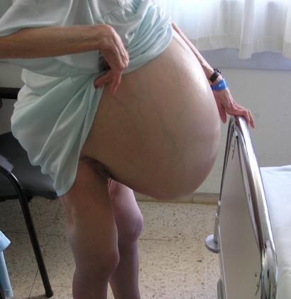
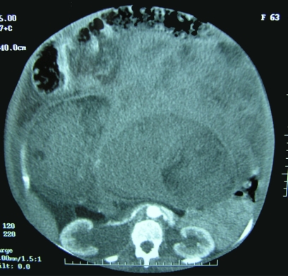
On examination, a hard mass that occupied the entire abdomen was palpated and there was edema in both lower extremities and cachexia involving the face and both upper extremities (Fig. 1). Abdominal computed tomography (CT) showed that the entire abdominal cavity was occupied by a heterogeneous mass with low-density areas (necrosis) and a scattered solid component that pushed aside all intestinal loops, resulting in moderate dilatation of the right renal excretory system in its 2 proximal thirds (Fig. 2).
FIG. 1. Appearance of the retroperitoneal sarcoma as a huge mass, which was accompanied by lower-extremity edema.
FIG. 2. Abdominal computed tomography shows a heterogeneous retroperitoneal mass occupying the entire abdominal cavity.
At laparotomy, we noted a liposarcomatous tumour occupying the entire abdominal cavity. We excised the tumour completely and performed a right-sided nephrectomy because the right ureter was involved, an appendectomy and a hysterectomy because of contact with the lesion and a cholecystectomy because of cholelithiasis.
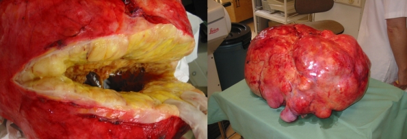
The pathological diagnosis was a mixoid liposarcoma, measuring 44.5 × 43 × 24 cm and weighing 31 kg. The resection margins were free of tumour cells (Fig. 3). The patient's postoperative course was uncomplicated. She received chemotherapy with doxorubicin and was disease-free on clinical examination and CT 2 years postoperatively.
FIG. 3. The excised surgical specimen, which was identified on pathological examination as a mixoid liposarcoma.
Discussion
Retroperitoneal sarcomas are usually asymptomatic. Their most typical manifestations are discomfort or nonspecific abdominal pain and a palpable abdominal mass. These tumours occur most frequently in men, usually in the fifth or sixth decade of life. The most appropriate diagnostic tests to determine their size and interrelation with neighbouring organs are abdominal CT and magnetic nuclear resonance imaging. The treatment of choice is complete surgical removal with tumour-free margins, which usually requires the resection of adjoining abdominal or retroperitoneal organs.2 The most commonly resected structures are kidney, ureter and large bowel, but resection of gallbladder, female reproductive organs, small bowel, stomach, adrenal glands, spleen, pancreas and vascular structures is sometimes required.
Large tumour size should not be regarded as a contraindication to surgical resection, which is currently the only potentially curative treatment. Preoperative bowel preparation is advisable in all cases. En bloc resection of the tumour and all involved organs is the procedure of choice. Curative resection is difficult when the tumour compromises vital structures such as the infrarenal aorta and inferior vena cava, but it has been demonstrated that these can be successfully resected.2 The most frequent and serious postoperative complications are hemorrhage and anastomotic leakage resulting from bowel resection.
There are several histologic types of retroperitoneal sarcomas: mixoid (which is the most common), well-differentiated, pleomorphic, mixed and undifferentiated. These tumours are locally aggressive and have a high recurrence rate (40%–80%), but metastases are rare. Some studies have shown that radiotherapy is useful for local control; however, in contrast to intestinal sarcomas, the presence of the abdominal viscera in the retroperitoneum limits the use of radiotherapy. Chemotherapy does not improve survival or the likelihood of metastasis. The prognostic factor associated with long-term survival is the radicality of the surgical resection, not tumour size.1,2
Retroperitoneal sarcomas, owing to their slow growth and retroperitoneal origin, can become very large. We have only been able to find 2 published cases of a retroperitoneal sarcoma larger than the one we report here,3,4 the mean size of these tumours being between 10 and 15 kg.
Competing interests: None declared.
Accepted for publication Nov. 20, 2007
Correspondence to: Dr. A. Morandeira, Department of General and Digestive Surgery, C/ Sant Joan S/N, Reus (Tarragona) 43201, Spain; antoniomorandeira@hotmail.com
References
- 1.Lewis JJ, Leung D, Woodruff JM, et al. Retroperitoneal soft-tissue sarcoma: analysis of 500 patients treated and followed at a single institution. Ann Surg 1998;228:355-65. [DOI] [PMC free article] [PubMed]
- 2.Bradley JC, Caplan R. Giant retroperitoneal sarcoma: a case report and review of the management of retroperitoneal sarcomas. Am Surg 2002;68:52-6. [PubMed]
- 3.Yol S, Tavli S, Tavli L, et al. Retroperitoneal and scrotal giant liposarcoma: report of a case. Surg Today 1998;28:339-42. [DOI] [PubMed]
- 4.McCallum OJ, Burke JJ, Childs AJ, et al. Retroperitoneal liposarcoma weighing over one hundred pounds with review of the literature. Gynecol Oncol 2006;103:1152-4. [DOI] [PubMed]


