Abstract
Several reports suggest that a chronic inflammatory process plays a key role in coronary artery plaque instability and subsequent occlusive thrombosis. In a previous study, we found that TLR4 (Toll-like receptor 4) mediates the synthesis of cytokines in circulating monocytes of patients with AMI (acute myocardial infarction); however, it remains unclear whether TLRs are expressed at the site of the ruptured plaque in these patients. The aim of the present study was to determine whether TLR2 and TLR4 are expressed at the site of ruptured plaques in patients with AMI and to compare this with systemic levels. The study included 62 patients with AMI, 20 patients with SA (stable angina) and 32 subjects with a normal coronary angiogram (control). Local samples from the site of the ruptured plaque were taken from patients with AMI using aspiration catheterization. Systemic blood samples from the aorta were taken from patients with AMI and SA and controls. Systemic levels of TLR2 and TLR4 were higher in patients with AMI than in patients with SA and controls. In patients with AMI, local TLR4 levels were higher than systemic levels. There was no significant difference in TLR2 levels between local and systemic samples. TLR4 immunostaining was positive in infiltrating macrophages in ruptured plaque material. Cardiac events were observed in 16 patients with AMI at the time of the 6-month follow-up study. Local and systemic levels of TLR4 were higher in patients with AMI with cardiac events than in those without. These results indicate an increase in monocytic TLR4 expression not only in the systemic circulation, but also at the site of plaque rupture. In conclusion, expression of both systemic and local plaque TLR4 may be one of the mechanisms responsible for the pathogenesis of AMI.
Keywords: atherosclerosis, inflammation, myocardial infarction, ruptured plaque, thrombosis, Toll-like receptor (TLR)
Abbreviations: CT, threshold cycle; FCS, fetal calf serum; GAPDH, glyceraldehyde-3-phosphate dehydrogenase; HSP, heat-shock protein; LPS, lipopolysaccharide; MFI, mean fluorescence intensity; MI, myocardial infarction; AMI, acute MI; MyD88, myeloid differentiation factor 88; PBMC, peripheral blood mononuclear cell; PCI, percutaneous coronary intervention; RT, reverse transcription; SA, stable angina; TIMI, thrombolysis in MI; TLR, Toll-like receptor; TNF-α, tumour necrosis factor-α
INTRODUCTION
Clinical and histopathological studies suggest that a chronic inflammatory process plays a key role in coronary artery plaque instability and subsequent occlusive thrombosis [1,2]. In an autopsy study [3], ruptured and vulnerable plaques from patients who died of AMI [acute MI (myocardial infarction)] had greater inflammatory macrophages and lymphocytes than stable plaques from patients with SA (stable angina), which suggests that activation of an immune response is involved in the progression of ruptured and vulnerable plaques.
Currently ten TLRs (Toll-like receptors) have been reported in mammalian species and these appear to recognize distinct pathogen-associated molecular patterns controlling innate immune responses [4]. Methe et al. [5] and our previous study [6] have demonstrated that activation of TLR4 in circulating monocytes is related to the downstream release of inflammatory cytokines in patients with AMI. These studies suggest a potential pathophysiological link between TLR4 signalling and the immune response in coronary atherosclerosis. TLR4 has been shown to be required not only for the bacterial endotoxin-induced inflammatory responses, but also for non-bacterial ligands, such as oxidative stress, fatty acids and HSP (heat-shock protein) [7–9]. TLR2 and TLR4 immunostaining is frequently co-localized with NF-κB (nuclear factor κB), which is a common downstream pathway of TLR2 and TLR4 signals, in atherosclerotic plaques [10]. There is cross-talk between TLR2 and TLR4, through which TLR2 expression is regulated by TLR4 expression [11,12]. It has been reported that there is an increase in both TLR2 and TLR4 levels in PBMCs (peripheral blood mononuclear cells) obtained from patients with coronary artery disease [13]. A histological study has shown that TLR4 but not TLR2 is expressed in macrophage-infiltrated coronary artery plaques obtained at autopsy [14]; however, it has not been determined whether TLR2 and TLR4 are expressed at the site of the culprit coronary artery in patients with AMI. The aim of the present study was to determine whether TLR2 and TLR4 are expressed at the site of ruptured plaques in patients with AMI and to compare this with systemic levels.
MATERIALS AND METHODS
Patients
The present study included 62 consecutive patients with first Q-wave AMI undergoing PCI (percutaneous coronary intervention) and stenting with aspiration catheterization, 20 patients with SA and 34 subjects with normal coronary angiographic findings. Inclusion criteria for AMI in the study were (i) continuous chest pain lasting >30 min, (ii) arrival at our hospital within 12 h of the onset of chest pain, (iii) ST-segment elevation ≥0.1 mV in two or more contiguous leads on 12-lead ECG, and (iv) an angiographically detected culprit lesion with TIMI (thrombolysis in MI) flow grade ≤2. In all patients with AMI, myocardial damage was confirmed by a troponin T level above the upper limit of normal (0.1 μg/ml), and a 2-fold increase in the creatinine kinase-MB isoenzyme. Exclusion criteria were (i) a culprit lesion in the left main coronary artery, (ii) a history of prior AMI, or (iii) cardiogenic shock, heart failure or infectious illness at the time of PCI. SA was diagnosed on the basis of the presence of (i) a history of typical chest pain on effort, (ii) lasting unchanged for more than 3 months and not associated with rest angina, (iii) documenting exercise-induced myocardial ischaemia, (iv) angiographically proven coronary artery disease, and (v) the exclusion parameters described for AMI. Controls were obtained from 34 subjects with suspected SA on the basis of symptoms and/or minor ECG changes. The resulting coronary angiographic finding and close clinical examination failed to show any evidence of SA, and these subjects were thus designated as controls.
The study protocol was approved by our hospital Ethics Committee, and informed consent was obtained from all subjects.
Sampling of blood and plaque material
Systemic blood samples were taken first from the aortic arch prior to PCI. Local samples at the site of occlusion were immediately obtained using aspiration catheterization (TVAC; Nipro) after crossing the occlusive lesion. Solid plaque materials were separated from liquid blood using a microfilter (pore filter size 40 μm). Liquid blood samples were used for monocyte isolation as local blood samples. Solid plaque material was also used for immunohistochemistry. In patients with SA and controls, systemic blood samples were taken from the aortic arch at the time of coronary angiography.
Cell preparation
PBMCs were isolated from local and systemic blood samples obtained from all subjects by Ficoll-Paque density gradient centrifugation and lymphocyte separation solution (Nacalai Tesque). To exclude platelet contamination, isolated PBMCs were washed and centrifuged twice with PBS containing 5 mmol/l EDTA at 400 g for 15 min at 20 °C [15]. Monocytes were isolated from PBMCs by adherence to a plastic dish (120 min, 37 °C). Monocytes were detached from the plastic dish by incubation in ice-cold PBS, washed three times with PBS and then resuspended at a final concentration of 1×10⁶ cells/ml in RPMI 1640 (Sigma–Aldrich).
Real-time RT (reverse transcription)-PCR
Total RNA was extracted from isolated monocytes by the acid guanidinium thiocyanate/phenol chloroform method [16] and were treated with DNase I (Gibco).
The published sequences for human TLR2 (forward primer, 5′-GCCAGGCGGCTGCTC-3′; reverse primer, 5′-TTGCAACACCAAACACTGGG-3′; and TaqMan probe, 5′-CGTTCTCTCAGGTGACTGCTCGGAGTTC-3′) [17] and TLR4 (forward primer, 5′-TGATTGTTGTGGTGTCCCA-3′; reverse primer, 5′-TGTCCTCCCACTCCAGGTAA-3′; and TaqMan probe, 5′-TCCTGCAGAAGGTGGAGAAGACCCT-3′) [18] were used for construction of primers and TaqMan probe. mRNA of the housekeeping gene GAPDH (glyceraldehyde-3-phosphate dehydrogenase) was amplified using TaqMan GAPDH control reagents as an internal control (PE Biosystems).
cDNA was synthesized and amplified from 100 ng of total RNA and 10-fold serial dilutions of human control RNA (PE Biosystem) by RT-PCR using a TaqMan EZ RT-PCR kit (PE Biosystem). The cDNA products were synthesized at 60 °C for 30 min and amplified with 40 cycles of PCR, with each cycle consisting of denaturation at 94 °C for 20 s, and annealing and extension at 62 °C for 1 min. A quantitative PCR method was developed using a 5-nuclease assay with detection on an ABI PRISM 7700 sequence detector (PE Biosystems). Amplifications were performed three times for each RNA sample. Each PCR run also included triplicate wells of each RNA sample. The ratio between the copy numbers of TLR2, TLR4 and GAPDH represented the normalized TLR2 and TLR4 levels for each sample and could be compared with those of other samples. To account for PCR amplification of contaminating genomic DNA, a control without RT was included.
Flow cytometric analysis
The amount of TLR2, TLR4 and CD14 on the monocyte cell surface was measured by FACS. Isolated monocytes were incubated with FITC-conjugated mouse anti-(human TLR2) and (human TLR4) antibodies (Santa Cruz Biotechnology) and a PerCP (peridinin chlorophyll protein)-conjugated CD14 antibody (Becton Dickinson). Isotype-matched irrelevant control IgG was used as a control (Becton Dickinson). TLR2 and TLR4 levels in CD14-positive cells were measured using a FACScan flow cytometer (Becton Dickinson) and are shown as MFIs (mean fluorescence intensities).
Immunohistochemistry
A monoclonal antibody to TLR2 (1:100 dilution; Apotech Biochem), a mouse monoclonal IgG2a against human TLR4 (1:100 dilution; Santa Cruz Biotechnology) and a monoclonal antibody to human CD68 (1:100 dilution; Dako) were used as primary antibodies. The tissue sections were deparaffined with xylene for 20 min and thoroughly dehydrated with serially diluted ethanol. After inhibition of endogenous peroxidase and blocking of non-specific reactions, primary antibodies were applied. Biotinylated mouse Ig was used as a secondary antibody. Peroxidase-labelled streptavidin (Histofine; MAX-PO kit; Nichiren) was applied and visualized using DAB (diaminobenzidine) as the chromogen. The specificity of the immunohistochemistry was confirmed by substituting the primary antibodies with a mouse IgG1 negative control (Dako) on control sections from thrombus material.
In vitro study
To calculate the generation capacities of TLR2 and TLR4, isolated monocytes were cultured in two culture media with and without HSP70 stimulation, which is a putative monocyte TLR ligand [9]. Monocytes were resuspended in either non-activating medium [RPMI 1640 with 10% (v/v) heat-inactivated FCS (fetal calf serum; Gibco), penicillin and streptomycin] or activating medium [RPMI 1640 with 2 μg/ml HSP70 (NSP-555; Stressgen Bioreagents), 10% (v/v) heat-inactivated FCS, penicillin and streptomycin]. Cells were then incubated in sterile polypropylene tubes (Becton Dickinson) for 24 h at 37 °C in 5% CO2. TLR2 and TLR4 MFIs were measured using the method described above. Cultured supernatant concentrations of TNF-α (tumour necrosis factor-α) were measured using the Bio-Plex system, which combines the principle of a sandwich immunoassay with Luminex fluorescent-bead-based technology (sensitivity 0.25 pg/ml; Bio-Rad Laboratories) [19].
Clinical follow-up study
Follow-up coronary angiography was carried out as a clinically driven event at least 6 months after PCI (mean follow-up, 191±10 days) to determine the recurrence of stenosis in all patients with AMI. Cardiac events were defined as target lesion revascularization (lesion segment with 5 mm margins from each end), target vessel revascularization (any intervention to the treated vessel), cardiac death and recurrent Q-wave MI.
Statistical analysis
Continuous variables are presented as means±S.D. Comparison of continuous variables was carried out using Student's t test and the non-parametric Mann–Whitney test. Statistical analysis of categorical variables was also carried out using χ2 analysis and Fisher exact analysis. A value of P<0.05 was considered statistically significant.
RESULTS
Baseline and clinical characteristics
Baseline characteristics of patients with AMI, patients with SA and controls are shown in Table 1.
Table 1. Baseline and clinical characteristics of study populations.
Values are means±S.D., or numbers (percentage). ACEI, angiotensin-converting enzyme inhibitors; ARB, angiotensin II type 1 receptor blockers; BMI, body mass index; CAD, coronary artery disease; DBP, diastolic blood pressure; HbA1c glycated haemoglobin; HDL, high-density lipoprotein; LDL, low-density lipoprotein; IFG, impaired fasting glucose; IGT, impaired glucose tolerance; SBP, systolic blood pressure. *P<0.05 compared with patients with AMI.
| Characteristic | Patients with AMI | Patients with SA | Controls |
|---|---|---|---|
| n | 62 | 20 | 34 |
| Age (years) | 64.0±13.4 | 64.6±12.2 | 62.1±10.1 |
| Male gender (n) | 44 (71%) | 15 (75%) | 27 (79%) |
| BMI (kg/m2) | 24.7±3.0 | 23.9±4.6 | 23.1±4.7 |
| Risk factors (n) | |||
| Hypertension | 46 (74%) | 15 (75%) | 27 (79%) |
| Hypercholesterolaemia | 41 (66%) | 13 (65%) | 23 (68%) |
| Diabetes or IGT/IFG | 37 (60%) | 11 (55%) | 20 (59%) |
| Smoking | 24 (39%) | 7 (35%) | 12 (35%) |
| Obesity | 27 (44%) | 8 (40%) | 15 (44%) |
| SBP (mmHg) | 127±15 | 25±18 | 24±11 |
| DBP (mmHg) | 71±11 | 72±14 | 69±13 |
| HDL-cholesterol (mg/dl) | 56±14 | 57±18 | 58±19 |
| LDL-cholesterol (mg/dl) | 111±30 | 109±39 | 107±41 |
| HbA1c (%) | 5.9±1.3 | 5.7±1.1 | 5.8±1.2 |
| Angiographic degree of CAD (n) | |||
| One-vessel disease | 37 (60%) | 13 (65%) | − |
| Two-vessel disease | 18 (29%) | 7 (35%) | − |
| Three-vessel disease | 7 (11%) | 0 | − |
| Culprit vessel (n) | − | ||
| Left anterior descending artery | 30 (48%) | 10 (50%) | − |
| Left circumflex artery | 8 (13%) | 3 (15%) | − |
| Right coronary artery | 24 (39%) | 7 (35%) | − |
| Medication (n) | − | ||
| Aspirin | 62 (100%) | 19 (95%) | 0* |
| Nitrates | 17 (27%) | 10 (50%) | 0* |
| β-Blocker | 15 (24%) | 6 (30%) | 5 (15%) |
| ACEI or ARB | 36 (58%) | 13 (65%) | 19 (56%) |
| Statins | 46 (74%) | 12 (60%) | 21 (62%) |
| Initial TIMI flow grade (n) | |||
| 0 | 52 (84%) | 0* | − |
| 1 | 10 (16%) | 2 (10%) | − |
| 2 | 0 | 18 (90%)* | − |
| 3 | 0 | 0 | − |
| Final TIMI flow grade (n) | |||
| 0 | 0 | − | − |
| 1 | 0 | − | − |
| 2 | 9 (15%) | − | − |
| 3 | 51 (85%) | − | − |
Angiographic findings after PCI with thrombus aspiration
All patients with AMI underwent stent implantation using a bare metal stent (Driver® coronary stent system; Medtronic). Liquid blood samples in the culprit coronary artery were obtained from all patients with AMI. Solid plaque materials were also obtained from 24 patients with AMI. For the final TIMI grade, the rate of TIMI3 was 85% in patients with AMI (Table 1).
Levels of TLR2 and TLR4
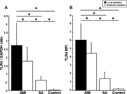
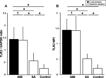
There was no difference in average CT (threshold cycle) of GAPDH between the AMI, SA and control groups (average CT of GAPDH, 20.76±1.73, 20.99±1.58 and 20.86±1.48 respectively; P=not significant). Spontaneous mRNA and protein (MFI) levels of TLR2 and TLR4 in systemic samples obtained from patients with AMI were higher than in patients with SA and controls (all P<0.01) (Figures 1 and 2). In patients with AMI, local levels of TLR4 were higher than in systemic samples (Figure 1), whereas there was no significant difference between local and systemic samples for TLR2 levels (Figure 2).
Figure 1. TLR4 levels in local and systemic samples.
(A) TLR4 mRNA and (B) TLR4 MFI. *P<0.05.
Figure 2. TLR2 levels in local and systemic samples.
(A) TLR2 mRNA and (B) TLR2 MFI. *P<0.05 and ¶P>0.05.
Immunohistochemistry for TLR2 and TLR4
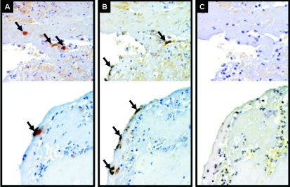
In solid plaque material removed from the site of the ruptured plaque, staining for TLR4 was localized in infiltrating macrophages (Figure 3A) and was positive in all solid plaque samples. Immunostaining of thrombus material showed CD68-positive macrophages (Figure 3B). TLR2 staining was not present in any of the samples (Figure 3C). There was no evidence of non-specific staining in solid plaque material obtained from patients with AMI.
Figure 3. Immunohistochemistry for TLR2, TLR4 and CD68 in thrombus samples obtained from patients with AMI.
(A) Immunostaining for TLR4 (brown staining, arrows) in infiltrating macrophages (magnification, ×200). (B) CD68-positive macrophages (brown staining, arrows) (magnification, ×200). (C) Immunostaining for TLR2 in thrombus samples obtained from patients with AMI (magnification, ×200).
Changes in TLR2 and TLR4 levels in response to HSP70
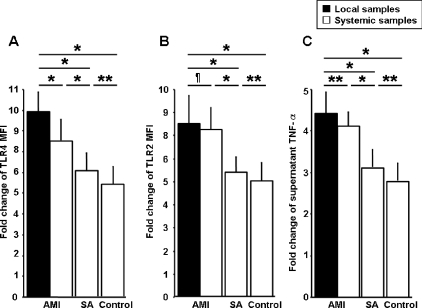
To quantify the effect of HSP70 stimulation on TLR expression, TLR levels in HSP70-stimulated monocytes were normalized to the corresponding levels in cultured monocytes without HSP70 stimulation, and levels were expressed as fold changes. The fold changes in TLR2 and TLR4 levels with HSP70 stimulation were higher in patients with AMI (both local and systemic samples) compared with patients with SA and controls (Figure 4). In patients with AMI, the fold change in TLR4 with HSP70 stimulation was significantly higher in local samples than in systemic samples (Figure 4A), whereas there was no significant difference between local and systemic samples for TLR2 (Figure 4B). The fold changes in supernatant TNF-α with HSP70 stimulation were higher in patients with AMI than in patients with SA and controls (Figure 4C). In patients with AMI, the fold change in supernatant TNF-α levels with HSP70 stimulation was significantly higher in local samples than in systemic samples (Figure 4C).
Figure 4. Cultured monocytes stimulated with HSP70.
Fold change in TLR4 MFI (A), TLR2 MFI (B) and supernatant TNF-α levels (C) with HSP70 stimulation. *P<0.01, **P<0.05 and ¶P>0.05.
Relationship between cardiac events and TLR2 and TLR4 levels
At the time of the 6-month follow-up study, cardiac events were observed in 16 out of 62 patients with AMI. These events consisted of target lesion revascularization in six patients, target vessel revascularization in seven patients, recurrent MI in two patients and cardiac death in one patient. When patients with AMI were divided into two subgroups, according to the presence or absence of cardiac events, both local and systemic levels of TLR4 were higher in patients with cardiac events than in those without [local TLR4 mRNA, 4.32±1.24 compared with 2.05±0.64 respectively (P<0.01); local TLR4 MFI, 8.33±2.01 compared with 5.14±0.78 respectively (P<0.01); systemic TLR4 mRNA, 2.67±0.89 compared with 1.41±0.47 respectively (P<0.01); and systemic TLR4 MFI, 4.99±1.50 compared with 4.23±1.19 respectively (P=0.03)]. In contrast, there were no significant differences in TLR2 levels between patients with cardiac events than those without [local TLR2 mRNA, 1.39±0.48 compared with 1.47±0.48 (P=0.58); local TLR2 MFI, 1.54±0.40 compared with 1.40±0.31 (P=0.20); systemic TLR2 mRNA, 1.52±0.61 compared with 1.36±0.50 (P=0.24); and systemic TLR2 MFI, 1.58±0.47 compared with 1.36±0.44 (P=0.26)].
DISCUSSION
The major findings of the present study are: (i) in patients with AMI, monocyte levels of TLR4, but not TLR2, were higher in local samples surrounding ruptured plaques than in systemic samples; (ii) TLR4 immunostaining was observed in infiltrating macrophages in ruptured plaque material, but TLR2 immunostaining was not; (iii) the in vitro study suggests that HSP70-stimulated levels of TLR4 in local samples were higher than in systemic samples; and (iv) local and systemic levels of TLR4 were higher in patients with AMI with cardiac events than in those without.
It has become evident that atherosclerosis is an inflammatory disease involving an immune response during its initiation and progression [20]. Studies have demonstrated that the TLR signalling pathway is activated by endogenous ligands, such as oxidative stress, fatty acids and HSPs [7–9]. Epidemiological studies also suggested that oxidative stress increases in subjects with many risk factors for atherosclerosis, such as diabetes, hypertension and smoking [21]. Some reports have demonstrated that expression of TLR4 was mainly localized in infiltrating macrophages in atherosclerotic lesions, suggesting a close link between the progression of coronary atherosclerosis and TLR4 signalling [10,14]. The present study has shown that mRNA and protein levels of TLR2 and TLR4 in circulating monocytes were higher in patients with AMI prior to PCI than in patients with SA and controls. Furthermore, in patients with AMI, the local levels of TLR4 at the site of ruptured plaque were higher than systemic levels. In agreement with the concept of an increase in local TLR4 expression, immunostaining demonstrated the localization of TLR4 in infiltrating macrophages in ruptured plaque material occluding the culprit coronary artery. The increase in local TLR4 levels in the culprit coronary artery in patients with AMI is a novel and unexpected finding. An apoE (apolipoprotien E)-deficient mouse model has reported that a loss of TLR4 and its adaptor molecule MyD88 (myeloid differentiation factor 88) decreased the severity of atherosclerosis and altered atherosclerotic plaque formation [22]. Furthermore, Bjorkbacka et al. [23] demonstrated, using MyD88-null mice, that TLR4 deficiency was associated with alterations in coronary plaque composition, which decreased both lipid and macrophage content and markedly decreased the expression of pro-inflammatory factors, including pro-inflammatory cytokines and chemokines. An experimental model has shown that activated macrophages within plaques are capable of degrading the extracellular matrix by secretion of MMP 9 (matrix metalloproteinase 9), which can be stimulated by TLR4 activation and which induces plaque degradation and rupture [24,25]. It has also been reported that activated TLR4 signalling induces the expression of apoptotic molecules of the Fas death pathway [26]. From these observations, it has been suggested that the expression of TLR4 in infiltrating macrophages in coronary arteries may be an important factor underlying coronary plaque destabilization and rupture.
In the present study, TLR2 levels did not differ between local and systemic samples in patients with AMI. Immunohistochemical findings showed that TLR2 immunostaining was not present in any plaque samples. This suggests that circulating monocytes may be a major cellular source of TLR2 expression in patients with AMI.
We have shown that TLR2 and TLR4 levels in circulating monocytes were higher in patients with AMI than in patients with SA and controls. Our previous study [27] demonstrated that circulating monocytes release HSP70, which is a potent endogenous ligand of monocyte TLR4 signalling, in response to myocardial ischaemic damage. Asea et al. [28] have reported that the release of HSP70, as a common endogenous ligand of both TLR2 and TLR4, in response to ischaemic myocardium may activate TLR2 and TLR4 signalling in circulating monocytes. TLR4 signalling up-regulates TLR2 in LPS (lipopolysaccharide)-stimulated macrophages, suggesting that the cross-talk between TLR2 and TLR4 works as a positive feedback loop [29]. TLR4/TLR2 cross-talk in activating a positive-feedback signal may lead to an amplification of monocyte activation and immune response via cytokine production. Our in vitro study has shown that recombinant HSP70 did indeed induce the expression of both TLR4 and TLR2 and the downstream release of TNF-α from monocytes. The increase in the levels of these molecules was greater in patients with AMI than in patients with SA or controls. These findings indicate that both TLR2 and TLR4 signals in monocytes may be up-regulated in patients with AMI compared with patients with SA and controls. It is therefore speculated that the monocyte TLR4 signal in the response to ischaemic myocardial injury may mediate the TLR2 signal via a positive feedback loop and be involved in the activation of the immune response in patients with AMI.
An important finding of our 6-month follow-up study was that systemic and local levels of TLR4 were higher in patients with AMI with cardiac events than in those without. TLR4 is associated with the initiation of an inflammatory response and production of chemokines and cytokines [30,31]. Increased pro-inflammatory cytokines, in particular TNF family members, have been shown to predict long-term incidence of adverse cardiovascular events in patients with AMI [32]. Although the present study could not confirm whether the local TLR signal affects the systemic TLR4 signal, monoyctic activation of both systemic and local TLR4 signals may be associated with the incidence of cardiac events in patients with AMI. A mouse model of MI has recently shown that inhibition of TLR4 with its antagonist attenuates the inflammatory response to MI, as shown by a significant decrease in infarct size and expression of inflammatory mediators [33]. It has been reported that a new benzisothiazole derivative, which inhibits TLR4 signal transduction, suppressed LPS-induced up-regulation of cytokines, adhesion molecules and procoagulant activity in human vascular endothelial cells and peripheral mononuclear cells, suggesting that this compound may inhibit the progression of atherosclerosis [34]. TLR4 signalling may therefore represent a significant target molecule for the design of specific inhibitors as a novel therapeutic agent to AMI.
A limitation of the present study is the small number of patients with AMI with recurrent MI and cardiac death compared with those with coronary revascularization in the follow-up study. This may be due to the exclusion from the study of patients with severe AMI, such as a culprit lesion in the left main coronary artery, a history of prior MI, cardiogenic shock or heart failure. In addition, the present study was limited by a small number of subjects and a lack of time-course data relating to TLR levels. Further studies will therefore be needed to establish a causal relationship between TLR4 levels and cardiac events in patients with AMI.
The results of the present study indicate an increase in monocytic TLR4 expression not only in the systemic circulation, but also at the site of plaque rupture. In conclusion, expression of both systemic and local plaque TLR4 may be one of the mechanisms responsible for the pathogenesis of AMI.
Acknowledgments
This study was supported by a grant from the Keiryokai Research Foundation (No. 98), and the Open Translational Research Center, Advanced Medical Science Center, Iwate Medical University, Iwate, Japan.
References
- 1.Falk E., Shah P. K., Fuster V. Coronary plaque disruption. Circulation. 1995;92:657–671. doi: 10.1161/01.cir.92.3.657. [DOI] [PubMed] [Google Scholar]
- 2.Van der Wal A. C., Becker A. E., van der Loos C. M., Das P. K. Site of intimal rupture or erosion of thrombosed coronary atherosclerotic plaques is characterized by an inflammatory process irrespective of the dominant plaque morphology. Circulation. 1994;89:36–44. doi: 10.1161/01.cir.89.1.36. [DOI] [PubMed] [Google Scholar]
- 3.Mauriello A., Sangiorgi G., Fratoni S., et al. Diffuse and active inflammation occurs in both vulnerable and stable plaques of the entire coronary tree: a histopathologic study of patients dying of acute myocardial infarction. J. Am. Coll. Cardiol. 2005;45:1585–1593. doi: 10.1016/j.jacc.2005.01.054. [DOI] [PubMed] [Google Scholar]
- 4.Takeda K., Kaisho T., Akira S. Toll-like receptors. Annu. Rev. Immunol. 2003;21:335–376. doi: 10.1146/annurev.immunol.21.120601.141126. [DOI] [PubMed] [Google Scholar]
- 5.Methe H., Kim J. O., Kofler S., Weis M., Nabauer M., Koglin J. Expansion of circulating Toll-like receptor 4-positive monocytes in patients with acute coronary syndrome. Circulation. 2005;111:2654–2661. doi: 10.1161/CIRCULATIONAHA.104.498865. [DOI] [PubMed] [Google Scholar]
- 6.Satoh M., Shimoda Y., Maesawa C., et al. Activated toll-like receptor 4 in monocytes is associated with heart failure after acute myocardial infarction. Int. J. Cardiol. 2006;109:226–234. doi: 10.1016/j.ijcard.2005.06.023. [DOI] [PubMed] [Google Scholar]
- 7.Asehnoune K., Strassheim D., Mitra S., Kim J. Y., Abraham E. Involvement of reactive oxygen species in Toll-like receptor 4-dependent activation of NF-κB. J. Immunol. 2004;172:2522–2529. doi: 10.4049/jimmunol.172.4.2522. [DOI] [PubMed] [Google Scholar]
- 8.Lee J. Y., Sohn K. H., Rhee S. H., Hwang D. Saturated fatty acids, but not unsaturated fatty acids, induce the expression of cyclooxygenase-2 mediated through Toll-like receptor 4. J. Biol. Chem. 2001;276:16683–16689. doi: 10.1074/jbc.M011695200. [DOI] [PubMed] [Google Scholar]
- 9.Vabulas R. M., Ahmad-Nejad P., Ghose S., Kirschning C. J., Issels R. D., Wagner H. HSP70 as endogenous stimulus of the Toll/interleukin-1 receptor signal pathway. J. Biol. Chem. 2002;277:15107–15112. doi: 10.1074/jbc.M111204200. [DOI] [PubMed] [Google Scholar]
- 10.Edfeldt K., Swedenborg J., Hansson G. K., Yan Z. Q. Expression of toll-like receptors in human atherosclerotic lesions: a possible pathway for plaque activation. Circulation. 2002;105:1158–1161. [PubMed] [Google Scholar]
- 11.Faure E., Thomas L., Xu H., Medvedev A., Equils O., Arditi M. Bacterial lipopolysaccharide and IFN-γ induce Toll-like receptor 2 and Toll-like receptor 4 expression in human endothelial cells: role of NF-κB activation. J. Immunol. 2001;166:2018–2024. doi: 10.4049/jimmunol.166.3.2018. [DOI] [PubMed] [Google Scholar]
- 12.Fan J., Frey R. S., Malik A. B. TLR4 signaling induces TLR2 expression in endothelial cells via neutrophil NADPH oxidase. J. Clin. Invest. 2003;112:1234–1243. doi: 10.1172/JCI18696. [DOI] [PMC free article] [PubMed] [Google Scholar]
- 13.Ashida K., Miyazaki K., Takayama E., et al. Characterization of the expression of TLR2 (toll-like receptor 2) and TLR4 on circulating monocytes in coronary artery disease. J. Atheroscler. Thromb. 2005;12:53–60. doi: 10.5551/jat.12.53. [DOI] [PubMed] [Google Scholar]
- 14.Xu X. H., Shah P. K., Faure E., et al. Toll-like receptor-4 is expressed by macrophages in murine and human lipid-rich atherosclerotic plaques and upregulated by oxidized LDL. Circulation. 2001;104:3103–3108. doi: 10.1161/hc5001.100631. [DOI] [PubMed] [Google Scholar]
- 15.Pawlowski N. A., Kaplan G., Hamill A. L., Cohn Z. A., Scott W. A. Arachidonic acid metabolism by human monocytes. Studies with platelet-depleted cultures. J. Exp. Med. 1983;158:393–412. doi: 10.1084/jem.158.2.393. [DOI] [PMC free article] [PubMed] [Google Scholar]
- 16.Chomczynski P., Sacchi N. Single-step method of RNA isolation by acid guanidinium thiocyanate-phenol-chloroform extraction. Anal. Biochem. 1987;162:156–159. doi: 10.1006/abio.1987.9999. [DOI] [PubMed] [Google Scholar]
- 17.Medzhitov R., Preston-Hurlburt P., Janeway C. A., Jr A human homologue of the Drosophila Toll protein signals activation of adaptive immunity. Nature. 1997;388:394–397. doi: 10.1038/41131. [DOI] [PubMed] [Google Scholar]
- 18.Rock F. L., Hardiman G., Timans J. C., Kastelein R. A., Bazan J. F. A family of human receptors structurally related to Drosophila Toll. Proc. Natl. Acad. Sci. U.S.A. 1998;95:588–593. doi: 10.1073/pnas.95.2.588. [DOI] [PMC free article] [PubMed] [Google Scholar]
- 19.de Jager W., te Velthuis H., Prakken B. J., Kuis W., Rijkers G. T. Simultaneous detection of 15 human cytokines in a single sample of stimulated peripheral blood mononuclear cells. Clin. Diagn. Lab. Immunol. 2003;10:133–139. doi: 10.1128/CDLI.10.1.133-139.2003. [DOI] [PMC free article] [PubMed] [Google Scholar]
- 20.Libby P. Inflammation in atherosclerosis. Nature. 2002;420:868–874. doi: 10.1038/nature01323. [DOI] [PubMed] [Google Scholar]
- 21.Keaney J. F, Jr, Larson M. G., Vasan R. S., et al. Obesity and systemic oxidative stress: clinical correlates of oxidative stress in the Framingham Study. Arterioscler. Thromb. Vasc. Biol. 2003;23:434–439. doi: 10.1161/01.ATV.0000058402.34138.11. [DOI] [PubMed] [Google Scholar]
- 22.Michelsen K. S., Wong M. H., Shah P. K., et al. Lack of Toll-like receptor 4 or myeloid differentiation factor 88 reduces atherosclerosis and alters plaque phenotype in mice deficient in apolipoprotein E. Proc. Natl. Acad. Sci. U.S.A. 2004;101:10679–10684. doi: 10.1073/pnas.0403249101. [DOI] [PMC free article] [PubMed] [Google Scholar]
- 23.Bjorkbacka H., Kunjathoor V. V., Moore K. J., et al. Reduced atherosclerosis in MyD88-null mice links elevated serum cholesterol levels to activation of innate immunity signaling pathways. Nat. Med. 2004;10:416–421. doi: 10.1038/nm1008. [DOI] [PubMed] [Google Scholar]
- 24.Gough P. J., Gomez I. G., Wille P. T., Raines E. W. Macrophage expression of active MMP-9 induces acute plaque disruption in apoE-deficient mice. J. Clin. Invest. 2006;116:59–69. doi: 10.1172/JCI25074. [DOI] [PMC free article] [PubMed] [Google Scholar]
- 25.Okamura Y., Watari M., Jerud E. S., et al. The extra domain A of fibronectin activates Toll-like receptor 4. J. Biol. Chem. 2001;276:10229–10233. doi: 10.1074/jbc.M100099200. [DOI] [PubMed] [Google Scholar]
- 26.Fukui M., Imamura R., Umemura M., Kawabe T., Suda T. Pathogen-associated molecular patterns sensitize macrophages to Fas ligand-induced apoptosis and IL-1β release. J. Immunol. 2003;171:1868–1874. doi: 10.4049/jimmunol.171.4.1868. [DOI] [PubMed] [Google Scholar]
- 27.Satoh M., Shimoda Y., Akatsu T., Ishikawa Y., Minami Y., Nakamura M. Elevated circulating levels of heat shock protein 70 are related to systemic inflammatory reaction through monocyte Toll signal in patients with heart failure after acute myocardial infarction. Eur. J. Heart Failure. 2006;8:810–815. doi: 10.1016/j.ejheart.2006.03.004. [DOI] [PubMed] [Google Scholar]
- 28.Asea A., Rehli M., Kabingu E., et al. Novel signal transduction pathway utilized by extracellular HSP70: role of toll-like receptor (TLR) 2 and TLR4. J. Biol. Chem. 2002;277:15028–15034. doi: 10.1074/jbc.M200497200. [DOI] [PubMed] [Google Scholar]
- 29.Fan J., Li Y., Vodovotz Y., Billiar T. R., Wilson M. A. Hemorrhagic shock-activated neutrophils augment TLR4 signaling-induced TLR2 upregulation in alveolar macrophages: role in hemorrhage-primed lung inflammation. Am. J. Physiol. Lung Cell. Mol. Physiol. 2006;290:L738–L746. doi: 10.1152/ajplung.00280.2005. [DOI] [PubMed] [Google Scholar]
- 30.Smiley S. T., King J. A., Hancock W. W. Fibrinogen stimulates macrophage chemokine secretion through toll-like receptor 4. J. Immunol. 2001;167:2887–2894. doi: 10.4049/jimmunol.167.5.2887. [DOI] [PubMed] [Google Scholar]
- 31.Vink A., Schoneveld A. H., van der Meer J. J., et al. In vivo evidence for a role of toll-like receptor 4 in the development of intimal lesions. Circulation. 2002;106:1985–1990. doi: 10.1161/01.cir.0000032146.75113.ee. [DOI] [PubMed] [Google Scholar]
- 32.Valgimigli M., Ceconi C., Malagutti P., et al. Tumor necrosis factor-α receptor 1 is a major predictor of mortality and new-onset heart failure in patients with acute myocardial infarction: the Cytokine-Activation and Long-Term Prognosis in Myocardial Infarction (C-ALPHA) study. Circulation. 2005;111:863–870. doi: 10.1161/01.CIR.0000155614.35441.69. [DOI] [PubMed] [Google Scholar]
- 33.Shimamoto A., Chong A. J., Yada M., et al. Inhibition of Toll-like receptor 4 with eritoran attenuates myocardial ischemia-reperfusion injury. Circulation. 2006;114(Suppl. 1):I270–I274. doi: 10.1161/CIRCULATIONAHA.105.000901. [DOI] [PubMed] [Google Scholar]
- 34.Nakamura M., Shimizu Y., Sato Y., et al. Toll-like receptor 4 signal transduction inhibitor, M62812, suppresses endothelial cell and leukocyte activation and prevents lethal septic shock in mice. Eur. J. Pharmacol. 2007;569:237–243. doi: 10.1016/j.ejphar.2007.05.013. [DOI] [PubMed] [Google Scholar]


