A 35-year-old woman was referred for diagnostic evaluation of stitching pain under her left breast. The pain was not related to exercise. There were no cardiovascular risk factors. Physical examination, routine blood testing and chest X-ray were normal. Her ECG showed abnormal ST segments in III and aVF, and down-sloping ST segments with negative T waves in V3 to V4.
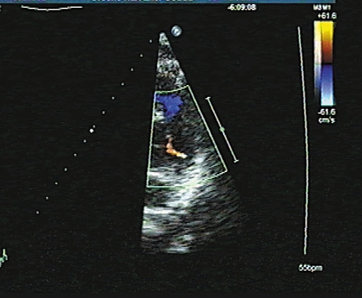
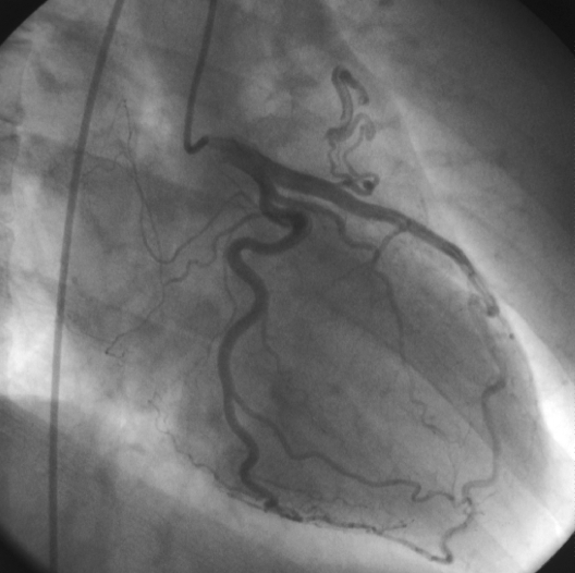
Echocardiography showed minor dilatation of the right atrium, and a diastolic flow in the parasternal short axis over the pulmonary valve (figure 1). There were no signs of atrial or ventricular septal defects. The echo aroused suspicion of a congenital abnormality. A right- and left-sided heart catheterisation was performed. Pressures were normal. Oxygen saturation from the right atrium, ventricle, pulmonary artery and aorta showed no evidence of an intracardiac shunt. Coronary angiography revealed an anomalous coronary fistula which originated between the proximal and distal segment of the left anterior descending branch (LAD), and seemed to drain into one of the atria (figure 2).
Figure 1 .
Parasternal short-axis view showing an abnormal flow over the pulmonary valve with colour Doppler echocardiography.
Figure 2 .
Coronary angiography in right anterior oblique view: a congenital coronary artery fistula from the proximal LAD, probably draining in the left atrium.
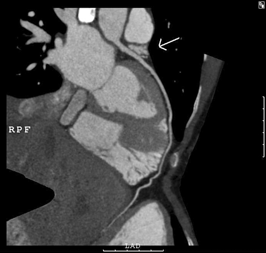
To visualise the exact course, multidetector computed tomography (MDCT) angiography was performed. The CT images showed a small, tortuous side artery from the LAD coursing cranially; the exact termination, however, could not be seen (figure 3). Based on the compilation of the results it was concluded that this fistula probably drained into the left atrium.
Figure 3 .
Multidetector CT image showing the LAD with a tortuous side artery coursing cranially.
Because the fistula did not seem to have any haemodynamic consequences, it was decided that closure of this abnormality was not indicated.
Congenital coronary artery fistulae (CAF) account for 0.08 to 0.4% of all congenital heart diseases.1,2 Clinical presentation ranges from angina pectoris, myocardial infarction, fatigue, orthopnoea, arrhythmias, stroke to endocarditis.1,2 However, the majority are asymptomatic and CAF are found incidentally at coronary angiography.2,3 Most fistula, 60%, arise from the right coronary artery (RCA), while 35% arise from the LAD. The fistula terminates in the right side of the heart in 90% of the cases.2
Coronary angiography is considered the gold standard for detection of a CAF. But rather frequently it is not possible to evaluate the exact course of a CAF at conventional angiography. Nowadays, new imaging techniques such as MDCT or magnetic resonance angiography are increasingly being used for visualisation of coronary anatomy with quite impressive results.3-6
By combining the results of the different imaging modalities we are now able to make an optimal compilation of the course of a congenital artery fistula.
Acknowledgements
We would like to thank Dr M.J.M. Cramer, Utrecht University Medical Center, and Dr E.R. Holman, Leiden University Medical Center for their support.
In this section a remarkable ‘image’ is presented and a short comment is given.
We invite you to send in images (in triplicate) with a short comment (one page at the most) to Bohn Stafleu van Loghum, PO Box 246, 3990 GA Houten, e-mail: l.jagers@bsl.nl.
‘Moving images’ are also welcomed and (after acceptance) will be published as aWeb Site Feature and shown on our website: www.cardiologie.nl
This section is edited by M.J.M. Cramer and J.J. Bax.
References
- 1.Gillebert C, Van Hoof R, Van de Werf F, Piessens J, De Geest H. Coronary artery fistula in an adult population. Eur Heart J 1986;7:437-43. [DOI] [PubMed] [Google Scholar]
- 2.Said SAM, Van der Werf T. Dutch survey of coronary artery fistulas in adults: Congenital solitary fistulas. Int J Cardiol 2006;106:323-32. [DOI] [PubMed] [Google Scholar]
- 3.Tan KT, Reed D, Howe J, Challenor V, Gibson M, McGann G. CT vs conventional angiography in unselected patients with suspected coronary heart disease. Int J Cardiol 2007;121:125-6. [DOI] [PubMed] [Google Scholar]
- 4.Utsunomiya D, Nishiharu T, Urata J, Ino M, Nakao K, Awai K, et al. Coronary arterial malformation depicted at multi-slice CT angiography. Int J Cardiovasc Imaging 2006;22:547-51. [DOI] [PubMed] [Google Scholar]
- 5.Schmid M, Achenbach S, Ludwig J, Baum U, Anders K, Pohle K, et al. Visualisation of coronary artery anomalies by contrast enhanced multidetector row spiral computed tomography. Int J Cardiol 2006;111:430-5. [DOI] [PubMed] [Google Scholar]
- 6.Schussler JM, Grayburn PA. Non-invasive coronary angiography using multislice computed tomography. Heart 2007;93:290-7. [DOI] [PMC free article] [PubMed] [Google Scholar]


