

Abstract
Radiolabeled anti-carcinoembryonic antigen (CEA) antibodies have the potential to give excellent images of a wide variety of human tumors, including tumors of the colon, breast, lung, and medullar thyroid. In order to realize the goals of routine and repetitive clinical imaging with anti-CEA antibodies, it is necessary that the antibodies have high affinity for CEA, low cross reactivity and uptake in normal tissues, and low immunogenicity. The humanized anti-CEA antibody hT84.66-M5A (M5A) fulfills these criteria with an affinity constant >1010 M−1, no reactivity with CEA cross-reacting antigens found in normal tissues, and >90% human protein sequence. A further requirement for routine clinical use of radiolabeled antibodies is a versatile method of radiolabeling that allows the use of multiple radionuclides that differ in their radioemissions and half-lives. We describe a versatile bifunctional chelator, DO3A-VS (1, 4, 7-tris(carboxymethyl)-10-(vinylsulfone)-1, 4, 7, 10-tetraazacyclododecane) that binds a range of radiometals including 111In for gamma-ray imaging and 64Cu for Positron Emission Tomography (PET), and which can be conjugated with negligible loss of immunoreactivity either to sulfhydryls (SH) in the hinge region of lightly reduced immunoglobulins or surface lysines (NH) of immunoglobulins.
Methods
Athymic mice peripherally xenografted with CEA-positive human colon tumors (LS-174T) were injected with 111In-labeled or 64Cu-labeled SH-DO3A-VS-M5A, NH-DO3A-VS-M5A, or DOTA-M5A and sacrificed at various time points for biodistribution measurements. Other mice injected with 64Cu-labeled SH-DO3A-VS-M5A or NH-DO3A-VS-M5A were imaged serially with small animal PET from 1 to 48 h post injection and then sacrificed for biodistribution measurements.
Results
Virtually identical biodistributions were obtained for SH- and NH-DO3A-VS-M5A or DOTA-M5A whether radiolabeled with 111In or 64Cu. Rapid tumor uptake of radiolabel was observed, reaching 40% injected dose/gram or more by 48 h. Importantly, excellent PET images of tumor were obtained as early as 22 h after injection of 64Cu-labeled SH- or NH-DO3A-VS-M5A.
Conclusions
Based on our correlative studies comparing the kinetics of radiolabeled anti-CEA antibodies in murine models with those in man, we predict that 64Cu-labeled intact, humanized antibodies can be used to image CEA positive tumors in the clinic.
Keywords: carcinoembryonic antigen, bifunctional chelate, radioimmunoimaging, positron emission tomography
In order to be effective tumor imaging agents, radiolabeled anti-tumor antibodies must demonstrate both high tumor uptake and high tumor to normal tissue ratios (1). Monoclonal antibodies (150 kDa) have a relatively slow blood clearance (second phase half-life t1/2 β = 48–72 h), allowing ample time for high accumulation in tumor, but suffer from low tumor to normal tissue contrast due to slow clearance from the vasculature. In order to improve the dynamic biodistribution, researchers have focused their attention on engineering recombinant antibody fragments. For example, single chain (sc) Fv recombinant antibodies (25 kDa) are rapidly cleared from the blood (t1/2 β = 0.5–2.0 h), resulting in very high tumor to blood ratios, but suffer from low accumulation of radioactivity in tumor. For this reason, intermediate-sized antibody fragments have been engineered and studied intensively (2). We have shown that in order to predict optimal tumor imaging, one must account for both tumor uptake and tumor to normal tissue contrast using pharmacokinetic analytic indicators such as the Imaging Figure of Merit (1). In spite of the advancement of engineered antibody fragments, the fact remains that most new antibodies with clinical potential originate as intact immunoglobulins. Conversion of IgGs to optimized recombinant fragments is time consuming and fraught with many problems, including the potential loss of immunoreactivity, loss of the original high affinity for target antigen, and difficulty in obtaining high production yields in culture. Therefore, we have investigated the development of methods that allow the direct imaging of tumors using intact immunoglobulins.
Another major consideration in developing a versatile radioimmunoimaging agent is the choice of radionuclide. While most intact antibodies can be easily and inexpensively radioiodinated, a potential drawback to radioiodinated antibodies is that they may be rapidly metabolized and dehalogenated in tissues, including the tumor, especially if the antigen undergoes internalization after binding to cells. Even in the case of anti-CEA antibodies that undergo negligible internalization, we and others have observed higher tumor uptake and persistence at the tumor site for radiometal- compared to radioiodine-labeled antibodies (3). For this reason we have favored the use of radiometals for labeling antibodies, but there are still drawbacks including high normal tissue accumulation in the kidney and liver (4). Furthermore, to take advantage of the wide range of radiometals available, the antibody must be conjugated to a bifunctional chelator. Although much progress has been made in this area (5–8), the search for a truly versatile bifunctional chelator continues. In this report, we describe such a bifunctional chelator (DO3A-VS), based on the stable metal ion binding properties of the tetraazamacrocyle ring with pendant carboxymethy groups and the chemically reactive linker (vinylsulfone, VS) that can be conjugated to either the easily reduced disulfides in the immunoglobulin hinge region or surface lysines of intact antibodies. We demonstrate the versatility of DO3A-VS in labeling a humanized anti-CEA antibody hT84.66-M5A and imaging tumors in a murine xenograft model using both 111In for gamma-ray imaging and 64Cu for PET imaging. The PET images obtained for 64Cu-labeled M5A show excellent tumor to normal tissue ratios as early as 24 h post injection, suggesting that intact antibodies radiolabeled with 64Cu can be useful for imaging CEA-positive tumors in man.
PET imaging with the glucose analogue [18F]fluorodeoxyglucose (FDG) has, in recent years, become the modality of choice for assessing extent of disease in many types of cancer, including those in which CEA expression is common. PET/FDG is also increasingly used for assessment of residual tumor viability and recurrence following treatment. Nonetheless, there remains a potential role for tumor-targeted PET imaging agents (immuno-PET, (9)) such as 64Cu anti-CEA MAbs both due to the limitations of FDG (including uptake in regions of inflammation, high accumulation in bladder, and a tendency for low to moderate uptake in less aggressive tumors) and the need to assess tumor targeting and biodistribution in radioimmunotherapy with these antibodies. The use of radiometal positron emitters for immuno-PET is especially attractive since they can be chelated to antibody-chelate conjugates in a kit form. Among the current choices are 86Y with a half-life of 14.7 h (10), 89Zr with a 3.3 d half-life (11), and 64Cu with a 12.7 h half-life (12). Although 86Y has the potential advantage of being coupled to 90Y beta particle therapy, its availability and prompt gamma-emissions make its use limited. 86Zr is attractive in terms of its long half-life and good spatial imaging, but has a potential problem with a 909 keV gamma emission. Appropriate BFCs for this metal ion have only recently been desctibed (11). 64Cu was chosen for the studies reported here based on its wide availability, good spatial resolution, and relatively short half-life, limiting its potential imaging dose to patients.
MATERIALS AND METHODS
Materials, Cell Culture, Antibody, Immunoreactivity and Mass Spectrometry
DO3A tri-t-butyl ester and NHS-DOTA were obtained from Macrocyclics, Inc. (Dallas TX) The synthesis of DO3A-VS was previously described (13). SATA (N-succinimidyl-S-acetylthioacetate ) was purchased from Pierce Biotechnology (Rockford, IL). All other reagents were obtained from Aldrich-Sigma (St. Louis, MO). LS-174T cells were purchased from ATCC and maintained in sterile growth media consisting of Eagle’s Minimal Essential Media 1X (EMEM) (Cellgro, Herndon, VA) supplemented with 10% heat inactivated Fetal Bovine Serum (FBS) (Omega Scientific, Tarzana, CA), and 1% L-Glutamine, 10mM Sodium Pyruvate, 0.1mM Non-Essential AA. The production and purification of anti-CEA hT84.66-M5A has been previously described (14). Chelate conjugated antibodies were radiolabeled either with 111In (Mallinckrodt, St. Louis. MO, 74-259 MBq/mg of protein) or with 64Cu (Washington University, St. Louis, MO, 148-351 MBq/mg of protein). Percent labeling was measured by ITLC or by size exclusion chromatography (SEC) on a TSK column (1 × 30 cm, 1 ml/min, TOSOH Bioscience) or a Superdex 75 column (1 × 30 cm, 0.5 mL/min, Pharmacia Biotech). Radiolabeled antibody was purified by SEC on the same columns (in saline, fraction sizes of 1.0 and 0.5 mL, respectively). Immunoreactivity was determined by incubating the purified radiolabeled sample with a 20-fold excess by mass of CEA or a 2-domain recombinant version of CEA, NA3 (15) at 37°C for 15 min and then running the sample over a Superose 6HR10/30 column (GE Healthcare). Immunoreactivity was calculated as percentage total radioactivity that shifted to complexes of higher molecular weight. MALDI-MS was performed on a Perkin-Elmer-Sciex prOTOF 2000 using sinapinic acid [10 mg/mL in TFA/water/acetonitrile (0.1/50/50 v/v/v)] as a matrix. Prior to mixing with matrix, the sample was desalted on a Sephadex G25 spin column (100–200 mg in a 250 μL pipette tip) that was equilibrated in water.
Conjugation and Radiolabeling
SH-DO3A-VS-M5A
For conjugation of DO3A-VS to reduced cysteine hinge residues, M5A antibody (2 mg of 6.5 mg/mL, 13 nmole in PBS pH 7.5) was mixed with 144 μL of PBS and 2 μL of TCEP (tris-carboxyethyl phosphine, 200 mM, 400 nmole) and reduced for 2 h at 37 °C under argon on a rotator. Excess TCEP was removed on a Sephadex G25 spin column (1 mL, in PBS, 1200 rpm, 2 min), reacted with 3.6 μL of DO3A-VS (100 mg/mL in water, 774 nmole) under argon for 2 h at RT on a rotator, and dialyzed vs pH 7.0 0.25 M ammonium acetate containing 1g/L Chelex for 5 d with five changes of buffer. Aliquots were removed to confirm (a) complete reduction by SDS gel electrophoresis and (b) reaction with DO3A-VS by MALDI-TOF-MS and IEF gel electrophoresis.
NH-DO3A-VS-M5A
For conjugation of DO3A-VS to surface lysine residues, 2 mg (13 nmole) of M5A (6.5 mg/mL in PBS) was mixed with 100 μL of PBS plus 6.2 μL of DO3A-VS (100 mg/mL in water, 1.3 μmoles), the pH was adjusted to 9.0 with 0.1 NaOH, and the mixture was reacted for 18 h at RT on a rotator. The sample was dialyzed vs 0.25 M ammonium acetate as described above.
SATA-DO3A-VS-M5A
For conjugation of DO3A-VS to SATA generated surface sulfhydryls, 2 mg (13 nmole) of M5A (6.5 mg/mL in PBS) was mixed with 100 μL of PBS plus 2 μL of SATA (0.2 M in DMF, 400 nmole), reacted for 2 h at RT on a rotator, and then treated with 6 μL of hydroxylamine hydrochloride (0.2 M, 1200 nmole) to remove the acetyl protecting groups. The product was purified on a Superdex 200 column (Amersham, 1 × 30 cm, in PBS, 0.5 mL/min) and the antibody peak fraction (at 25 min) collected, concentrated to 0.4 mL on a Amicon Ultra-4, and reacted with 3.6 μL of DO3A-VS (100 mg/mL, 0.77 μmoles) for 2 h at RT on a rotator. The sample was dialyzed vs 0.25 M ammonium actetate as above.
NHS-DOTA-M5A
For conjugation of DOTA to surface lysine residues, M5A was conjugated at a molar ratio of 20:1 chelate:antibody with DOTA-NHS-ester. Briefly, 10.5 mg (70 nmole) of M5A (7 mg/mL) was dialyzed against PBS+chelex pH 7.2 and 0.1M NaHCO3+chelex pH 8.5 in a Slide-A-Lyzer cassette (Pierce, Rockford, IL). 150 μL of DOTA-NHS-ester (7.6 mg/mL in water, 1.4 μmoles) was added and reacted for 2 h at RT on a rotator. The sample was dialyzed vs 0.25 M ammonium acetate as described above.
Conjugates were concentrated to 3–5 mg/ml in a Amicon Ultra-4 10,000 cut off (Millipore, Billerica, MA) and sterile filtered.Conjugates were characterized by both SDS and IEF gel electrophoresis (see Results). The antibody conjugates were buffered with 0.25M ammonium acetate, pH 7, except for M5A-NHS-DOTA that was buffered with 0.25M Sodium Acetate, pH 7.2. Also M5A-NHS-DOTA was more concentrated (7.7 mg/ml) than the other conjugates (3–5 mg/ml).
For labeling with 111In no additional buffer was added to the reaction mixture except for NHS-DOTA-M5A, where 0.25M Sodium Acetate, pH 6.5 was added. This mixture was incubated for 45–60 minutes at 43°C and the reaction was terminated by the addition of one-ninth the reaction volume of 10mM DTPA to achieve a final concentration of 1mM. The radiolabeled product was then purified on a size exclusion column. For labeling with 64Cu, 0.1M ammonium citrate, pH 5.5 was added to the reaction mixture. This mixture was incubated for 45 minutes at 43°C and the reaction was terminated by the addition of one-ninth the reaction volume of 10mM DTPA to achieve a final concentration of 1 mM. The radiolabeled product was then purified on a size exclusion column.
Serum stability studies
The DO3A-VS NH and SH M5A antibody conjugates were radiolabeled with 64Cu at a specific activity of 11 μC/μg (80 μg of antibody) using 0.87 mC of 64CuCl2 for 45 min at 43 oC in ammonium citrate (0.1 M, pH 5.5). The radiolabeled samples were purified on a TSK G2000 SW size exclusion column (7.5 mm × 30 cm) and the peak IgG fractions pooled and counted. One aliquot (ca 10,000,000 cpm) was incubated with human serum (15 μL sample plus 150 μL human serum) at 37 °C and aliquots (200,000 cpm uncorrected for decay) removed at various times for analysis by SEC (Pharmacia Superose 6, 1 × 30 cm). A second aliquot (10 μC, 1–2 μg) was injected into a nude mouse, blood removed at various times, and the serum fraction analyzed by SEC as above.
Xenograft Model, Biodistributions and PET imaging
Female, athymic nu/nu mice (Charles River), 10–12 weeks old, were injected with LS-174T human colon cancer cells (5 × 10⁶) s.c. in the flank. Tumors were established within 7–10 days post injection. Athymic mice bearing LS-174T xenografts were injected via a tail vein with 200 μl of 133–148 kBq/1.7–2.2 μg 111In or 333–370 kBq/2–2.4 μg of 64Cu labeled DO3AVS-conjugates. Five mice were sacrificed at various time points (see Results) and dissected for biodistribution measurements. Samples (blood, liver, spleen, kidneys, lungs, tumor and carcass) were weighed with an analytical balance and assayed for radioactivity using a calibrated gamma counter. Measured activities were corrected for radiodecay after injection, and percentage of injected activity dose per gram of tissue (%ID/g) was calculated for each specimen.
Tumor-bearing mice (22–26 g) were injected i.v. with 64Cu-labeled antibody and imaged beginning at 1.0–1.7 h, 4.5–5.5 h, 21–26 h and 45–48 h with a small-animal PET scanner (microPET Model R4; Siemens/CTIMI, Knoxvillle, TN). Injected activity and protein load ranged from 42 to 127 kBq/g and from 0.3 to 0.6 μg/g, respectively. Shortly before scanning, mice were anesthetized with isoflurane, secured in a prone position on a thin cardboard platform attached across the scanner bed and centered in the instrument’s field of view. Scan duration was 20 min for the two earliest time points and 60 min for the two later time points. The microPET’s laser alignment tool was used to position the base of the tail approximately 4.0 cm from the axial center of the 8.0 cm-long field of view in each scan; mice were placed in approximately the same orientation for each scan by aligning them with body outlines traced on the cardboard platforms prior to the scans at 1 h.
Immediately after completion of the final scan, a blood sample (0.2 cc) was obtained by cardiac puncture, the mouse was sacrificed, and the tumor and various major organs were excised, weighed, and counted. Measured activities were corrected for radiodecay after injection, and percentage of injected activity dose per gram of tissue (%ID/g) was calculated for each specimen. Tumor weights at time of sacrifice ranged from 126 to 227 mg.
Image processing and analysis were performed with the standard microPET software. Scan data were sorted into two-dimensional sinograms using the Fourier rebinning method and corrected for intrascan radiodecay, detector non-uniformity and random coincidence noise. Images were reconstructed by the iterative ordered subsets expectation maximization (OSEM) method (4 iterations, 16 subsets). Volumetric regions of interest (VOIs) were defined to include the hottest 10–15% of voxels in the images of heart and the hottest 40–50% of voxels in the images of tumor. For liver, VOIs were drawn to lie well within the organ boundaries while excluding the region containing the large central blood vessels. Image activity densities were decay corrected to time of injection to obtain relative time-activity curves (TACs). Absolute TACs for tumor, liver and blood (in units of %ID/g) were obtained by normalizing the final time points of the PET-derived TACs to the direct tissue assays. Blood TACs were equated with heart TACs normalized to the blood samples taken just before sacrifice.
RESULTS AND DISCUSSION
DO3AVS-M5A Conjugates
In developing a bifunctional chelator for conjugation to intact immunoglobulins, our criteria were rapid synthesis, high and stable metal ion binding to a variety of radiometals, and an ability to conjugate to either sulfhydryl or amino groups present on the immunoglobulin molecule. Indeed, we had such a bifunctional chelator (DO3A-VS) in hand that previously had been shown to have high metal binding stability when conjugated to an antibody (13). Our interest in the use of vinyl sulfone as a bifunctional linker stems from its ability to react with primary surface amines at pH 9, as well as with thiols at acidic to neutral pH (13). In addition, we found that the thiol conjugates were chemically more stable than the often-used maleimide thiol conjugates (8). The most accessible and/or reactive disulfides of immunoglobulins are located in the hinge region away from the antigen combining sites, and require only mild reduction of the antibody for generation of free sulfhydryls, and they can be targeted by DO3A-VS under very specific reaction conditions (pH 5.0- 7.0 after mild reduction). Many studies have shown that modification of the hinge sulfhydryl groups has little or no effect on the antigen binding properties of an antibody (16–19), making this an ideal approach for site-specific conjugation (Figure 1). Nonetheless, the insertion of any derivative into the hinge region requires careful documentation of retention of antigen-binding activity. In addition, we have shown that vinyl sulfone-Cys-DOTA conjugates can react with surface lysine residues on antibodies at pH 9.0 (13). Thus, by control of pH and/or prior reduction of the antibody, one can conjugate DO3A-VS either to hinge region sulfhydryls or surface lysine residues.
Figure 1. Schematic model of an immunoglobulin (IgG1) showing reduction of hinge sulfhydryls.
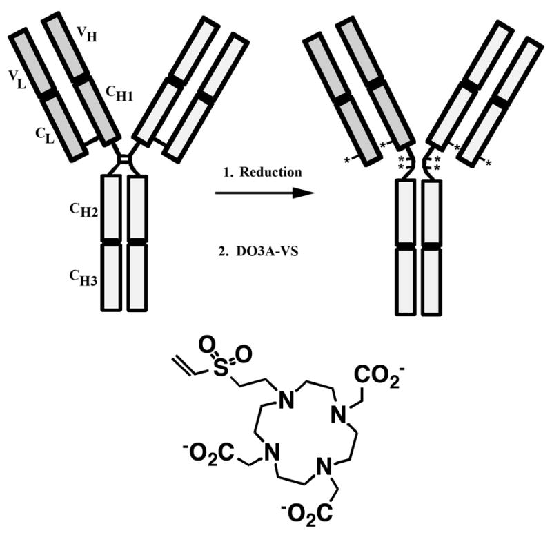
(*). In the second step, the sulfhydryls are reacted with DO3A-VS.
Based on the concepts outlined above, the bifunctional reagent DO3A-VS was reacted either with the hinge region sulfhydryls (SH) or the surface lysines (NH) of hT84.66-M5A (M5A) and immunoreactivity and specific isotope incorporation were measured (Table 1). From these studies we determined that both SH- and NH-DO3A-VS-M5A retained high immunoreactivity and had isotope incorporation activities comparable to those published for other DOTA-antibody conjugates (13). We determined the relative degree of conjugation of DO3A-VS to M5A by IEF (Figure 2), an analysis that showed a shift to more acidic pI (pH) with increasing amounts of chelate substitution. Based on this analysis, conjugation of DO3A-VS to surface lysines was more extensive than to reduced sulfhydryls in the hinge region. To further characterize the degree of modification of the antibody with DO3A-VS, MALDI-TOF analysis was performed on the SH modified sample. The results shown in Figure 3 reveal that both the light chain and heavy chain have been extensively modified (0.7 DO3A-VS per light chain and 3.3 DO3A-VS per heavy chain). Thus, the total number of chelates per 150 kDa immunoglobulin was eight.
Table 1.
Radiolabeling of DO3AVS-M5A conjugates.1
| Conjugate | Labeling Buffer | Ratio (μCi/ug) | % labeling | S.A. (μCi/ug) | % IR |
|---|---|---|---|---|---|
| SH-DO3AVS, In-111 | None | 2.1 | 99.8 | 2.1 | 100 |
| SH-DO3AVS, Cu-64 | 0.1M Ammonium Citrate, pH 5.55 | 4.2 | 100 | 4.2 | 100 |
| NH-DO3AVS, In-111 | None | 2.2 | 83.8 | 1.8 | 100 |
| NH-DO3AVS, Cu-64 | 0.1M Ammonium Citrate, pH 5.5 | 9.5 | 50 | 4.8 | 100 |
| SATA-DO3AVS, In-111 | None | 2.9 | 97 | 2.8 | 100 |
| NH-NHS-DOTA, In-111 | 0.25M Sodium Acetate, pH 6.5 | 7.1 | 99.3 | 7.1 | 100 |
| NH-NHS-DOTA, Cu-64 | 0.1M Ammonium Citrate, pH 5.5 | 8.3 | 99 | 8.2 | 100 |
Reaction conditions include radiolabeling buffer with pH. Ratio refers to the μCi of isotope to μg of protein. Percent labeling efficiency was determined by HPLC purification. Specific activity (S.A.) was calculated by multiplying the labeling ratio with percent labeling. Percent immunoreactivity (%IR) was determined by in vitro incubation with 20 times mass excess of CEA (except for SH-DO3AVS, In-111, which was tested against 20 times mass excess of NA3).
Figure 2. Characterization of DO3A-VS-M5A conjugates by IEF gel electrophoresis.

1: IEF standards (see pI values to the left). 2, 5, and 7: unconjugated M5A. 3: M5A after reduction with TCEP. 4: SH-DO3A-VS-M5A. 6: NH-DO3A-VS-M5A. 8: SATA-DO3A-VS-M5A.
Figure 3. MALDI-TOF-MS analysis of SH-DO3A-VS-M5A.

A reduced M5A. The peak at 23,893 is the light chain, the peak at 50,672 is the heavy chain and the peaks labeled L2 and HL are light chain and heavy-light chain dimers, respectively. B: SH-DO3A-VS-M5A. The light chain has two peaks, one unmodified at m/z 23,893, and one increased by 462 mass units corresponding to the addition of one DO3A-VS. Based on peak heights the degree of modification for the light chain is 0.67. The heavy chain has increased by 1517 mass units corresponding to the addition of an average 3.3 DO3A-VS. Note: The light chain has one “hinge” cysteine and the heavy chain three hinge cysteine residues in an IgG1 molecule.
In order to compare these conjugates to a more traditional DOTA conjugate, M5A was also reacted with a commercially available bifunctional chelator, DOTA-NHS. This DOTA-conjugate also labeled well with 111In and 64Cu and retained high immunoreactivity (Table 1). MALDI-TOF analysis of this conjugate also revealed an average of eight chelates/immunoglobulin (data not shown). Other methods for conjugating DOTA to antibodies include that developed by Meares and coworkers, where lysines on the antibody were first reacted with iminothiolane and then conjugated to the bifunctional chelator BAT (20). To approximate this approach, we first reacted surface lysines with SATA, then with hydroxylamine to deprotect the lysine-coupled protected thiol groups, followed with DO3A-VS. This conjugate also gave high levels of isotope incorporation and retained its high immunoreactivity (Table 1), but contained up to 10% dimers due to sulfhydryl crosslinking (Figure 4).
Figure 4. SEC analysis of radiolabeled M5A conjugates.

A SH-DO3A-VS-M5A radiolabeled with 111In (black) or 64Cu (blue) before and after the addition of NA3 (black dotted) or CEA (blue dotted). B: NH-DO3A-VS-M5A radiolabeled with 111In (black) or 64Cu (blue) before and after the addition of CEA (black or blue dotted). C: SATA-DO3A-VS-M5A radiolabeled with 111In (black) before and after the addition of CEA (black dotted). D: DOTA-NHS-M5A radiolabeled with 111In (black) or 64Cu (blue) before and after the addition of CEA (back or blue dotted). Radioactivity (arbitrary units) shown on the y-axis.
Prior to proceeding to biodistibution studies, we analyzed each of the radiolabeled conjugates by SEC before and after the addition of CEA to determine if the sample was homogeneous (single peak of the correct size) and if it was fully immunoreactive (shifted to higher molecular weight by the addition of a two-domain recombinant form of CEA, NA3, or human CEA). The results shown in Figure 4 demonstrate that the SH- and NH-DO3A-VS-M5A as well as the DOTA-NHS-M5A conjugates were free of aggregates, while the SATA-DO3A-VS-M5A conjugate contained up to 10% aggregates, but importantly, all four conjugates were 100% immunoreactive. For three of the conjugates tested, the results were equivalent whether labeled with 111In or 64Cu.
Since the serum stability of 64Cu-labeled DOTA-based BFCs has been questioned (21, 22), it was necessary to test both their in vitro and in vivo serum stabilities. When either SH or NH conjugated 64Cu-labeled DO3A-VS-M5A was incubated with human serum at 37 °C and analyzed by SEC, 100% of the radioactivity remained in the IgG peak over a time course of 70 h after which too few remaining counts remained for analysis (Figure 5A and 5B). In addition, when both radiolabeled conjugates were injected into mice, blood removed, and the serum fractions analyzed by SEC, 100% of the radioactivity was found in the IgG fraction over the time course of the analysis (Figure 5C and 5D). These analyses demonstrate that no significant transchelation of 64Cu occurs in serum either in vitro or in vivo for both conjugates. These data are similar to that reported by Lewis et al. (23) who showed that both 64Cu labeled biotin DOTA and a 64Cu labeled DOTA-antibody conjugate were 100% stable in serum for up to 48 hr. We conclude that the so-called instability of 64Cu DOTA or TETA based conjugates refers to metabolism of the radiolabeled chelates or their conjugates in the liver where uptake of the radiolabel into SOD has been documented (24, 25). The redistribution of radiolabels into other proteins after metabolism is a well recognized phenomenon as illustrated by the metabolic redistribution of radioiodine from radioiodinated proteins into thyroglobulin.
Figure 5. Serum stability analysis of 64Cu-labeled M5A conjugates.

64Cu labeled DO3A-VS-M5A was incubated with human serum and analyzed over time by SEC. The major peak at 42 min corresponds to the elution time for IgG. A: SH-DO3A-VS-M5A. B: NH-DO3A-VS-M5A. The sample was also injected into nude mice, blood removed over time, and the serum fraction analyzed by SEC. C: SH-DO3A-VS-M5A. D: NH-DO3A-VS-M5A.
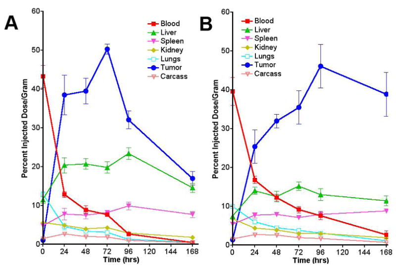
Biodistributions of Radiolabeled M5A Conjugates
SH- and NH-conjugated DO3A-VS-M5A were radiolabeled with 111In or 64Cu, and biodistibutions were performed in nude mice bearing CEA positive LS-174T xenografts. The results shown in Figure 6 demonstrate high tumor uptake, reaching a maximum of 40–50% ID/g at 48–96 h. It is noteworthy that, overall, the dynamic biodistributions are similar between SH- and NH- conjugates whether radiolabeled with 111In or 64Cu both in terms of kinetics of tumor and normal tissue uptake and the ratios of tumor to normal tissue uptake. The one exception may be higher tumor uptake for 64Cu-labeled NH-DO3A-VS-M5A, but in this experiment higher blood levels track with the higher tumor uptake, leading to similar tumor to blood ratios compared to the other radiolabeled conjugates. We conclude from this study that SH- and NH-DO3A-VS conjugates give equivalent biodistributions whether radiolabeled with 111In or 64Cu.
Figure 6. Biodistributions of 111In- and 64Cu-SH- and NH-DO3A-VS-M5A in nude mice bearing LS174T xenografts.

The average %ID/g (± sem.) of five animals per time point is shown. A: 111In-labeled SH-DO3A-VS-M5A. B: 64Cu-labeled SH-DO3A-VS-M5A. C: 111In-labeled NH-DO3A-VS-M5A. D: 64Cu-labeled NH-DO3A-VS-M5A.
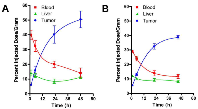
As mentioned above, we were interested in comparing the SH- and NH-DO3A-VS-M5A conjugates with (a) DO3A-VS conjugated to surface sulfhydryls introduced into M5A by reaction of surface lysines with SATA, and (b) the more conventional DOTA-NHS conjugated antibody. The results shown in Figure 7 demonstrate roughly equivalent tumor uptakes for both conjugates compared to each other and to those shown in Figure 6, but have interesting differences in the liver clearances. Over the period 0–96 h, the SATA conjugate had almost twice the liver uptake of the SH- and NH-DO3A-VS-M5A conjugates, while the DOTA-NHS conjugate had about a 25–50% increase in liver uptake compared to SH- and NH-DO3A-VS-M5A. The higher liver uptake for the SATA conjugate is likely due to the presence of aggregates that are absent in the other three conjugates (see Figure 4). However, the increase in liver uptake observed for the DOTA-NHS conjugate is harder to explain and implies that the SH- and NH-DO3A-VS conjugates may have improved imaging properties over those of the conventional DOTA-NHS conjugate.
Figure 7. Biodistributions of 111In- SATA-DO3A-VS-M5A and DOTA-NHS-M5A in nude mice bearing LS174T xenografts.
The average %ID/g (± sem) of five animals per time point is shown. A: 111In-labeled SATA-DO3A-VS-M5A. B: 111In-labeled DOTA-NHS-M5A.
64Cu PET Imaging of SH- and NH-DO3A-VS-M5A
Based on the above biodistribution results, we predicted that tumors would be visualized as early as 24 h with 64Cu-labeled conjugates for whole antibody, since tumor-to-blood ratios were > 1.0 by that time. [Due to the constraint on injected activity because of radiation dose considerations, 24 h is a practical upper time limit on 64Cu (t½13 h) imaging in humans.] The dynamic biodistributions of radiolabel in the LS174T tumor/nude mouse model following injection of 64Cu SH- and NH-DO3A-VS-M5A are illustrated via serial microPET images in Figure 8. The kinetics of the radiolabel were very similar for both forms of 64Cu M5A antibody. At 1 and 5 h, the activity remained primarily intravascular, with tumor beginning to appear at the second time point. Tumor was the primary site of localization by 22h, and tumor-to-normal tissue contrast was further increased by 45h. Time-activity curves (TACs) derived from the microPET experiments (Figure 9) are quantitatively similar to those obtained in the direct-assay biodistribution experiments for 64Cu SH- and NH-DO3A-VS-M5A (Figures 6C and 6D).
Figure 8. Dynamic biodistribution of 64Cu-labeled hT84.66-M5A MAbs in athymic mice peripherally xenografted with CEA-positive LS-174T colon tumors.

(A) SH-DO3A-VS-M5A; (B) NH-DO3A-VS-M5A. Images are PET anterior-view maximum intensity projections (MIPs) normalized to reflect radiodecay-corrected relative image intensity per unit injected activity. Labels shown in red, yellow and green show directly-measured %ID/g in blood (heart), liver and tumor, respectively. Tumor weights at time of sacrifice were 126 and 155 mg in (A) and (B) respectively.
Figure 9. Time-activity curves derived from serial microPET studies.
Data are average %ID/g (± sem.) for three (first 3 time points for SH-DO3A-VS-M5A) or two (NH-DO3A-VS-M5A and final time point for SH-DO3A-VS-M5A) mice.
Note that, in spite of the very small sample sizes (n = 2 or 3 per data point), the standard deviations of the microPET time-activity curves (Figure 9) are similar to those of the standard biodistribution experiments, where n = 5 per data point (Figure 6). This illustrates a major advantage of serial quantitative imaging over the standard biodistribution approach, since it allows the determination of an entire TAC for a single mouse.
CONCLUSIONS
Although much progress has been made in engineering radiolabeled antibodies for tumor imaging, many challenges still exist. In this study, we have returned to the use of intact antibodies because they arise at the earliest stage of clinical antibody development and can be produced in large quantities. Since recombinant engineering of antibodies is a time consuming process and fraught with many obstacles, it is important to determine at the earliest point possible if the antibody of interest can indeed provide high targeting of tumor relative to normal tissue. If it can, then one could potentially use it at this stage of development as an in vivo diagnostic agent.
Until recently, radioimaging with antibodies was limited to the use of planar or SPECT imaging modalities, which have relatively poor spatial resolution and are not quantitative. Although PET has the potential to solve this problem, the need to match the isotope half-life with the blood clearance time of the antibody has limited most immuno-PET studies to antibody fragments labeled with short half-life radionuclides such as 18F. The newly available, 12.7 h half-life positron emitter 64Cu offers a potential solution to this problem because it can be used to radiolabel whole antibody-chelate conjugates in a kit format. Based on the animal studies shown here, intact anti-CEA MAbs may deliver sufficient radiolabel to tumor to allow PET imaging as early as 22 h after injection. The improvement in PET images over those obtained with planar or SPECT imaging of 123I or 111In labeled antibodies is obvious; however, the ability to image tumors with low normal tissue background with whole 64Cu-labeled antibodies in the mouse model came as a surprise, because most of our experience has been with planar gamma images. We attribute the majority of this improvement to not only the 3D nature and relatively high resolution of the microPET images, but also to the change in chelates, where DO3A-VS conjugates show less liver uptake that the more conventional DOTA-NHS conjugates. The ability to measure dynamic biodistributions in individual mice with micro-PET/64Cu-labeled-MAb, as illustrated here, confers greater precision for a given sample size or, alternatively, permits substantial reduction in the number of mice required.
The practical advantages of using DO3A-VS-whole antibody conjugates include a versatile chemistry allowing conjugation at thiols in the mildly reduced hinge region or to surface amino groups in immunoglobulins, as well as, maintaining the inherent metal ion binding and stability of the macrocyclic ring. Although reaction of DO3A-VS at the hinge region requires mild reduction of the antibody and removal of excess reducing agent, this is easily accomplished by the use of a spin-column and the resulting hinge thiols remain in the reduced state for up to 72 h before re-oxidation occurs. Reaction of DO3A-VS at lysine residues requires brief exposure of the antibody to pH 9, conditions which, in our hands, have not compromised antigen-binding activity of several antibodies. Thus, DO3A-VS may be conjugated to antibodies under several conditions offering the researcher alternatives to the conventional NHS-active ester derivatives of DOTA that are inherently unstable and therefore less predictable in batch to batch conjugations. In addition, the quality of the PET images obtained within 24 h after injection indicates that PET/64Cu-DO3A-VS-M5A should be evaluated for clinical use.
Acknowledgments
This research was supported by NCI grant CA43904.
References
- 1.Williams LE, Wu AM, Yazaki PJ, Liu A, Raubitschek AA, Shively JE, Wong JY. Numerical selection of optimal tumor imaging agents with application to engineered antibodies. Cancer Biother Radiopharm. 2001;16:25–35. doi: 10.1089/108497801750095989. [DOI] [PubMed] [Google Scholar]
- 2.Kenanova V, Wu AM. Tailoring antibodies for radionuclide delivery. Expert Opin Drug Deliv. 2006;3:53–70. doi: 10.1517/17425247.3.1.53. [DOI] [PubMed] [Google Scholar]
- 3.Yazaki PJ, Wu AM, Tsai SW, Williams LE, Ikler DN, Wong JY, Shively JE, Raubitschek AA. Tumor targeting of radiometal labeled anti-CEA recombinant T84.66 diabody and T84.66 minibody: comparison to radioiodinated fragments. Bioconjug Chem. 2001;12:220–8. doi: 10.1021/bc000092h. [DOI] [PubMed] [Google Scholar]
- 4.Li L, Yazaki PJ, Anderson AL, Crow D, Colcher D, Wu AM, Williams LE, Wong JY, Raubitschek A, Shively JE. Improved biodistribution and radioimmunoimaging with poly(ethylene glycol)-DOTA-conjugated anti-CEA diabody. Bioconjug Chem. 2006;17:68–76. doi: 10.1021/bc0502614. [DOI] [PubMed] [Google Scholar]
- 5.Milenic DE, Garmestani K, Chappell LL, Dadachova E, Yordanov A, Ma D, Schlom J, Brechbiel MW. In vivo comparison of macrocyclic and acyclic ligands for radiolabeling of monoclonal antibodies with 177Lu for radioimmunotherapeutic applications. Nucl Med Biol. 2002;29:431–42. doi: 10.1016/s0969-8051(02)00294-9. [DOI] [PubMed] [Google Scholar]
- 6.Gansow OA. Newer approaches to the radiolabeling of monoclonal antibodies by use of metal chelates. Int J Rad Appl Instrum B. 1991;18:369–81. doi: 10.1016/0883-2897(91)90063-q. [DOI] [PubMed] [Google Scholar]
- 7.Lewis MR, Raubitschek A, Shively JE. A facile, water-soluble method for modification of proteins with DOTA. Use of elevated temperature and optimized pH to achieve high specific activity and high chelate stability in radiolabeled immunoconjugates. Bioconjugate Chem. 1994;5:565–576. doi: 10.1021/bc00030a012. [DOI] [PubMed] [Google Scholar]
- 8.Lewis MR, Shively JE. Maleimidocysteineamido-DOTA derivatives: new reagents for radiometal chelate conjugation to antibody sulfhydryl groups undergo pH-dependent cleavage reactions. Bioconj Chem. 1998;9:72–86. doi: 10.1021/bc970136v. [DOI] [PubMed] [Google Scholar]
- 9.Verel I, Visser GW, van Dongen GA. The promise of immuno-PET in radioimmunotherapy. J Nucl Med. 2005;46(Suppl 1):164S–71S. [PubMed] [Google Scholar]
- 10.Garmestani K, Milenic DE, Plascjak PS, Brechbiel MW. A new and convenient method for purification of 86Y using a Sr(II) selective resin and comparison of biodistribution of 86Y and 111In labeled Herceptin. Nucl Med Biol. 2002;29:599–606. doi: 10.1016/s0969-8051(02)00322-0. [DOI] [PubMed] [Google Scholar]
- 11.Borjesson PK, Jauw YW, Boellaard R, de Bree R, Comans EF, Roos JC, Castelijns JA, Vosjan MJ, Kummer JA, Leemans CR, Lammertsma AA, van Dongen GA. Performance of immuno-positron emission tomography with zirconium-89-labeled chimeric monoclonal antibody U36 in the detection of lymph node metastases in head and neck cancer patients. Clin Cancer Res. 2006;12:2133–40. doi: 10.1158/1078-0432.CCR-05-2137. [DOI] [PubMed] [Google Scholar]
- 12.Wu AM, Yazaki PJ, Tsai S, Nguyen K, Anderson AL, McCarthy DW, Welch MJ, Shively JE, Williams LE, Raubitschek AA, Wong JY, Toyokuni T, Phelps ME, Gambhir SS. High-resolution microPET imaging of carcinoembryonic antigen-positive xenografts by using a copper-64-labeled engineered antibody fragment. Proc Natl Acad Sci U S A. 2000;97:8495–500. doi: 10.1073/pnas.150228297. [DOI] [PMC free article] [PubMed] [Google Scholar]
- 13.Li L, Tsai SW, Anderson AL, Keire DA, Raubitschek AA, Shively JE. Vinyl sulfone bifunctional derivatives of DOTA allow sulfhydryl- or amino-directed coupling to antibodies. Conjugates retain immunoreactivity and have similar biodistributions. Bioconjug Chem. 2002;13:110–5. doi: 10.1021/bc015535b. [DOI] [PubMed] [Google Scholar]
- 14.Yazaki PJ, Sherman MA, Shively JE, Ikle D, Williams LE, Wong JY, Colcher D, Wu AM, Raubitschek AA. Humanization of the anti-CEA T84.66 antibody based on crystal structure data. Protein Eng Des Sel. 2004;17:481–9. doi: 10.1093/protein/gzh056. [DOI] [PubMed] [Google Scholar]
- 15.You YH, Hefta LJ, Yazaki PJ, Wu AM, Shively JE. Expression, purification, and characterization of a two domain carcinoembryonic antigen minigene (N-A3) in pichia pastoris. The essential role of the N-domain. Anticancer Res. 1998;18:3193–201. [PubMed] [Google Scholar]
- 16.Feehally J, Allen AC. Pathogenesis of IgA nephropathy. Ann Med Interne (Paris) 1999;150:91–8. [PubMed] [Google Scholar]
- 17.Baranowska-Kortylewicz J, Kassis AI. Labeling of immunoglobulins with bifunctional, sulfhydryl-selective, and photoreactive coumarins. Bioconjug Chem. 1993;4:300–4. doi: 10.1021/bc00022a009. [DOI] [PubMed] [Google Scholar]
- 18.Wright JK, Engel J, Jaton JC. Selective reduction and proteolysis in the hinge region of liganded and unliganded antibodies: identical kinetics suggest lack of major conformational change in the hinge region. Eur J Immunol. 1978;8:309–14. doi: 10.1002/eji.1830080505. [DOI] [PubMed] [Google Scholar]
- 19.Sanderson RJ, Hering MA, James SF, Sun MM, Doronina SO, Siadak AW, Senter PD, Wahl AF. In vivo drug-linker stability of an anti-CD30 dipeptide-linked auristatin immunoconjugate. Clin Cancer Res. 2005;11:843–52. [PubMed] [Google Scholar]
- 20.Kukis DL, Diril H, Greiner DP, DeNardo SJ, DeNardo GL, Salako QA, Meares CF. A comparative study of copper-67 radiolabeling and kinetic stabilities of antibody-macrocycle chelate conjugates. Cancer. 1994;73:779–86. doi: 10.1002/1097-0142(19940201)73:3+<779::aid-cncr2820731306>3.0.co;2-3. [DOI] [PubMed] [Google Scholar]
- 21.Boswell CA, McQuade P, Weisman GR, Wong EH, Anderson CJ. Optimization of labeling and metabolite analysis of copper-64-labeled azamacrocyclic chelators by radio-LC-MS. Nucl Med Biol. 2005;32:29–38. doi: 10.1016/j.nucmedbio.2004.09.004. [DOI] [PubMed] [Google Scholar]
- 22.Boswell CA, Sun X, Niu W, Weisman GR, Wong EH, Rheingold AL, Anderson CJ. Comparative in vivo stability of copper-64-labeled cross-bridged and conventional tetraazamacrocyclic complexes. J Med Chem. 2004;47:1465–74. doi: 10.1021/jm030383m. [DOI] [PubMed] [Google Scholar]
- 23.Lewis MR, Wang M, Axworthy DB, Theodore LJ, Mallet RW, Fritzberg AR, Welch MJ, Anderson CJ. In vivo evaluation of pretargeted 64Cu for tumor imaging and therapy. J Nucl Med. 2003;44:1284–92. [PubMed] [Google Scholar]
- 24.Bryan JN, Jia F, Mohsin H, Sivaguru G, Miller WH, Anderson CJ, Henry CJ, Lewis MR. Comparative uptakes and biodistributions of internalizing vs. noninternalizing copper-64 radioimmunoconjugates in cell and animal models of colon cancer. Nucl Med Biol. 2005;32:851–8. doi: 10.1016/j.nucmedbio.2005.05.006. [DOI] [PubMed] [Google Scholar]
- 25.Bass LA, Wang M, Welch MJ, Anderson CJ. In vivo transchelation of copper-64 from TETA-octreotide to superoxide dismutase in rat liver. Bioconjug Chem. 2000;11:527–32. doi: 10.1021/bc990167l. [DOI] [PubMed] [Google Scholar]