Abstract
Here we describe an association between α3β1 integrin and transmembrane-4 superfamily (TM4SF) protein CD151. This association is maintained in relatively stringent detergents and thus is remarkably stable in comparison with previously reported integrin–TM4SF protein associations. Also, the association is highly specific (i.e., observed in vitro in absence of any other cell surface proteins), and highly stoichiometric (nearly 90% of α3β1 associated with CD151). In addition, α3β1 and CD151 appeared in parallel on many cell lines and showed nearly identical skin staining patterns. Compared with other integrins, α3β1 exhibited a considerably higher level of associated phosphatidylinositol-4-kinase (PtdIns 4-kinase) activity, most of which was removed upon immunodepletion of CD151. Specificity for CD151 and PtdIns 4-kinase association resided in the extracellular domain of α3β1, thus establishing a novel paradigm for the specific recruitment of an intracellular signaling molecule. Finally, antibodies to either CD151 or α3β1 caused a ∼88–92% reduction in neutrophil motility in response to f-Met-Leu-Phe on fibronectin, suggesting an functionally important role of these complexes in cell migration.
INTRODUCTION
The integrin family of heterodimeric receptors provides structural links between the extracellular environment and the cytoskeleton-signaling network and thus helps to regulate cell migration, differentiation, cell cycle progression, apoptosis, phagocytosis, ECM assembly, and metalloproteinase activity (Brown, 1991; Stetler-Stevenson et al., 1993; Huttenlocher et al., 1995; Schwartz et al., 1995; Assoian, 1997; Frisch and Ruoslahti, 1997). Many studies of integrin-mediated signaling have focused on tyrosine phosphorylations (i.e., pp60Src, pp125FAK, pp72Syk) and activation of MAP kinases and Rho family guanosine triphosphatases (Schwartz et al., 1995). In addition, integrins can colocalize with several important cytoplasmic and cytoskeletal signaling molecules (Miyamoto et al., 1995). For example, insulin receptor substrate-1 (IRS-1) interacts with αvβ3 upon insulin stimulation (Vuori and Ruoslahti, 1994), thus linking it to growth factor receptor-bound protein 2 (Grb2) and phosphatidylinositol 3-kinase (PtdIns 3-kinase). Also, the adaptor proteins, Shc and Grb2, were recruited to a subclass of β1 integrins possibly via an extracellular or transmembrane association with caveolin (Wary et al., 1996). At least 13 different proteins have been suggested to associate directly with various integrin cytoplasmic domains (Shattil and Ginsberg, 1997), but more studies are needed before the implications of these findings can be properly appreciated.
Integrins may not only associate with intracellular proteins, but may also engage in lateral associations with other transmembrane proteins such as CD47 (Lindberg et al., 1996), CD147/EMMPRIN (Berditchevski et al., 1997a), and transmembrane 4 superfamily (TM4SF) proteins (Wright and Tomlinson, 1994; Hemler et al., 1996; Maecker et al., 1997). The TM4SF proteins include 10 central members, each containing putative small (20–27 amino acids) and large (75–130 amino acids) extracellular domains and four putative hydrophobic transmembrane domains (Wright and Tomlinson, 1994; Maecker et al., 1997; Tachibana et al., 1997). Similar to integrins, these molecules play key roles in the regulation of cellular proliferation, development, motility, and tumor cell growth and metastasis. The TM4SF proteins may function as “adaptors” or “molecular facilitators,” organizing various cell-surface proteins into large multimeric complexes on the cell surface. In this regard, TM4SF proteins can specifically coimmunoprecipitate with an array of cell surface proteins, including the diphtheria toxin receptor, the B cell CD19–CD21–Leu-13 complex, major histocompatibility complex (MHC) class II, CD2, CD4, and CD8, as well as the α3β1, α4β1, α6β1, and αIIβ3 integrins (Wright and Tomlinson, 1994; Hemler et al., 1996; Maecker et al., 1997).
Also, TM4SF proteins may provide a bridge between particular β1 integrins and a type II PtdIns 4-kinase (Berditchevski et al., 1997b). Type II PtdIns 4-kinases are activated by nonionic detergent and inhibited by both adenosine and mAb 4C5G (Graziani et al., 1992; Wong and Cantley, 1994; Wong et al., 1998). PtdIns 4-kinases convert PtdIns into phosphatidylinositol-4-phosphate (PtdIns-4-P), a highly relevant intermediate in multiple phosphatidylinositide-signaling pathways. For example, PtdIns-4-P can be converted to phosphatidylinositol-4,5-bisphosphate (PtdIns-4,5-P2), thus providing a substrate for phospholipase C during growth factor induced signaling (Berridge, 1987). Alternatively, PtdIns-4-P and PtdIns-4,5-P2 can be phosphorylated by PtdIns 3-kinase, to produce PtdIns-3,4-P2 and PtdIns-3,4,5-P3, which are potent regulators of several biological functions. In addition to serving as substrates, PtdIns-4-P and PtdIns-4,5-P2 themselves can interact with actin-binding proteins and thus regulate actin polymerization (Janmey, 1993).
Thus far, it has been somewhat difficult to evaluate the meaning of integrin–TM4SF–PtdIns 4-kinase protein interactions because, 1) TM4SF proteins associate with so many different proteins, 2) only a small fraction of coimmunoprecipitating integrin appears to be associated with TM4SF proteins, and 3) associations have been observed exclusively under conditions of very gentle cell lysis using mild detergents. To further complicate matters, TM4SF proteins can form complexes with each other, thus hindering the distinction between direct and indirect associations.
In this report, we demonstrate that α3β1 has substantially more associated PtdIns-4-kinase activity than any other integrin tested. This occurs largely because of the association of α3β1 integrin with the TM4SF protein CD151, also known as PETA-3 or SFA-1 (Fitter et al., 1995; Hasegawa et al., 1996), which links the integrin to PtdIns 4-kinase. The α3β1–CD151 association is seen even in relatively stringent detergent conditions and occurs at a considerably higher stoichiometry than any other TM4SF–integrin, TM4SF–TM4SF, TM4SF–other protein, or integrin–other protein associations yet described. Finally, both anti-CD151 and anti-α3 antibodies almost completely inhibit neutrophil chemotactic migration under agarose, suggesting that CD151–α3β1 and/or CD151–α3β1–PtdIns 4-kinase complexes play key roles in cell motility.
MATERIALS AND METHODS
Cell Lines
The T cell line Molt-4 and erythroleukemic cell line K562 were maintained in RPMI-1640 media supplemented with 10% FBS and antibiotics. Untransfected K562 or mock-transfected K562 cells (K562-neo) express α5β1 integrin, but no other detectable β1 integrins. K562 cells transfected with human integrin α2, α3, α4, and α6 cDNAs have been described elsewhere (Berditchevski et al., 1995; Mannion et al., 1996) and were maintained in RPMI supplemented with 10% FBS, antibiotics, and 1 mg/ml geneticin (GIBCO BRL, Gaithersburg, MD). Molt-4-pZeo-CD151 cells were established by Molt-4 transfection with CD151 cDNA (generated by reverse transcriptase-PCR) in the expression plasmid pZeoSV (Invitrogen, San Diego, CA). Molt-4-pZeo cells were transfected with vector alone. Chinese hamster ovary (CHO) cells were cotransfected with human α3 and human β1 cDNAs as described previously (Weitzman et al., 1995), and cells were maintained in α-minus minimum essential medium containing 10% dialyzed FBS, 0.5 mg/ml geneticin, and antibiotics. The fibrosarcoma cell line HT1080 and epidermoid carcinoma cell line A431 were propagated in DMEM supplemented with 10% FBS and antibiotics.
Antibodies
Anti-integrin mAbs utilized were anti-α2, A2-IIE10 (Bergelson et al., 1994); anti-α3, A3-IVA5 and A3-X8 (Weitzman et al., 1993); anti-α5, A5-PUJ2 (Pujades et al., 1996); anti-α6, A6-ELE (Lee et al., 1995) and A6-BB (our unpublished results); anti-αv, P3G8 (Wayner et al., 1991); and anti-β1, TS2/16 (Hemler et al., 1984). Other mAbs were anti-MHC class I, W6/32 (Barnstable et al., 1978); anti-CD9, BU16 (Biogenesis, Poole, England) and C9-BB (Berditchevski et al., 1996); anti-CD63, 6H1 (Berditchevski et al., 1995); anti-CD81, M38 (Fukudome et al., 1992); anti-CD98, 8A6 (our unpublished results); anti-CD151 mAbs, SFA1–2B4 (Hasegawa et al., 1996) and 14A2 (Fitter et al., 1995); anti-PtdIns 4-kinase, 4C5G (Endemann et al., 1991); and negative control antibodies, P3 (Lemke et al., 1978), J2A2 (Hemler and Strominger, 1982), 187.1 (Yelton et al., 1981), and 4.6.19 (Weitzman et al., 1997). The anti-CD151 mAb 5C11 was produced as previously described (Berditchevski et al., 1997a).
Purification of 5C11 Antigen
K562 cells (10 g) were lysed in 1 l phosphate buffer, pH 7.2, containing 2% n-octyl glucoside, 2 mM MgCl2, 2 mM PMSF, 2 μg/ml aprotinin, and 2 μg/ml leupeptin. Solubilized proteins were sequentially incubated in batch with 5 g protein A Sepharose beads (Pharmacia Biotech, Piscataway, NJ) and with irrelevant mAb 4.6.19 coupled to Sepharose 4B for 16 h. The precleared lysate was then incubated in batch with mAb 5C11 conjugated to Sepharose 4B beads (3 ml of packed beads) for 5 h. After washing with 500 ml lysis buffer, bound protein was eluted using 100 mM glycine, pH 3.0. Fractions of 0.5 ml were collected and immediately neutralized with 0.1 volume of 1 M Tris-HCl, pH 9.0. Eluted fractions were analyzed by SDS-PAGE followed by silver staining. 5C11 antigen was prepared for amino-terminal sequencing as described previously (Berditchevski et al., 1997a).
Flow Cytometry
Cells were incubated with negative control mAb or specific mAb, washed three times, and then incubated with fluorescein isothiocyanate (FITC)-conjugated goat anti-mouse immunoglobulin (Ig). Stained cells were analyzed using a FACScan (Becton Dickinson, Mountain View, CA). Fluorescence with negative control mAb was subtracted to give specific mean fluorescence intensity (MFI) units.
Immunoprecipitation
Metabolic labeling was performed by incubating cells for 4 h to overnight in methionine-free DMEM supplemented with 1 mCi/flask [35S]-methionine (DuPont NEN, Boston, MA) and 5% dialyzed FBS at 37°C, 5% CO2. Cell lines were lysed for 1 h in immunoprecipitation buffer (150 mM NaCl, 5 mM MgCl2, and 25 mM HEPES, pH 7.5) supplemented with detergent, 1 mM phenylmethylsulfonyl fluoride, 20 μg/ml aprotinin, 10 μg/ml leupeptin, 2 mM NaF, and 100 μM Na3VO4. The detergents Brij 99 (Acros, Pittsburgh, PA), Brij 96 (Sigma Chemical, St. Louis, MO), Triton X-100 (Sigma), SDS (GIBCO BRL), and n-Octyl-β-d-glucopyranoside (Sigma) were used in this study. Insoluble material was cleared from lysates by centrifugation at 12,000 rpm for 15 min, and lysate was incubated with protein A-Sepharose (Pharmacia Biotech) precoupled with 187.1 antibody for 1 h at 4°C to eliminate nonspecific binding material. For TM4SF protein immunodepletion experiments, lysates were incubated four separate times, for 1 h at 4°C, with 6H1 mAb or 5C11 mAb directly conjugated to CnBr-activated Sepharose (Pharmacia Biotech). Mock immunodepletions were carried out with Sepharose alone. Lysates were then incubated with specific mAbs coupled to protein A-Sepharose for 1 h at 4°C. Immune complexes bound to protein A-Sepharose were washed four times in the appropriate lysis buffer, and proteins were eluted from the Sepharose using 0.1 M glycine, pH 2.7, and neutralized with 1 M Tris, pH 8.0. Immune complexes were either resolved by nonreducing SDS-PAGE or used for reimmunoprecipitation studies.
For reimmunoprecipitation, immune complexes were incubated for 30 min at 4°C in ∼0.5 ml of the appropriate lysis buffer supplemented with 0.5% SDS, before reimmunoprecipitation with 5C11 mAb directly coupled to CnBr-activated Sepharose. Immune complexes were washed four times in lysis buffer and eluted from Sepharose as described above. 35S-labeled proteins were detected by exposure of dried gels to O-Xar film (Eastman Kodak, Rochester, NY) at −70°C.
β1 Integrin Immunoblotting
Proteins resolved by SDS-PAGE were electophoretically transferred to a nitrocellulose membrane (Schleicher & Schuell, Keene, NH), and the membrane was blocked for 1 h at room temperature with PBS containing 0.05% Tween 20 (PBST) and 5% dry milk. Blots were incubated for 2 h with biotinylated-TS2/16 mAb (0.5 μg/ml), washed four times with PBST, and incubated an additional hour with 1:3000 diluted extravidin-peroxidase (Sigma). After extensive washing with PBST, β1 was visualized using Renaissance Chemiluminescent Reagent (DuPont NEN).
Immunofluorescence Staining
Cryostat sections (6 μm) of normal human abdominal skin were kindly provided by Dr. N. Hotchin, University of Birmingham. Sections were fixed in precooled (−20°C) acetone for 3 min, air dried, and incubated in PBS containing 20% heat-inactivated goat serum. Sections were stained with a combination of mAbs to α3 integrin (A3-X8, IgG1) and to TM4SF proteins CD9 (BU16, IgG2a) or CD151 (14A2, IgG2a). Sections were subsequently washed three times with PBS, and staining was visualized with a mixture of rhodamine-conjugated goat anti-mouse IgG1 and fluorescein-conjugated goat anti-mouse IgG2a antibodies. These antibodies showed no cross-reactivity with inappropriate Ig isotypes. The slides were mounted with Dabco and examined using a Nikon fluorescence microscope.
Lipid Kinase Assays
Lipid kinase assays were essentially carried out as previously described (Berditchevski et al., 1997b). After immunoprecipitation, immune complexes were washed four times in lysis buffer and one time in 10 mM HEPES + 5 mM MgCl2 before phosphoinositide kinase reactions were performed directly on protein A-Sepharose beads. Briefly, the reaction mixture included 20 mM HEPES (pH 7.5), 10 mM MgCl2, 50 μM ATP (Pharmacia Biotech), 0.3% Triton X-100, 10–15 μCi [32P]-ATP (DuPont NEN), and 200 μg/ml sonicated l-α-phosphatidylinositol (PtdIns, Avanti Polar Lipids, Alabaster, AL) as a substrate. For some experiments, 5 μg mAb 4C5G or 200 μM adenosine (Sigma) were added to inhibit PtdIns 4-kinase reactions, as expected for type II PtdIns 4-kinase (Graziani et al., 1992). Reactions were carried out for 5 min at room temperature and stopped with 2 M HCl. Lipids were extracted with 1:1 (vol/vol) chloroform-methanol, and the organic layer was resolved by TLC on potassium oxalate-treated Silica gel 60 aluminum sheets (EM Science, Darmstadt, Germany). Standard PtdIns-phosphate products were generated as previously described (Berditchevski et al., 1997b). β-Emitting radioactivity corresponding to PtdIns-P was quantitated using a Betascope 603 Blot Analyzer (Betagen, Waltham, MA). Lipid kinase activity is expressed as counts per min within a defined area representing the PtdIns-4-[32P] spot. Specific lipid kinase activity was obtained by subtracting cpm measured in control IgG immunoprecipitates (P3 or J2A2) from cpm measured in test immunoprecipitates. Background control cpm values were typically <2% of values obtained using α3 integrin immunoprecipitates. Removal of lipid kinase activity upon immunodepletion of TM4SF proteins was calculated as follows: % reduction = (1 − cpm from immunodepleted lysate/cpm from mock-precleared lysate) × 100.
Neutrophil Migration
Human granulocytes from the blood of healthy volunteers were isolated by gradient centrifugation on Ficoll-Hypaque (Sigma), followed by erythrocyte sedimentation with 3% dextran (Derevianko et al., 1997). After removal of contaminating erythrocytes by hypotonic lysis, cells were resuspended in ice-cold MEM (Life Technologies) and visually enumerated. Polymorphonuclear leukocyte (PMN) purity and viability was consistently 95%. For migration assays, two-well chambered slides (Lab-Tek Permanox Chambered Slides, Fisher Scientific, Fair Lawn, NJ) were pretreated with purified, endotoxin-free human fibronectin (Collaborative Research, Bedford, MA) at 6 μg/ml. Two milliliters were placed in each well and the slides were incubated at 37°C for 30 min, after which soluble β-glucan, 0.05 μg/ml (Betafectin, Alpha Beta Technology, Worcester, MA), was included for an additional 30 min in the fibronectin coating solution. β-Glucan is an established activator of immune effector cells (Czop et al., 1978). Before use, wells were washed with PBS and allowed to air dry. Next, 1% agarose (Seakem GTG, FMC Bioproducts, Rockland, ME) was boiled in sterile, endotoxin-free isotonic saline, diluted 1:1 with MEM, and then distributed into the precoated chambered slides. Monoclonal antibodies (5–10 μg/ml) were incorporated into the agarose. Using a plastic template and beveled punch, three 2-mm wells were created, each separated by a distance of 2 mm. The central well received 20 μl of neutrophils in MEM at 2 × 10⁷ cells/ml: 10 μl of 10 nM f-Met-Leu-Phe (fMLP) (Sigma) were placed in the left well and 10 μl of PBS in the right well (negative control). The slides were incubated for 4 h at 37°C with 7% CO2 and then formalin fixed for 10 min. After removal of the agarose, the cells were stained with 2% crystal violet for 5 min. Migration was assessed via Microprojector magnification (Bausch & Lomb, Rochester, NY). One millimeter magnified represents ∼0.03 mm actual distance.
RESULTS
PtdIns 4-Kinase Preferentially Associates with α3β1
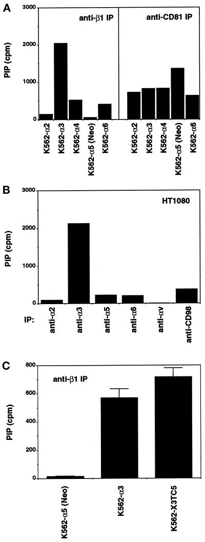
It was shown previously that α3β1 integrin is linked indirectly to PtdIns 4-kinase, possibly through TM4SF proteins such as CD81 and CD63 (Berditchevski et al., 1997b). Here we show that in K562 (Figure 1A, left panel) and HT1080 (Figure 1B) cells, PtdIns 4-kinase activity was predominantly associated with α3β1, as determined by quantitation of PtdIns-4-P production. As anticipated, no PtdIns 4-kinase activity was coimmunoprecipitated with α2β1, α5β1, or αV integrins, since those integrins generally do not associate with TM4SF proteins. Unexpectedly, relatively little kinase activity was coimmunoprecipitated with α4β1 or α6β1 (Figure 1, A and B) even though those integrins are known to associate with both CD63 and CD81 (Berditchevski et al., 1996; Mannion et al., 1996). Results similar to those in Figure 1, A and B, were also obtained using A431 cells (our unpublished results). In control experiments, comparable levels of lipid kinase activity were coimmunoprecipitated with CD81 from all K562 transfectants (Figure 1A, right panel), and lipid kinase activity was not associated with the very highly expressed transmembrane protein, CD98 (Figure 1B).
Figure 1.
Among β1 integrins, α3β1 has the most associated PtdIns 4-kinase activity. (A) K562 transfectants were lysed in 1% Brij 99 and immunoprecipitated with anti-β1 (left panel) or anti-CD81 (right panel) antibodies (TS2/16 and M38, respecely). Immunoprecipitates were then assayed for specific lipid kinase activity as described in MATERIALS AND METHODS. Data are representative of three separate experiments. (B) Lipid kinase activity was measured in immunoprecipitates from HT1080 cells. HT1080 cells express approximately 138, 167, 91, 100, 55, and 245 MFI units of α2, α3, α5, α6, αv, and CD98, respectively. Data are representative of four separate experiments. (C) K562 cells transfected with wild-type α3 or chimeric α3 (X3TC5) were lysed in 1% Brij 99 and immunoprecipitated with anti-β1, before determining lipid kinase activity. Data are presented as the mean ± SD from three separate immunoprecipitations and are representative of two experiments.
The strong preference of PtdIns 4-kinase for α3β1 over α5β1 was seen again in Figure 1C. In the same experiment, chimeric α3 (X3TC5) still showed substantial association with PtdIns 4-kinase activity, despite replacement of the transmembrane and cytoplasmic domains of α3 with those of α5. Because specificity for intracellular PtdIns 4-kinase resides in the ectodomain of the α3 chain, the association must be indirect. In subsequent studies we undertook a search for transmembrane linker protein(s) that would perhaps 1) link PtdIns 4-kinase to α3β1 with specificity for the α3 ectodomain, and 2) show appropriate selectivity for α3β1, compared with other integrins.
Identification of CD151, a TM4SF Protein Associating with α3β1 Integrin
To identify integrin-associated proteins on the cell surface, we had previously developed a monoclonal antibody production and screening strategy (Berditchevski et al., 1997a; Tachibana et al., 1997). Mice were immunized with immune complexes containing α3β1 and associated proteins that had been purified from HT1080 fibrosarcoma cells. Next, hybridoma clones were screened to select for mAbs that 1) bound to the surface of HT1080 cells, 2) did not bind directly to integrin α3 or β1 subunits themselves, but 3) coimmunoprecipitated integrin-like proteins from biotinylated HT1080 cells lysed in nonstringent detergent conditions. Among the several antibodies previously selected to recognize integrin-associated proteins, mAb 5C11 coimmunoprecipitated multiple proteins from biotinylated HT1080 cells lysed in mild detergent conditions (Berditchevski et al., 1997a). The 5C11 mAb was subsequently shown to recognize directly a protein of ∼27–29 kDa from K562 cells.
To identify the protein recognized by mAb 5C11, a 5C11-immunoaffinity column was utilized to purify 13 pmol of this protein from 10 g of K562 cells. Amino-terminal amino acid analysis revealed a nine-residue sequence (G-E-F-N-E-K-[K/I]-T-[T/Y]) that closely matches the published sequence (M-G-E-F-N-E-K-K-T-T) of the human CD151/SFA-1/PETA-3 protein, a 27–29 kDa TM4SF protein (Fitter et al., 1995; Hasegawa et al., 1996). To confirm that mAb 5C11 recognizes CD151, we showed that mAb 5C11 strongly stained the surface of CD151-transfected Molt-4 cells (MFI = 38), with only background staining of untransfected (MFI = 5) or vector-transfected Molt-4 cells (MFI = 3) (our unpublished results). An established anti-CD151 mAb, SFA1–2B4 (Hasegawa et al., 1996), confirmed the presence of CD151 in CD151-transfected Molt-4 cells (MFI = 47). mAb 5C11 also selectively bound to CD151-transfected CHO cells.
CD151–α3β1 Association Is Specific and Unusually Stable
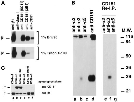
Associations of several TM4SF proteins with specific β1 integrins have previously been demonstrated using mild detergent conditions. Indeed, under mild cell lysis conditions (1% Brij 96) immunoprecipitations of TM4SF proteins CD151 and CD81, from HT1080 cells, yielded β1 integrin as detected by Western blotting (Figure 2A, top panel). A mAb against MHC class I antigen did not coimmunoprecipitate β1 integrin, even though the MHC-I protein was present on HT1080 cells. Remarkably, when cells were lysed under more stringent detergent conditions (1% Triton X-100), the association between CD151 and β1 integrin was maintained, even though association between CD81 and integrin was abolished (Figure 2A, lower panel). Association between β1 integrins and the TM4SF protein CD63, previously observed in nonstringent conditions (Berditchevski et al., 1995), was also abolished under 1% Triton conditions (see Figure 3B below). The anti-CD151 mAb SFA1–2B4 also coimmunoprecipitated β1 integrin (Figure 2A, lower panel). The decreased level of β1 is at least partly due to the lower yield of CD151 obtained using mAb SFA1–2B4 under these conditions.
Figure 2.
CD151 specifically associates with α3β1. (A) HT1080 fibrosarcoma cells were lysed with either 1% Brij 96 (upper panel) or 1% Triton X-100 (lower panel) before immunoprecipitation with the indicated antibodies. The immune complexes were resolved by nonreducing SDS-PAGE, immunoblotted with anti-β1 mAb TS2/16, and developed by chemiluminescence. HT1080 cells express approximately 325, 33, 94, and 68 MFI units of β1, MHC class I, CD151, and CD81, respectively. (B) Metabolically labeled HT1080 cells were lysed in 1% Brij 96 supplemented with 0.2% SDS and immunoprecipitated with the indicated antibodies. Immune complexes were eluted from protein A-Sepharose at low pH (pH 2.7) and either directly analyzed (lanes a–d) or reimmunoprecipitated with 5C11-Sepharose (lanes e–g) in 1% Brij 96 containing 0.5% SDS, as described in MATERIALS AND METHODS. Fourfold more immunoprecipitated material was loaded in lane d compared with lanes a–c. Immunoprecipitates were resolved by 12% nonreducing SDS-PAGE, and proteins were detected by autoradiography. (C) K562 cells transfected with the indicated α chain cDNAs or vector-alone (lane e) were lysed in 1% Triton X-100, and lysates were immunoprecipitated with antibodies against CD151 (5C11, upper panel) or β1 (TS2/16, lower panel). Immune complexes were resolved by SDS-PAGE and immunoblotted with anti-β1 mAb TS2/16. K562-X3TC5 cells (lane c) are transfected with a chimeric α3 containing the α3 extracellular domain and α5 transmembrane and cytoplasmic domains.
Figure 3.
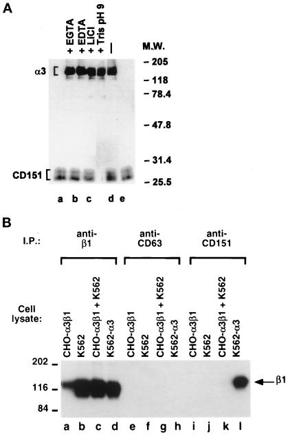
CD151–α3β1 complexes are biochemically stable and form before cell lysis. (A) Metabolically labeled HT1080 cells were lysed and immunoprecipitated with 5C11 mAb in 2% n-octyl-β-d-glucopyranoside alone (lane e) or supplemented with 10 mM EGTA (lane a), 10 mM EDTA (lane b), 1 M LiCl (lane c), or 100 mM Tris pH9.0 (lane d). (B) The cell lines K562, K562-α3, and CHO-α3β1 (containing human α3 and β1) were lysed in 1% Triton X-100, mixed 1:1 with K562 cell lysate or lysis buffer alone, and subsequently immunoprecipitated with antibodies against β1 (TS2/16), CD63 (6H1), or CD151 (5C11). Immune complexes were resolved by 8% nonreducing SDS-PAGE and immunoblotted with an antibody against β1 (TS2/16).
The CD151 protein preferentially associates with α3β1, compared with other β1 integrins, as seen (Figure 2B) using metabolically labeled HT1080 cells, and a different set of relatively stringent cell lysis conditions (1% Brij 96 + 0.2% SDS). Upon immunoprecipitation of α3 (lane b), but not α2 (lane a) or α5 (lane c), a protein was coimmunoprecipitated (lane b), that resembled 28 kDa CD151 (lane d). To verify this result, immune complexes were dissociated, and then CD151 was directly reimmunoprecipitated from an α3 immunoprecipitate (lane f), but not from α2 or α5 immunoprecipitates (Figure 2B, lanes e and g).
For reciprocal demonstration of specific CD151–α3β1 association, we utilized K562-integrin transfectants, again lysed under relatively stringent conditions (1% Triton X-100). As indicated (Figure 2C, upper panel), immunoprecipitates of CD151 contained the integrin β1 subunit, only when CD151 was immunoprecipitated from K562-α3 cells (lane b), but not from K562 cells containing α2, α4, α5, or α6 subunits (lanes a, d, e, and f). Also, anti-CD151 mAb coimmunoprecipitated β1 from K562-X3TC5 cells (lane c), in which a chimeric integrin α subunit contains the α3 extracellular domain and α5 transmembrane and cytoplasmic domains (lane c). Thus, CD151 association is specified by the α3 ectodomain. Control immunoprecipitation of β1, followed by Western blotting of β1 (Figure 2C, lower panel) revealed that all cell lysates contained substantial amounts of integrin β1 subunit. Under milder detergent lysis conditions (1% Brij 96 or 1% Brij 99), CD151 did associate with α6β1, but still did not associate with α2 or α5 integrins (our unpublished results).
To further analyze the CD151–α3β1 interaction, we altered the ionic strength, pH, and divalent cation levels of metabolically labeled HT1080 cell lysates, before CD151 immunoprecipitation (Figure 3A). When ionic strength was increased by adding 1 M LiCl (lane c) or pH was elevated by adding 100 mM Tris at pH 9.0 (lane d), there was minimal change in the amount of α3 coimmunoprecipitated with CD151. Chelation of divalent cations by adding 10 mM EGTA (lane a) or 10 mM EDTA (lane b) also had minimal effect on α3 integrin coimmunoprecipitation. Thus, CD151 association with α3β1 does not resemble ligand binding to α3β1, which is strongly divalent cation dependent. Consistent with this, a point mutation within α3 (W220A) that completely eliminated α3β1 interactions with laminin-5 (Krukonis, Dersch, and Isberg, unpublished data), had no effect on association with CD151 (our unpublished results).
To examine the possibility that CD151–α3β1 complexes could be forming post cell lysis, a lysate-mixing experiment was carried out. K562 cell lysate, containing human CD151 but not α3β1, was mixed with CHO-α3β1 cell lysate, that contains transfected human α3 and β1 but not human CD151. Then, human CD151 was immunoprecipitated, but no β1 integrin was coimmunoprecipitated as detected by Western blotting (Figure 3B, lane k). Thus, α3β1 had not associated with human CD151 post cell lysis, or exchanged human CD151 for endogenous hamster CD151. However, when human CD151 and α3β1 were both originally present in the same cell before lysis (i.e., in K562-α3 cells), then β1 integrin was coimmunoprecipitated with anti-human CD151 mAb (lane l). This association was specific, because immunoprecipitation of a control TM4SF protein, CD63, was unable to coimmunoprecipitate β1 under these relatively stringent detergent conditions (lane h). In other control experiments, CD151 immunoprecipitation did not yield β1 if human CD151 was absent (in CHO-α3β1 cells, lane i) or if α3β1 was absent (in K562 cells, lane j). Also, β1 was readily detectable in lysates of CHO-α3β1, K562, and K562-α3 cells (lanes a–d), and α3 was present at substantial levels in CHO-α3β1 and K562-α3 cells, as determined by flow cytometry.
α3β1–CD151 Association Occurs on the Cell Surface and at High Stoichiometry
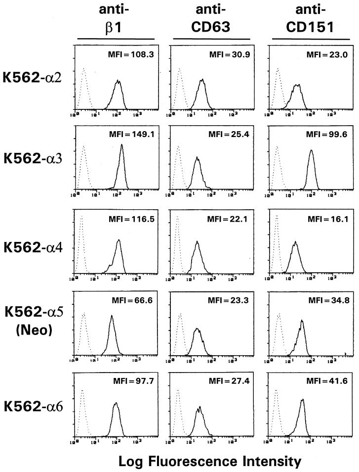
The anti-CD151 mAb 5C11 was initially selected for its ability to coprecipitate cell surface-biotinylated integrin (Berditchevski et al., 1997a), indicating that CD151–α3β1 association occurs on the cell surface. Now we show that ectopic expression of α3β1 on the surface of α3-transfected K562 cells was accompanied by a nearly threefold increase (MFI = 35 → MFI = 100) in the surface levels of CD151 (Figure 4). The increase in CD151 expression was detected using two different antibodies to CD151 including mAb 5C11 (Figure 4) and mAb SFA1.2B4 (our unpublished results). This effect was specific for α3, because transfection of α2, α4, or α6 did not markedly increase CD151 levels (MFI = 23, 16, and 42, respectively). In addition, there was specificity at the level of TM4SF proteins, since α3 transfection did not alter the cell surface expression of CD63. At present, we have not done the reciprocal experiment (i.e., examine the effect of CD151 transfection on α3β1 levels) because we have yet to identify a potential host cell line that is α3β1-positive and CD151-negative (see Table 1 and DISCUSSION). Nonetheless, our results indicate that cell surface α3β1 expression promotes increased surface CD151 expression.
Figure 4.
Transfection of α3 increases the cell surface expression of CD151. K562 cells transfected with α2, α3, α4, or α6 cDNA, or vector alone (K562-α5(Neo)) were stained with the negative control antibody, P3 (dotted lines), or with antibodies against β1 (TS2/16, first column), CD63 (6H1, second column) or CD151 (5C11, third column) before analysis by flow cytometry. Specific mean fluorescence intensity (MFI) values are indicated. Except for K562-α5(Neo) (in which α5β1 is the only β1 integrin), transfectants express similar levels of the respective α chains. The transfected α chains represent between 72 and 94% of total β1 integrin in these transfectants.
Table 1.
Comparison of α3β1, CD151, and CD9 expression levels on various cell types
| Cell line designation | Tissue derivation | Expressiona
|
||
|---|---|---|---|---|
| α3 | CD151 | CD9 | ||
| Nalm-6 | B cell derived | − | − | +++ |
| OCI-Ly8 | B cell derived | − | − | ++ |
| Molt-4 | T cell derived | − | + | + |
| K562 | Erythroleukemia | − | ++ | − |
| Jurkat | T cell derived | + | + | + |
| Peer | T cell derived | + | ++ | ND |
| RD | Rhabdomyosarcoma | + | ++ | + |
| NT-2 | Embryonic carcinoma | + | + | + |
| SSC25 | Squamous cell carcinoma | ++ | ++ | − |
| HT1080 | Fibrosarcoma | +++ | +++ | − |
| CAKI-1 | Kidney carcinoma | +++ | +++ | − |
| MDA-MB-231 | Breast carcinoma | +++ | +++ | − |
| A431 | Epidermoid carcinoma | +++ | +++ | ++ |
Cell surface expression levels of α3, CD151, and CD9 were determined by flow cytometry, as described in MATERIALS AND METHODS. Specific MFI values were 0–2 (−), 3–20 (+), 21–100 (++), or >101 (+++).
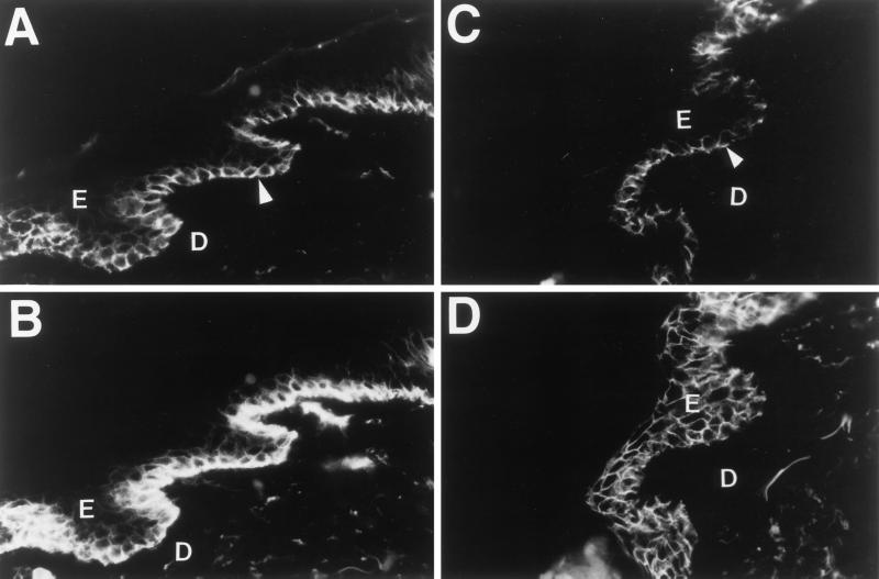
To gain further information about the potential relevance of CD151–α3β1 complexes, we examined the distribution of CD151 and α3 in human skin sections by immunofluorescent staining. As expected, α3 was detected in the basal layer of keratinocytes of the epidermis (Figure 5, A and C, arrows). Moreover, CD151 expression was also restricted to the basal layer of keratinocytes (Figure 5B), colocalizing with the α3 signal. In contrast, another TM4SF protein, CD9, was observed throughout the stratified squamous epithelium of the epidermis (Figure 5D and Berditchevski et al., 1996).
Figure 5.
CD151 and α3β1 codistribute in the basal layer of keratinocytes of human epidermis. Sections of human skin were stained with mAbs against α3 (A and C), CD151 (B), or CD9(D), as described in MATERIALS AND METHODS. Arrowheads point to the basal keratinocytes. E, epidermis; D, dermis. No staining was observed using secondary antibodies alone (our unpublished results).
To estimate α3β1/CD151 stoichiometry, we lysed metabolically labeled HT1080 cells under relatively stringent detergent conditions (1% Triton X-100), and then removed essentially all CD151 by extensive immunodepletion (Figure 6C). Importantly, nearly all α3 integrin was removed at the same time. Quantitative analysis (see legend) revealed that CD151 depletion resulted in removal of ∼100% of CD151-associated integrin α chain, ∼90% labeled α3 protein subsequently precipitated by anti-α3 mAb A3-IVA5 (Figure 6A), and ∼70% of labeled α3 protein precipitated by rabbit anti-α3 polyclonal antibody (our unpublished results). We suspect that, compared with mAb anti-α3, polyclonal anti-α3 antibody recognizes more α3 precursor protein, some of which may be unassociated with CD151 and thus not removed by anti-CD151 mAb. Immunodepletion of CD151 failed to remove any integrin α2 (Figure 6A) or TM4SF CD81 (Figure 6B). These results indicate that most of the α3β1 is specifically associated with CD151.
Figure 6.
Analysis of α3β1-CD151 stoichiometry. Metabolically labeled HT1080 cells were lysed in 1% Triton X-100, and lysates were cleared using anti-CD151 5C11-conjugated Sepharose (+) or unconjugated Sepharose (−). Lysates were subsequently immunoprecipitated with the indicated antibodies, and immune complexes were resolved by nonreducing SDS-PAGE before detecting integrin α chains (A), CD81 (B), or CD151 (C) by autoradiography. The percent reduction in the levels of integrin immunoprecipitated upon CD151 immunodepletion was determined by measuring the amount of 35S-methionine corresponding to the respective proteins in a Betascope 603 Blot Analyzer. At least 400 counts of 35S-methionine were detected in all positive (i.e., nondepleted) lanes during 1 h; and background (< 10%) was subtracted before dividing CD151 depleted/nondepleted counts.
CD151 May Link PtdIns 4-Kinase to α3β1
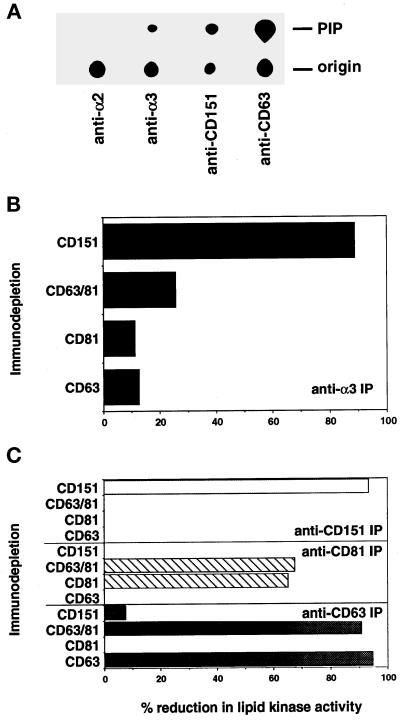
Because CD151 associated stably and preferentially with the α3β1 integrin (Figures 2 and 6), with dependence on the α3 ectodomain (Figure 2C), it appeared to be an ideal candidate to link α3β1 to PtdIns 4-kinase. Indeed, from lysates of HT1080 cells, similar levels of PtdIns 4-kinase activity were coimmunoprecipitated with both anti-α3 mAb and anti-CD151 mAb (Figure 7A), as seen by the production of PtdIns-4-P. In comparison to CD151 and α3β1, negligible PtdIns 4-kinase activity coprecipitated with α2β1, and a substantial amount of PtdIns 4-kinase activity was associated with CD63 (Figure 7A). In α3-negative K562 cells, CD151 still associated with a substantial level of PtdIns 4-kinase. The roughly estimated ratio of CD151-associated kinase activity/CD151 expression level (in arbitrary kinase activity units/CD151 MFI units) was ∼0.46 in K562-α3 cells, and ∼0.55 in K562-mock cells. Thus, α3β1 is clearly not required for CD151 association with PtdIns 4-kinase, and PtdIns 4-kinase is likely more proximal to CD151 than to α3β1. The enzymatic activity associated with CD151 was blocked by specific inhibitors of type II PtdIns 4-kinase (i.e., adenosine and the mAb 4C5G), as seen previously for lipid kinase activity associated with other TM4SF proteins (Berditchevski et al., 1997b).
Figure 7.
PtdIns 4-kinase is present in CD151–α3β1 complexes. (A) HT1080 cells were lysed in 1% Brij 99 and immunoprecipitated with the indicated antibodies. Immunoprecipitates were assayed for kinase activity as described in MATERIALS AND METHODS. The monoclonal antibodies A2-IIE10 (α2), A3-IVA5 (α3), 5C11 (CD151), and 6H1 (CD63) were used for immunoprecipitation. (B and C) Cell lysates were immunodepleted using unconjugated Sepharose beads, or beads conjugated with anti-CD151 mAb 5C11, anti-CD63 mAb 6H1, anti-CD81 mAb M38, or combined anti-CD63/anti-CD81 mAbs, before immunoprecipitation of α3 (B) or CD63, CD81, and CD151 (C). Immunoprecipitates were assayed for lipid kinase activity, and results are expressed as percent reduction of specific lipid kinase activity resulting from immunodepletion with CD151, CD63, CD81, or CD63/CD81 combined. Results each represent the mean of 4–5 experiments (1 using A431 cells, 3–4 using HT1080 cells).
Upon immunodepletion of CD151 from HT1080 or A431 cell lysates, approximately 89% of the lipid kinase activity associated with α3 was removed (Figure 7B). In contrast, only ∼12%, 11%, and 25% of α3-associated PtdIns 4-kinase activity was removed upon immunodepletion of CD63, CD81, or both CD63 and CD81, respectively (Figure 7B). Approximately 95%, 93%, and 65% of the PtdIns 4-kinase activity associated with CD63, CD151, and CD81 was removed by prior immunodepletion of the respective protein (Figure 7C), thus demonstrating the efficiency of immunodepletion. Furthermore, immunodepletion of CD151 had a negligible effect on the activity associated with CD63 (8% reduction), demonstrating that activities associated with these two TM4SF proteins are mutually exclusive.
Roles for CD151 and α3β1 in Cell Motility
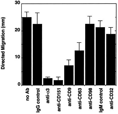
To ascertain the functional importance of α3β1–CD151 complexes, we examined the effects of anti-CD151 monoclonal antibody perturbation on α3β1-dependent PMN motility. As shown elsewhere, the polysaccharide, β-glucan, stimulates directed motility of PMN in response to fMLP on a fibronectin matrix. That motility was entirely blocked by an anti-α3β1 mAb (A3-X8), but was not blocked by antibodies to α2, α4, α5, α6, αM, or β2 integrins (Reichner and Harler, unpublished data). As shown here (Figure 8), antibodies to α3 and CD151 markedly reduced the directed migration of PMN (by 89% and 92%, respectively). This inhibition was not due to potential effects on Fc receptors, since isotype-matched control IgG and antibodies directly recognizing Fcγ (type II receptor (CD32) had no effect on PMN chemotaxis. Antibodies to CD98, another neutrophil surface protein, also had no inhibitory effect. Importantly, reduction in cell motility is not due to mAb-induced neutrophil homotypic aggregation — our anti-α3 and CD151 antibodies had no effect on that parameter. Interestingly, antibodies to two other TM4SF proteins, CD9 and CD63, showed substantial reductions in PMN chemotaxis (68% and 44%, respectively) but were not as potent as the anti-CD151 antibody.
Figure 8.
Inhibition of PMN chemotaxis by antibody perturbation of α3β1 and CD151. PMN migration in response to fMLP was measured on fibronectin in the presence of β-glucan. mAbs A3-X8 (α3), 5C11 (CD151), C9-BB(CD9), 6H1 (CD63), 8A6 (CD98), and 4.6.19 (CD32) were incorporated into the agarose, as described in MATERIALS AND METHODS. Isotype-matched IgG and IgM served as controls. None of these antibodies triggered neutrophil homotypic aggregation. Migration distances, calculated from Microprojector-magnified images, are presented as mean ± SD from quadruplicate samples. Data are representative of three separate experiments. Background random migration values (no fMLP) ranged between 3–6 mm. mAbs against α3 and CD151 significantly inhibited PMN motility (p < 0.0001).
DISCUSSION
Identification of CD151
The mAb 5C11 was obtained as previously described, after injection of a mouse with α3β1 integrin complexes, and specific selection for monoclonal antibodies to integrin-associated proteins (Berditchevski et al., 1997a). The protein recognized by mAb 5C11 is CD151, based on size, amino terminal sequence, and 5C11 reactivity with CD151-transfected cells. CD151, also known as PETA-3 (Fitter et al., 1995) and SFA-1 (Hasegawa et al., 1996), is a TM4SF protein broadly expressed in many tissues and on many cell types, including endothelial, epithelial, and muscle cells (Sincock et al., 1997). The finding that immunization of a mouse with α3β1 complexes yielded a monoclonal antibody to the TM4SF protein CD151 is consistent with the previously documented association of α3β1 with other TM4SF proteins including CD9, CD63, CD81, and NAG-2 (Berditchevski et al., 1995, 1996; Nakamura et al., 1995; Jones et al., 1996; Tachibana et al., 1997).
Association of α3β1 with CD151 Is Stable and Specific
Reciprocal coimmunoprecipitation experiments not only confirmed α3β1–CD151 association, but also revealed that the complex was unusually stable since it was maintained in relatively stringent detergent conditions as compared with other integrin–TM4SF protein associations. These include buffers containing 1% NP40 + 0.5% DOC + 0.1% SDS (our unpublished results), as well as 1% Triton X-100, or 1% Brij 96 + 0.2% SDS. In contrast, those same detergent conditions caused α3β1 to dissociate from CD81, CD63, and CD9, as shown here and elsewhere (Berditchevski et al., 1996). In addition, α3β1–CD151 association is insensitive to high concentrations of LiCl.
The unusually stable α3β1–CD151 association was also highly specific. In relatively stringent detergent conditions, CD151 did not associate with α2β1, α4β1, α5β1, and α6β1; and α3β1 did not associate with CD9, CD63, and CD81. Furthermore, CD151 and α3β1 coprecipitations yielded no other labeled cell surface proteins (e.g., Figures 2B and 3A). This is in marked contrast to previous direct immunoprecipitations (e.g., Berditchevski et al., 1995, 1996; Mannion et al., 1996) that yielded several additional proteins. In addition, we utilized dithiobis(succinimidyl propionate) to obtain strong covalent cross-linking of α3β1 to the CD151 protein (our unpublished results). Together these data suggest that CD151 complexes directly to α3β1, with no other supporting proteins required.
In milder detergents, α6β1 association was preserved, consistent with previously documented association of α6β1 with other TM4SF proteins (Berditchevski et al., 1995, 1996; Schmidt et al., 1996; Tachibana et al., 1997). Importantly, other experiments showed that α3β1–CD151 complexes were not formed post cell lysis and did not resemble typical integrin–ligand complexes. The association between α3β1 and CD151 cannot simply be a misinterpretation due to an artefact of mAb cross-reactivity. Our anti-CD151 mAb 5C11 does not recognize human α3β1 transfected into CHO cells (Berditchevski et al., 1997a), and it does not reimmunoprecipitate α3β1 under conditions in which it does directly reimmunoprecipitate CD151 (e.g., see Figure 2B). Furthermore, mAb 5C11 continues to recognize CD151 on K562 cells even when α3 is absent, and a second anti-CD151 mAb (SFA1–2B4) also coimmunoprecipitated α3β1.
Although unique for the integrin–TM4SF protein interactions, firm association of TM4SF proteins with other cell surface molecules has been previously characterized for uroplakins, distant members of the TM4SF. Uroplakin Ia and uroplakin Ib are principal components of the “asymmetric unit membrane” at the luminal surface of urinary bladder epithelium and form tight complexes with uroplakin II and uroplakin III, respectively (Wu et al., 1995b).
Nearly All α3β1 Is Associated with CD151
In addition to being stable and specific, α3β1–CD51 complexes occurred at high stoichiometry, with at least 70–90% of cell surface α3β1 being removed upon anti-CD151 immunodepletion. In previous studies of integrin–TM4SF protein association, stoichiometry was difficult to assess due to the mild detergents utilized. Nonetheless, not more than ∼5% of any particular integrin was previously estimated by coimmunoprecipitation analysis to be complexed with TM4SF proteins (Berditchevski et al., 1995, 1996; Mannion et al., 1996). Notably, ectopic expression of α3β1 in K562 cells selectively promoted the appearance of CD151 (but not CD63). We have not yet been able to identify (or create by transfection) a cell line expressing α3β1 that does not also express CD151 (Table 1), although CD151 can appear without α3 (e.g., on K562 cells). In this regard, the expression of α3 on various cell lines correlates well with the coexpression of CD151, but not with CD9, another α3-associated TM4SF protein (Table 1). Thus, we hypothesize that CD151 association might be required for α3β1 expression. This issue should be resolved definitively upon the preparation of a CD151 knockout mouse.
Consistent with their close association in cell lines in vitro, CD151 and α3 integrin showed very similar patterns of staining of the basal layers of skin epidermis, with the most intense staining in the deepest basal cell layer. These results, showing for the first time a direct comparison of CD151 and α3 in skin, are consistent with previous α3 (Carter et al., 1990; Berditchevski et al., 1996; Dipersio et al., 1997), and CD151 (Sincock et al., 1997) skin staining results. Although other TM4SF proteins, such as CD9, CD63, and CD81, are present in skin, these proteins are homogeneously expressed throughout the stratified layers of epidermis (Figure 5 and Berditchevski et al., 1996), and thus clearly differ from CD151 staining. Therefore, among TM4SF proteins tested, CD151 alone shows a distribution in skin epithelium identical to that of α3.
CD151 Links PtdIns 4-Kinase to α3β1
Here we show, in three different cell lines, that much more PtdIns 4-kinase activity is found associated with α3β1 than any other integrin tested. This indirect association, involving an intracellular enzyme (PtdIns 4-kinase) and the α3 ectodomain, obviously requires a transmembrane linker protein. Other integrins (e.g., α6β1 and α4β1) did not show much associated PtdIns 4-kinase activity, even though they do associate with TM4SF proteins such as CD81 and CD63. Thus, we must modify our previous suggestion that TM4SF proteins, such as CD63 and CD81, may link α3β1 to PtdIns 4-kinase (Berditchevski et al., 1997b). Instead, our search for a linker protein has led us to another TM4SF protein, called CD151. Evidence that CD151 acts as a linker between PtdIns 4-kinase and α3β1 is as follows: First, CD151 association with α3β1 is specific and direct (see above). Second, CD151 associated with PtdIns 4 kinase, even in the absence of the α3β1 integrin. Thus, the PtdIns 4-kinase is more proximal to CD151, than to α3β1. Third, removal of the large subset of α3β1 molecules associated with CD151 caused the simultaneous removal of nearly all α3β1 capable of associating with PtdIns 4-kinase. Fourth, PtdIns 4-kinase association with α3β1 required the α3 ectodomain. This result is entirely consistent with CD151, which also requires the α3 ectodomain for association, being the critical transmembrane linker protein. Results shown here provide a new paradigm for integrin signaling. This may be the first demonstration of a mechanism whereby specific association of an intracellular signaling enzyme can be determined by the extracellular domain of an integrin. In a previous study, integrin cytoplasmic domains were shown not to be required for specific integrin association and signaling through intracellular Shc and Grb2. However, it was unclear whether specificity was mostly provided by integrin transmembrane or extracellular domains (Wary et al., 1996).
The PtdIns 4-kinase seen here is biochemically distinct from another type II PtdIns 4-kinase (PI4Kα, found in the endoplasmic reticulum) and from a wortmannin-sensitive PtdIns 4-kinase (PI4Kβ, found in Golgi and cytosol). Rather, it appears to correspond to the major type II PtdIns 4-kinase found in plasma membranes and lysosomes, but which has not yet been cloned (Graziani et al., 1992; Berditchevski et al., 1997b; Wong et al., 1998).
CD151 not only associates with α3β1 and PtdIns 4-kinase, but under mild detergent conditions it also associates with other TM4SF proteins (our unpublished results). Thus, a CD151 linker function possibly could explain the previously reported coimmunoprecipitation of α3β1 with CD9, CD63, CD81, and NAG-2 (Hemler et al., 1996; Maecker et al., 1997; Tachibana et al., 1997), but this remains to be determined. Regardless, not all integrin–TM4SF protein associations are dependent on CD151. In particular, associations of α4β1 with several TM4SF proteins must be independent of CD151, since they occur in CD151-negative cell lines (Mannion et al., 1996).
Potential Functional Relevance of α3β1–CD151–PtdIns 4-Kinase Complexes
As established elsewhere, the directed migration of PMNs on a fibronectin substrate, toward fMLP, is stimulated by the polysaccharide β-glucan. This migration is not blocked by mAb to α2, α4, α5, α6, αM, or β2 integrins, but is entirely inhibited by an anti-α3 mAb (Reichner and Harler, unpublished data). As shown here, anti-CD151 mAb consistently blocked this motility to the same extent as anti-α3 antibody (88–92% inhibition). Although α3β1 does not mediate strong cell adhesion to fibronectin, α3β1-dependent cell migration on fibronectin has been previously reported (Melchiori et al., 1995).
In another study, α3β1 and CD151 were readily coprecipitated from a differentiated neuronal cell line, and antibodies to both α3β1 and CD151 showed marked inhibition of neurite outgrowth. This effect was highly specific, since the antibodies only inhibited neurite outgrowth on laminin-5 (an α3β1 ligand), but not on laminin-1, and inhibition was not seen with an antibody to another TM4SF-associated integrin (α6β1), or to another TM4SF protein (CD9) (Stipp and Hemler, unpublished data). Together these results suggest that the α3β1–CD151–PtdIns 4-kinase complex may act as a functional unit to support cell migration.
Interestingly, the anti-α3β1 mAb A3-X8 antibody used here to inhibit neutrophil migration does not block cell adhesion (Weitzman et al., 1993). Likewise, mAb to CD151 failed to inhibit PMN adhesion to fibronectin (our unpublished observations). In addition, utilizing four different anti-CD151 mAbs to ligate and/or cross-link CD151, we have been unable to demonstrate any stimulatory or inhibitory effect on α3β1-mediated adhesion to laminin-5–containing matrices (our unpublished observations). These experiments were carried out using three different cell lines (K562-α3, a kidney epithelial cell line, and human saphenous vein endothelial cells). These findings are consistent with the inability of other TM4SF proteins to alter integrin-mediated adhesion (Hemler et al., 1996) and contribute to the accumulating evidence (e.g., Domanico et al., 1997; Hangan et al., 1997) that cell migration can be regulated independent of cell adhesion. We hypothesize that the α3β1 integrin, at the migrating edge of a cell, recruits CD151 and PtdIns 4-kinase to the same site. In this regard, it was previously established that α3β1, as well as other TM4SF proteins, localizes into cellular filopodia and lamellipodia (Berditchevski et al., 1997b). Possibly, anti-α3 and anti-CD151 mAb may disrupt positioning and/or critical lateral interactions at these sites, without affecting cell adhesion.
Our results add to the accumulating evidence that TM4SF–integrin complexes (Banerjee et al., 1997; Domanico et al., 1997) and TM4SF proteins in general (Hemler et al., 1996; Maecker et al., 1997) may play an important role in cell motility. Indeed, antibodies to other TM4SF proteins (CD9 and CD63) had some inhibitory effects on PMN chemotaxis, consistent with the established role of these proteins in cell migration (Ikeyama et al., 1993; Radford et al., 1997), although neither antibody inhibited PMN chemotaxis to the same extent as anti-CD151 antibody. We presume that the differential effects of these antibodies relate to the tighter association and higher stoichiometry of α3β1–CD151 complexes relative to α3β1–CD9 and α3β1–CD63 complexes.
The α3β1 integrin has been implicated in a wide variety of biological processes including kidney, lung, and skin development (Kreidberg et al., 1996; Dipersio et al., 1997), branching morphogenesis of mammary cells (Berdichevsky et al., 1994), tumorigenesis (Weitzman et al., 1996), wound healing (Larjava et al., 1993), extracellular matrix assembly (Wu et al., 1995a), and cell motility. On neutrophils, α3β1 may help to regulate phagocytosis (Gresham et al., 1996), bactericidal activity (Simms and D’amico, 1997), and apoptosis (Leuenroth et al., 1997). Here we have established that CD151 is present on neutrophils and other cells that express α3β1. Thus we predict that CD151, and possibly also PtdIns 4-kinase, may contribute to the biology of α3β1 in each of these cases. The PtdIns-4-P product of PtdIns 4-kinase may make multiple contributions to phosphoinositide signaling (see INTRODUCTION). For example, α3β1–CD151-associated PtdIns 4-kinase potentially could produce PtdIns-4-P, or ultimately PtdIns-4,5-P2. These are both established regulators of the actin cytoskeleton, which is needed for cell motility.
In summary, we demonstrate that complexes of α3β1 with CD151 are not only highly specific, and present in multiple cell types both in vitro and ex vivo, but also are more robust (i.e., more stable) than any previously described integrin interaction with a TM4SF protein or with any other integrin-associated protein. Furthermore, we show that α3β1 can associate with PtdIns 4-kinase to a greater extent than any other β1 integrin, largely due to a novel mechanism whereby the extracellular domain of α3 specifies, through a CD151 linker, association with intracellular PtdIns 4-kinase. Finally, we show that α3β1–CD151 and/or α3β1–CD151–PtdIns 4-kinase complexes play a major role in regulating cell motility.
Note added in proof. In a recent paper, another group has also shown that CD151 may influence cell motility and associate with α3β1 integrin (Yanez-Mo, M., Alfranca, A., Cabanas, C., Marazuela, M., Tejedor, R., Ursa, M.A., Ashman, L.K., de Landazuri, M.O., and Sanchez-Madrid, F. [1998]. J. Cell Biol. 141, 791–804).
ACKNOWLEDGMENTS
We thank Dr. Hitoshi Hasegawa and Dr. Leonie Ashman for kindly providing the mAbs, SFA1.2B4 and 14A2, respectively. We also thank Drs. Lewis C. Cantley and Christopher L. Carpenter for providing mAb 4C5G, Dr. N. Hotchin for providing human skin sections, Dr. David Batt for help with graphics, and Dr. Chris Stipp for critical review of the manuscript, as well as for making available unpublished data. Work was supported by National Institutes of Health grants GM-38903 and GM-46526 (to M.E.H.) and GM-51493 (to J.R.). R.L.Y was supported by a fellowship from the National Institute of Health (AI-09490).
REFERENCES
- Assoian RK. Anchorage-dependent cell cycle progression. J Cell Biol. 1997;136:1–4. doi: 10.1083/jcb.136.1.1. [DOI] [PMC free article] [PubMed] [Google Scholar]
- Banerjee SA, Hadjiargyrou M, Patterson PH. An antibody to the tetraspan membrane protein CD9 promotes neurite formation in a partially α3β1 integrin-dependent manner. J Neurosci. 1997;17:2756–2765. doi: 10.1523/JNEUROSCI.17-08-02756.1997. [DOI] [PMC free article] [PubMed] [Google Scholar]
- Barnstable CJ, Bodmer WF, Brown G, Galfré G, Milstein C, Williams AF, Zeigler A. Production of monoclonal antibodies to group A erythrocytes, HLA and other human cell surface antigens — new tools for genetic analysis. Cell. 1978;14:9–20. doi: 10.1016/0092-8674(78)90296-9. [DOI] [PubMed] [Google Scholar]
- Berdichevsky F, Alford D, D’Souza B, Taylor-Papadimitriou J. Branching morphogenesis of human mammary epithelial cells in collagen gels. J Cell Sci. 1994;107:3557–3568. doi: 10.1242/jcs.107.12.3557. [DOI] [PubMed] [Google Scholar]
- Berditchevski F, Bazzoni G, Hemler ME. Specific association of CD63 with the VLA-3 and VLA-6 integrins. J Biol Chem. 1995;270:17784–17790. doi: 10.1074/jbc.270.30.17784. [DOI] [PubMed] [Google Scholar]
- Berditchevski F, Chang S, Bodorova J, Hemler ME. Generation of monoclonal antibodies to integrin-associated proteins: Evidence that α3β1 complexes with EMMPRIN/basigin/OX47/M6. J Biol Chem. 1997a;272:29174–29180. doi: 10.1074/jbc.272.46.29174. [DOI] [PubMed] [Google Scholar]
- Berditchevski F, Tolias KF, Wong K, Carpenter CL, Hemler ME. A novel link between integrins, TM4SF proteins (CD63, CD81) and phosphatidylinositol 4-kinase. J Biol Chem. 1997b;272:2595–2598. doi: 10.1074/jbc.272.5.2595. [DOI] [PubMed] [Google Scholar]
- Berditchevski F, Zutter MM, Hemler ME. Characterization of novel complexes on the cell surface between integrins and proteins with 4 transmembranes (TM4 proteins) Mol Biol Cell. 1996;7:193–207. doi: 10.1091/mbc.7.2.193. [DOI] [PMC free article] [PubMed] [Google Scholar]
- Bergelson JM, St. John N, Kawaguchi S, Pasqualini R, Berdichevsky F, Hemler ME, Finberg RW. The I domain is essential for echovirus 1 interaction with VLA-2. Cell Adhes Commun. 1994;2:455–464. doi: 10.3109/15419069409004455. [DOI] [PubMed] [Google Scholar]
- Berridge MJ. Inositol triphosphate and diacylglycerol: two interacting second messengers. Annu Rev Biochem. 1987;56:159–193. doi: 10.1146/annurev.bi.56.070187.001111. [DOI] [PubMed] [Google Scholar]
- Brown EJ. Complement receptors and phagocytosis. Curr Opin Immunol. 1991;3:76–82. doi: 10.1016/0952-7915(91)90081-b. [DOI] [PubMed] [Google Scholar]
- Carter WG, Wayner EA, Bouchard TS, Kaur P. The role of integrins α2β1 and α3β1 in cell-cell and cell-substrate adhesion of human epidermal cells. J Cell Biol. 1990;110:1387–1404. doi: 10.1083/jcb.110.4.1387. [DOI] [PMC free article] [PubMed] [Google Scholar]
- Czop JK, Fearon DT, Austen KF. Opsonin-independent phagocytosis of activators of the alternative complement pathway by human monocytes. J Immunol. 1978;120:1132–1138. [PubMed] [Google Scholar]
- Derevianko A, D’amico R, Graeber T, Keeping H, Simms HH. Endogenous PMN-derived reactive oxygen intermediates provide feedback regulation on respiratory burst signal transduction. J Leukocyte Biol. 1997;62:268–276. doi: 10.1002/jlb.62.2.268. [DOI] [PubMed] [Google Scholar]
- Dipersio CM, Hodivala-Dilke KM, Jaenisch R, Kreidberg JA. Alpha-3-Beta-1 integrin is required for normal development of the epidermal basement membrane. J Cell Biol. 1997;137:729–742. doi: 10.1083/jcb.137.3.729. [DOI] [PMC free article] [PubMed] [Google Scholar]
- Domanico SZ, Pelletier AJ, Havran WL, Quaranta V. Integrin α6Aβ1 induces CD81-dependent cell motility without engaging the extracellular matrix migration substrate. Mol Biol Cell. 1997;8:2253–2265. doi: 10.1091/mbc.8.11.2253. [DOI] [PMC free article] [PubMed] [Google Scholar]
- Endemann GC, Graziani A, Cantley LC. A monoclonal antibody distinguishes two types of phosphatidylinositol 4-kinase. Biochem J. 1991;273:63–66. doi: 10.1042/bj2730063. [DOI] [PMC free article] [PubMed] [Google Scholar]
- Fitter S, Tetaz TH, Berndt MC, Ashman LK. Molecular cloning of cDNA encoding a novel platelet-endothelial cell tetra-span antigen, PETA-3. Blood. 1995;86:1348–1355. [PubMed] [Google Scholar]
- Frisch SM, Ruoslahti E. Integrins and aniokis. Curr Opin Cell Biol. 1997;5:701–706. doi: 10.1016/s0955-0674(97)80124-x. [DOI] [PubMed] [Google Scholar]
- Fukudome K, Furuse M, Imai T, Nishimura M, Takagi S, Hinuma Y, Yoshie O. Identification of membrane antigen C33 recognized by monoclonal antibodies inhibitory to human T-cell leukemia virus type 1 (HTLV-1)-induced syncytium formation: altered glycosylation of C33 antigen in HTLV-1-positive T cells. J Virol. 1992;66:1394–1401. doi: 10.1128/jvi.66.3.1394-1401.1992. [DOI] [PMC free article] [PubMed] [Google Scholar]
- Graziani A, Ling LE, Endemann G, Carpenter CL, Cantley LC. Purification and characterization of human erythrocyte phosphatidylinositol 4-kinase. Phosphatidylinositol 4-kinase and phosphatidylinositol 3-monophosphate 4-kinase are distinct enzymes. Biochem J. 1992;284:39–45. doi: 10.1042/bj2840039. [DOI] [PMC free article] [PubMed] [Google Scholar]
- Gresham HD, Graham IL, Griffin GL, Hsieh JC, Dong LJ, Chung AE, Senior RM. Domain-specific interactions between entactin and neutrophil integrins. G2 domain ligation of integrin α3β1 and E domain ligation of the leukocyte response integrin signal for different responses. J Biol Chem. 1996;271:30587–30594. doi: 10.1074/jbc.271.48.30587. [DOI] [PubMed] [Google Scholar]
- Hangan D, Morris VL, Boeters L, von Ballestrem C, Uniyal S, Chan BM. An epitope on VLA-6 (α6β1) integrin involved in migration but not adhesion is required for extravasation of murine melanoma B16F1 cells in liver. Cancer Res. 1997;57:3812–3817. [PubMed] [Google Scholar]
- Hasegawa H, Utsunomiya Y, Kishimoto K, Yanagisawa K, Fujita S. SFA-1, a novel cellular gene induced by human T-cell leukemia virus type 1, is a member of the transmembrane 4 superfamily. J Virol. 1996;70:3258–3263. doi: 10.1128/jvi.70.5.3258-3263.1996. [DOI] [PMC free article] [PubMed] [Google Scholar]
- Hemler ME, Mannion BA, Berditchevski F. Association of TM4SF proteins with integrins: relevance to cancer. Biochim Biophys Acta. 1996;1287:67–71. doi: 10.1016/0304-419x(96)00007-8. [DOI] [PubMed] [Google Scholar]
- Hemler ME, Sánchez-Madrid F, Flotte TJ, Krensky AM, Burakoff SJ, Bhan AK, Springer TA, Strominger JL. Glycoproteins of 210,000 and 130,000 m. w. on activated T cells: cell distribution and antigenic relation to components on resting cells and T cell lines. J Immunol. 1984;132:3011–3018. [PubMed] [Google Scholar]
- Hemler ME, Strominger JL. Monoclonal antibodies reacting with immunogenic mycoplasma proteins present in human hematopoietic cell lines. J Immunol. 1982;129:2734–2738. [PubMed] [Google Scholar]
- Huttenlocher A, Sandborg RR, Horwitz AF. Adhesion in cell migration. Curr Opin Cell Biol. 1995;7:697–706. doi: 10.1016/0955-0674(95)80112-x. [DOI] [PubMed] [Google Scholar]
- Ikeyama S, Koyama M, Yamaoko M, Sasada R, Miyake M. Suppression of cell motility and metastasis by transfection with human motility-related protein (MRP-1/CD9) DNA. J Exp Med. 1993;177:1231–1237. doi: 10.1084/jem.177.5.1231. [DOI] [PMC free article] [PubMed] [Google Scholar]
- Janmey PA. Phosphoinositides and calcium as regulators of cellular actin assembly and disassembly. Annu Rev Physiol. 1993;56:169–191. doi: 10.1146/annurev.ph.56.030194.001125. [DOI] [PubMed] [Google Scholar]
- Jones PH, Bishop LA, Watt FM. Functional significance of CD9 association with β1 integrins in human epidermal keratinocytes. Cell Adhes Commun. 1996;4:297–305. doi: 10.3109/15419069609010773. [DOI] [PubMed] [Google Scholar]
- Kreidberg JA, Donovan MJ, Goldstein SL, Rennke H, Shepherd K, Jones RC, Jaenisch R. α3β1 integrin has a crucial role in kidney and lung organogenesis. Development. 1996;122:3537–3547. doi: 10.1242/dev.122.11.3537. [DOI] [PubMed] [Google Scholar]
- Larjava H, Salo T, Haapasalmi K, Kramer RH, Heino J. Expression of integrins and basement membrane components by wound keratinocytes. J Clin Invest. 1993;92:1425–1435. doi: 10.1172/JCI116719. [DOI] [PMC free article] [PubMed] [Google Scholar]
- Lee RT, Berditchevski F, Cheng GC, Hemler ME. Integrin-mediated collagen matrix reorganization by cultured human vascular smooth muscle cells. Circ Res. 1995;76:209–214. doi: 10.1161/01.res.76.2.209. [DOI] [PubMed] [Google Scholar]
- Lemke H, Hammerling GJ, Hohmann C, Rajewsky K. Hybrid cell lines secreting monoclonal antibody specific for major histocompatibility antigens of the mouse. Nature. 1978;271:249–251. doi: 10.1038/271249a0. [DOI] [PubMed] [Google Scholar]
- Leuenroth S, Isaacson E, Lee C, Keeping H, Simms HH. Integrin regulation of polymorphonuclear leukocyte apoxis during hypoxia is primarily dependent on very late activation antigens 3 and 5. Surgery. 1997;122:153–162. doi: 10.1016/s0039-6060(97)90004-0. [DOI] [PubMed] [Google Scholar]
- Lindberg FP, Bullard DC, Caver TE, Gresham HD, Beaudet AL, Brown EJ. Decreased resistance to bacterial infection and granulocyte defects in IAP-deficient mice. Science. 1996;274:795–798. doi: 10.1126/science.274.5288.795. [DOI] [PubMed] [Google Scholar]
- Maecker HT, Todd SC, Levy S. The tetraspanin superfamily: molecular facilitators. FASEB J. 1997;11:428–442. [PubMed] [Google Scholar]
- Mannion BA, Berditchevski F, Kraeft S-K, Chen LB, Hemler ME. TM4SF proteins CD81 (TAPA-1), CD82, CD63 and CD53 specifically associate with α4β1 integrin. J Immunol. 1996;157:2039–2047. [PubMed] [Google Scholar]
- Melchiori A, Mortarini R, Carlone S, Marchisio PC, Anichini A, Noonan DM, Albini A. The α3β1 integrin is involved in melanoma cell migration and invasion. Exp Cell Res. 1995;219:233–242. doi: 10.1006/excr.1995.1223. [DOI] [PubMed] [Google Scholar]
- Miyamoto S, Teramoto H, Coso OA, Gutkind JS, Burbelo PD, Akiyama SK, Yamada KM. Integrin function: molecular hierarchies of cytoskeletal and signaling molecules. J Cell Biol. 1995;131:791–805. doi: 10.1083/jcb.131.3.791. [DOI] [PMC free article] [PubMed] [Google Scholar]
- Nakamura K, Iwamoto R, Mekada E. Membrane-anchored heparin-binding EGF-like growth factor (HB-EGF) and diphtheria toxin receptor-associated protein (DRAP27)/CD9 form a complex with integrin α3β1 at cell-cell contact sites. J Cell Biol. 1995;129:1691–1705. doi: 10.1083/jcb.129.6.1691. [DOI] [PMC free article] [PubMed] [Google Scholar]
- Pujades C, Teixid J, Bazzoni G, Hemler ME. Integrin cysteines 278 and 717 modulate VLA-4 ligand binding and also contribute to α4/180 formation. Biochem J. 1996;313:899–908. doi: 10.1042/bj3130899. [DOI] [PMC free article] [PubMed] [Google Scholar]
- Radford KJ, Thorne RF, Hersey P. Regulation of tumor cell motility and migration by CD63 in a human melanoma cell line. J Immunol. 1997;158:3353–3358. [PubMed] [Google Scholar]
- Schmidt C, Kunemund V, Wintergerst ES, Schmitz B, Schachner M. CD9 of mouse brain is implicated in neurite outgrowth and cell migration in vitro and is associated with the α6/β1 integrin and the neural adhesion molecule L1. J Neurosci Res. 1996;43:12–31. doi: 10.1002/jnr.490430103. [DOI] [PubMed] [Google Scholar]
- Schwartz MA, Schaller MD, Ginsberg MH. Integrins: emerging paradigms of signal transduction. Annu Rev Cell Dev Biol. 1995;11:549–599. doi: 10.1146/annurev.cb.11.110195.003001. [DOI] [PubMed] [Google Scholar]
- Shattil SJ, Ginsberg MH. Integrin signalling in vascular biology. J Clin Invest. 1997;100:1–5. doi: 10.1172/JCI119500. [DOI] [PMC free article] [PubMed] [Google Scholar]
- Simms HH, D’amico R. Studies on polymorphonuclear leukocyte bactericidal function III: the role of extracellular matrix proteins. J Surg Res. 1997;72:123–128. doi: 10.1006/jsre.1997.5184. [DOI] [PubMed] [Google Scholar]
- Sincock PM, Mayrohofer G, Ashman LK. Localization of the transmembrane 4 superfamily (TM4SF) member PETA-3 (CD151) in normal human tissues — comparison with CD9, CD63, and α5β1 integrin. J Histochem Cytochem. 1997;45:515–525. doi: 10.1177/002215549704500404. [DOI] [PubMed] [Google Scholar]
- Stetler-Stevenson WG, Aznavoorian S, Liotta LA. Tumor cell interactions with the extracellular matrix during invasion and metastasis. Annu Rev Cell Biol. 1993;9:541–573. doi: 10.1146/annurev.cb.09.110193.002545. [DOI] [PubMed] [Google Scholar]
- Tachibana I, Bodorova J, Berditchevski F, Zutter MM, Hemler ME. NAG-2, a novel transmembrane-4 superfamily (TM4SF) protein that complexes with integrins and other TM4SF proteins. J Biol Chem. 1997;272:29181–29189. doi: 10.1074/jbc.272.46.29181. [DOI] [PubMed] [Google Scholar]
- Vuori K, Ruoslahti E. Association of insulin receptor substrate-1 with integrins. Science. 1994;266:1576–1578. doi: 10.1126/science.7527156. [DOI] [PubMed] [Google Scholar]
- Wary KK, Mainiero F, Isakoff SJ, Marcantonio EE, Giancotti FG. The adaptor protein Shc couples a class of integrins to the control of cell cycle progression. Cell. 1996;87:733–743. doi: 10.1016/s0092-8674(00)81392-6. [DOI] [PubMed] [Google Scholar]
- Wayner EA, Orlando RA, Cheresh DA. Integrins αvβ3 and αvβ5 contribute to cell attachment to vitronectin but differentially distribute on the cell surface. J Cell Biol. 1991;113:919–929. doi: 10.1083/jcb.113.4.919. [DOI] [PMC free article] [PubMed] [Google Scholar]
- Weitzman JB, Chen A, Hemler ME. Investigation of the role of β1 integrins in cell-cell adhesion. J Cell Sci. 1995;108:3635–3644. doi: 10.1242/jcs.108.11.3635. [DOI] [PubMed] [Google Scholar]
- Weitzman JB, Hemler ME, Brodt P. Inhibition of rhabdomyosarcoma cell tumorigenicity by α3 integrin. Cell Adhes Commun. 1996;4:41–52. doi: 10.3109/15419069609010762. [DOI] [PubMed] [Google Scholar]
- Weitzman JB, Pasqualini R, Takada Y, Hemler ME. The function and distinctive regulation of the integrin VLA-3 in cell adhesion, spreading and homotypic cell aggregation. J Biol Chem. 1993;268:8651–8657. [PubMed] [Google Scholar]
- Weitzman JB, Pujades C, Hemler ME. Integrin α chain cytoplasmic tails regulate αantibody-redirected cell adhesion, independent of ligand binding. Eur J Immunol. 1997;27:78–84. doi: 10.1002/eji.1830270112. [DOI] [PubMed] [Google Scholar]
- Wong K, Cantley LC. Cloning and characterization of a human phosphatidylinositol 4-kinase. J Biol Chem. 1994;269:28878–28884. [PubMed] [Google Scholar]
- Wong K, Meyers R, Cantley LC. Subcellular locations of phosphatidylinositol 4-kinase isoforms. J Biol Chem. 1998;272:13236–13241. doi: 10.1074/jbc.272.20.13236. [DOI] [PubMed] [Google Scholar]
- Wright MD, Tomlinson MG. The ins and outs of the transmembrane 4 superfamily. Immunol Today. 1994;15:588–594. doi: 10.1016/0167-5699(94)90222-4. [DOI] [PubMed] [Google Scholar]
- Wu CY, Chung AE, McDonald JA. A novel role for α3β1 integrins in extracellular matrix assembly. J Cell Sci. 1995a;108:2511–2523. doi: 10.1242/jcs.108.6.2511. [DOI] [PubMed] [Google Scholar]
- Wu X-R, Medina JJ, Sun T-T. Selective interactions of UPIa and UPIb, two members of the transmembrane 4 superfamily, with distinct single transmembrane-domained proteins in differentiated urothelial cells. J Biol Chem. 1995b;270:29752–29759. doi: 10.1074/jbc.270.50.29752. [DOI] [PubMed] [Google Scholar]
- Yelton DE, Desaymard C, Scharff MD. Use of monoclonal anti-mouse immunoglobulin to detect mouse antibodies. Hybridoma. 1981;1:5–11. doi: 10.1089/hyb.1.1981.1.5. [DOI] [PubMed] [Google Scholar]


