Abstract
The small GTPase Rab5 has emerged as an important regulator of animal development, and it is essential for endocytic trafficking. However, the mechanisms that link Rab5 activation to cargo entry into early endosomes remain unclear. We show here that Drosophila Rabenosyn (Rbsn) is a Rab5 effector that bridges an interaction between Rab5 and the Sec1/Munc18-family protein Vps45, and we further identify the syntaxin Avalanche (Avl) as a target for Vps45 activity. Rbsn and Vps45, like Avl and Rab5, are specifically localized to early endosomes and are required for endocytosis. Ultrastructural analysis of rbsn, Vps45, avl, and Rab5 null mutant cells, which show identical defects, demonstrates that all four proteins are required for vesicle fusion to form early endosomes. These defects lead to loss of epithelial polarity in mutant tissues, which overproliferate to form neoplastic tumors. This work represents the first characterization of a Rab5 effector as a tumor suppressor, and it provides in vivo evidence for a Rbsn–Vps45 complex on early endosomes that links Rab5 to the SNARE fusion machinery.
INTRODUCTION
The transport of protein cargoes to the numerous compartments within cells requires the budding, movement, and fusion of membrane-bound vesicles. The myriad itineraries that vesicles follow require robust regulatory mechanisms to ensure specificity of delivery. One important site of regulation is at the fusion reaction itself. The core machinery that enables vesicle fusion consists of soluble N-ethylmaleimide-sensitive factor attachment protein receptor (SNARE) proteins, which are transmembrane proteins located on the donor and target membranes that each contribute one of the four α-helices found in an assembled SNARE complex. Formation of a fusion-competent complex requires the incorporation of an α-helix from each of the different subfamilies of SNARE motifs, the Qa-, Qb-, Qc-, and R-SNAREs (Fasshauer et al., 1998). Individual SNAREs within each subfamily are localized to distinct cellular compartments, suggesting that this distribution along with intrinsic SNARE pairing propensities may contribute to membrane fusion specificity (McNew et al., 2000; Bock et al., 2001; Chen and Scheller, 2001; Ungermann and Langosch, 2005). However, the properties of SNAREs alone seem insufficient to account for the specificity seen in vivo, indicating that other regulators are important to ensure the integrity of intracellular traffic.
Rab proteins play a key regulatory role in SNARE-mediated fusion events. Like SNAREs, these small GTPases show distinct intracellular localization patterns, and they are required for specific transport steps (Stenmark and Olkkonen, 2001). Rabs are thought to influence vesicle fusion by serving as molecular switches that, when activated, recruit additional factors—“Rab effectors”—to their site of action (Zerial and McBride, 2001; Grosshans et al., 2006). Although activated Rabs generally bind to many different proteins, only a subset of these are actually direct effectors of vesicle trafficking. Identification of trafficking effectors requires a demonstration that the Rab and the effector are required for the same transport step. Genetic analyses in yeast have identified such proteins, in which loss-of-function phenotypes mimic those of mutations in specific Rabs and SNAREs (Aalto et al., 1993; Tsukada et al., 1999; Seals et al., 2000). These trafficking effectors are structurally, and apparently functionally, diverse. Some effectors are thought to act as a physical “tether” to mediate attachment between an incoming vesicle and its target membrane, bringing them into proximity before vesicle fusion (Waters and Hughson, 2000; Whyte and Munro, 2002). Other effectors recruit proteins such as the Sec1/Munc-18 family (SM proteins), which bind and regulate the SNARE fusion complex itself (Carr et al., 1999); these modes may not be mutually exclusive. Because the mechanisms by which Rab activation controls membrane fusion are varied and unclear, a thorough understanding requires the identification of Rab trafficking effectors and the molecular interactions by which they link the Rabs to the SNARE complexes.
Although yeast genetics has pioneered the determination of Rab effectors that mediate most stages of intracellular transport, an important exception is plasma membrane-to-early endosome traffic. This is a particularly significant step in metazoan organisms, where the internalization of cell surface proteins into the endosomal pathway regulates many critical cell–cell interactions, including signaling and adhesion. Current knowledge of the mechanisms underlying cargo delivery to early endosomes derives from several different approaches in mammalian cells, including biochemical interactions and in vitro reconstitution of endosomal fusion reactions, which have demonstrated the central role of Rab5 in this event (Gorvel et al., 1991; Bucci et al., 1992; Stenmark et al., 1994; Barbieri et al., 1998). Intriguingly, these studies have also identified two effectors, EEA1 and Rabenosyn-5, which are recruited to endosomes by activated Rab5 and are associated, directly and indirectly, respectively, with SNAREs (McBride et al., 1999; Nielsen et al., 2000). The indirect association of Rabenosyn-5 with SNAREs is through Vps45, an SM protein that binds various syntaxins in vitro (Nielsen et al., 2000). Despite these interactions, functional studies have not demonstrated that these proteins are required for plasma membrane-to-early endosome transport in vivo; the identity of the Rab5 effector that mediates this trafficking step thus remains unresolved.
Drosophila has emerged as a valuable system to study endocytosis in vivo, in particular for the stage of early endosomal entry. Reverse genetics originally established that, as in mammalian cells, Drosophila Rab5 is required for this trafficking step (Wucherpfennig et al., 2003). Recently, a forward genetic screen identified mutations in a syntaxin, called Avalanche (avl), that cause a similar endocytic phenotype to that of Rab5 mutations (Lu and Bilder, 2005). The endocytic defects of Rab5 and avl imaginal disc cells lead to a loss of epithelial architecture, and mutant tissues show dramatic overgrowth to form tumor-like cell masses; this phenotype is termed “neoplastic.” To identify factors that link Rab5 activation to Avl-mediated vesicle fusion, we screened for new mutations that produced the same tumorous phenotype (Menut et al., 2007). In this study, we describe two previously uncharacterized genes, which encode the Drosophila proteins Rabenosyn (Rbsn) and Vps45, and we demonstrate that both are required for plasma membrane-to-early endosome trafficking. We have further used genetics, ultrastructural analysis and biochemical interactions to link Rab5 and Avl activities through Rbsn and Vps45. Our data are consistent with a model in which Rbsn, via Vps45 binding, functions as a Rab5 effector and tumor suppressor by mediating early endosomal fusion.
MATERIALS AND METHODS
Drosophila Genetics
40-3 and X-17 were generated on an FRT40A chromosome, whereas JJ-2 and GG-11 alleles were generated on FRT82B by using methanesulfonic acid ethyl ester mutagenesis (Menut et al., 2007). Animals referred to in this text as rbsn and Vps45 mutants are the null alleles rbsn40-3 and Vps45JJ-2. Other alleles used are avl1 and Rab52 (Lu and Bilder, 2005) and scrib2 (Bilder and Perrimon, 2000). The E(spl)mβ-LacZ chromosome was provided by J. Posakony (University of California, San Diego, La Jolla, CA). Mutant eye discs were generated using the eyFLP-cell lethal system as described in Menut et al. (2007). Germ line clones were produced using the ovoD1 system as described in Chou and Perrimon (1996), additionally using an ovoD1 FRT80 chromosome (Bänziger et al., 2006). All other stocks were obtained from Bloomington Stock Center (Bloomington, IN), including deficiencies used for complementation tests and genetic interactions [Df(1)Exel6254 (Parks et al., 2004) removing Syx16, Df(3L)BSC10 removing Syx13]. Experiments were performed at 25°C, with the exception of the Vps45 RNA interference (RNAi) genetic interactions, which were maintained at room temperature.
Molecular Biology
The 40-3, X-17, JJ-2, and GG-11 alleles were identified by sequencing polymerase chain reaction (PCR) products amplified from genomic DNA isolated from homozygous larvae; at least two independent reactions were sequenced for each allele. Wild-type and rbsn mutant extracts were prepared by boiling imaginal discs generated using the eyflp-cell lethal system in sample buffer. The Vps45 RNAi transgene was generated by cloning a 723-bp sequence (available upon request) from genomic clone BACR19N07 into a modified pWIZ vector in the tail-to-tail orientation. MBP-Rabenosyn and MBP-Vps45 were generated by cloning the full coding regions into the pMAL vector. Glutathione transferase (GST) fusion proteins were produced by cloning the entire coding region of Rbsn or the cytoplasmic domains of Syx1a (amino acids 1-268), Syx7 (amino acids 1-258), Syx13 (amino acids 1-258), and Syx16 (amino acids 1-329) into a pGEX vector. All fusion proteins were expressed and purified from bacteria by using standard protocols. Western blots were performed using standard techniques with antibodies against Rbsn (1:1000), α-Spectrin (Dubreuil et al., 1987; Developmental Studies Hybridoma Bank, University of Iowa, Iowa City, IA), MBP (1:10,000, New England Biolabs, Ipswich, MA), or GST (1:5000; gift from M. Welch, University of California, Berkeley, CA). Horseradish peroxidase-conjugated secondary antibodies were from Jackson ImmunoResearch Laboratories (West Grove, PA). Nonsubject lanes were removed for clarity. Vps45-V5 was generated by cloning the full-length Vps45 coding region into the pMT/V5-His-TOPO ® vector (Invitrogen, Carlsbad, CA).
In Vitro Binding Assays
Wild-type Rab2, Rab4, Rab5, and Rab11 coding regions (gift of J. Zhang and M. Scott, Stanford University, Palo Alto, CA) were cloned into a pGEX vector, and the GST fusion proteins purified and loaded with guanosine diphosphate (GDP) or guanosine 5′-O-(3-thio)triphosphate (GTPγS). Lysis and wash buffers used during purification contained 5 mM and 1 mM GDP, respectively, whereas nucleotide loading protocol was adapted from Lu and Settleman (1999). GTPases at a concentration of 1 μM were added to 10-fold molar excess MBP-Rbsn in binding buffer (50 mM HEPES, pH 7.5, 50 mM NaCl, 5 mM EDTA, 10 mM EGTA, and 1 mM dithiothreitol [DTT]), and washed in wash buffer (50 mM HEPES, pH 7.5, 50 mM NaCl, 30 mM MgCl2, and 1 mM DTT). GST-Syntaxins or GST-Rbsn at 1 μM were combined with MBP-Vps45 at 0.1 μM in binding buffer (20 mM HEPES, pH 7.4, 150 mM KoAc, 0.05% Tween 20, and 1 mM DTT) and washed in wash buffer (20 mM HEPES, pH 7.4, 250 mM KoAc, and 0.1% Triton X-100). Bound proteins were eluted by boiling in sample buffer and analyzed by SDS-polyacrylamide gel electrophoresis followed by immunoblotting.
Immunohistochemistry and Microscopy
Imaginal discs and ovaries were fixed and stained as described previously (Bilder et al., 2000) with tetramethylrhodamine B isothiocyanate-phalloidin (Sigma-Aldrich, St. Louis, MO) and primary antibodies: Avl (Lu and Bilder, 2005), Rbsn (generated in rabbits using a GST-Rbsn fusion protein; Pocono Rabbit Farm and Laboratory, Canadensis, PA), Crb (Tepass and Knust, 1993), atypical protein kinase C (aPKC) (Santa Cruz Biotechnology, Santa Cruz, CA), Notch (intracellular domain), Discs-Large-marked (Dlg), Mmp1 (Developmental Studies Hybridoma Bank), βgal (Cappel Laboratories, Durham, NC), and Syx16 (Xu et al., 2002). Secondary antibodies were from Invitrogen (Carlsbad, CA). Notch endocytosis assays were performed as described in Lu and Bilder (2005). S2 cells maintained at 25°C were transfected with Vps45-V5 (this work) and/or Rab5-YFP (gift of S. Eaton, Max Planck Institute of Molecular Cell Biology and Genetics, Dresden, Germany) by using Cellfectin (Invitrogen) according to the recommended protocol. Vps45-V5 expression was induced with 500 μM CuSO4 for 20 h. Cells were seeded on multitest slides (MP Biomedicals, Aurora, OH) then fixed and stained using the same protocol as in imaginal discs. Anti-V5 antibody was from Invitrogen (Carlsbad, CA). Confocal images shown are all single confocal cross sections, and they were collected on a TCS microscope (Leica, Wetzlar, Germany) by using 16×/numerical aperture (NA) 0.5 or 63×/NA1.4 oil lenses. Adult wings were mounted in Gary's Magic Mountant (Lawrence et al., 1986) and imaged using a Z16 APO microscope (Leica), with a Planapo 2.0× lens, fitted with a DFC300 FX camera. All images were edited with Adobe Photoshop 9.0 (Adobe Systems, Mount View, CA), including cropping regions of interest, and assembled using Adobe Illustrator 12.0.1 (Adobe Systems). For Notch, Crb, and Mmp1 static stains, mutant and wild-type discs were processed in the same tubes, and confocal settings were adjusted to maintain a linear intensity range for signals in wild-type and mutant discs.
Transmission Electron Microscopy
Ovaries containing stage 10 oocytes were dissected and immediately transferred to fixative containing 4% formaldehyde and 0.1% glutaraldehyde in 0.1 M phosphate buffer, pH 7.4. Single oocytes were embedded in 10% gelatin, infused with 2.3 M sucrose, and frozen in liquid nitrogen. Sections were cut at −110°C and picked up with a 1:1 mixture of 2.3 M sucrose and 2.0 M methyl cellulose. Sections were labeled with a rabbit antibody against yolk protein (Trougakos et al., 2001), followed by 10-nm protein A gold, and observed in a JEOL-1230 electron microscope (JEOL, Tokyo, Japan) at 60–80 kV. Images were recorded with a Morada digital camera using iTEM (Olympus SIS GmbH, Münster, Germany) software.
RESULTS
MENE(2L)-C Identifies a Novel Neoplastic Tumor Suppressor Gene
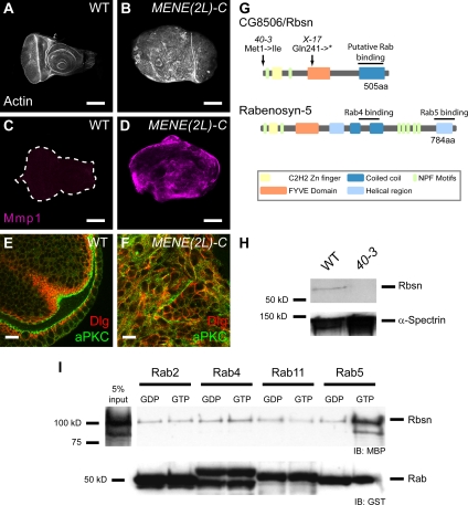
In a recent screen for mutations affecting epithelial polarity and proliferation in Drosophila, we identified many new complementation groups that control epithelial tissue architecture (Menut et al., 2007). This screen used the eyFLP/cell lethal system to generate eye imaginal discs composed predominantly of homozygous mutant cells in an otherwise heterozygous animal (hereafter “mutant eye discs”). In this assay, eye discs mutant for the MENE(2L)-C complementation group consist of rounded and dramatically disorganized masses of cells (Figure 1B, compare with Figure 1A). A similar phenotype is seen in the ovarian follicle cells. Although wild-type follicle cells form a monolayered epithelium, MENE(2L)-C mutant cells multilayer and often invade the germ cell cluster (Supplemental Figure S1). Staining for proteins normally localized to apical or lateral plasma membrane domains reveals that these domains are misspecified in MENE(2L)-C mutant cells. The normally apically restricted protein aPKC fails to remain distinct from Discs-Large-marked (Dlg) basolateral domains (Figure 1F, compare with Figure 1E), indicating that apicobasal polarity is disrupted in these mutants. MENE(2L)-C mutant eye discs also show strong upregulation of matrix metalloprotease 1 (Mmp1) expression (Figure 1D, compare with wild-type [WT] in 1C), which correlates with neoplastic transformation (Uhlirova and Bohmann, 2006; Menut et al., 2007; Srivastava et al., 2007). Finally, larvae with MENE(2L)-C mutant eye discs do not pupariate but continue to feed during an extended L3 stage; during this time, the eye discs grow to be significantly larger than wild-type eye discs. The polarity, proliferation, and gene expression phenotypes all resemble those seen in tissues mutant for previously characterized neoplastic tumor suppressor genes (nTSGs), including scribble (scrib) and Rab5 (Bilder et al., 2000; Lu and Bilder, 2005). However, complementation tests showed that MENE(2L)-C was not allelic to any known tumor suppressor gene. Collectively, these phenotypes therefore indicate that MENE(2L)-C disrupts a novel Drosophila neoplastic tumor suppressor gene.
Figure 1.
MENE(2L)-C mutations disrupt Drosophila Rabenosyn, which is a Rab5 effector and neoplastic tumor suppressor. Single confocal sections of WT (A) and entirely mutant MENE(2L)-C (B) eye discs stained with phalloidin to mark actin. MENE(2L)-C discs are larger than WT discs and lack their distinctive apical enrichment of actin filaments. MENE(2L)-C discs express Mmp1 (D; magenta), a marker correlated with neoplastic tissue, at significantly higher levels than WT discs (C). (E and F) Eye discs stained for aPKC to mark apical surfaces (green) and Dlg to mark basolateral surfaces (red). Wild-type cells have distinct aPKC and Dlg domains (E), whereas aPKC and Dlg are intermixed in MENE(2L)-C cells (F). (G) Locations of the lesions in the MENE(2L)-C40-3 (40-3) and MENE(2L)-CX-17 (X-17) alleles are indicated. Protein domains are labeled as colored boxes according to legend, compared with human Rabenosyn-5. (H) Western blot of wild-type and MENE(2L)-C40-3 mutant imaginal disc extracts probed with anti-Rbsn (top) or anti-α-Spectrin antibody as a loading control (bottom). (I) In vitro GST pull-down assays between recombinant MBP-Rbsn and GST-Rab proteins. Western blots against MBP (top) to detect bound Rbsn and GST (bottom) as a loading control. GST-Rab2 is included as a negative control, showing that Rbsn binds specifically only to Rab5-GTP. Bars, 100 μm (A–D) and 10 μm (E and F).
MENE(2L)-C Disrupts a Protein Related to Human Rabenosyn-5
To identify the gene disrupted by MENE(2L)-C alleles, we performed complementation tests with chromosomal deficiency stocks and found a small deficiency, Df(2L)Exel7034, that failed to complement the two extant MENE(2L)-C alleles. Sequencing of genes located within the genomic region deleted in Df(2L)Exel7034 revealed that each MENE(2L)-C allele carries a lesion in the gene CG8506, which encodes a 505 amino acid protein (Figure 1G). MENE(2L)-C40-3 is a missense mutation altering the initiating ATG to ATA; the next in-frame ATG is located at amino acid 116. MENE(2L)-CX17 is a nonsense mutation that introduces a stop codon at amino acid 241. Both alleles show identical phenotypes in imaginal discs as well as in follicle cell epithelia, and animals either homozygous for each allele or hemizygous over Df(2L)Exel7034 die before the second larval instar. In addition, antibodies raised against a GST-CG8506 fusion protein recognize a polypeptide of the expected molecular mass of 56 kDa in wild-type larval extracts; this polypeptide is absent from extracts of MENE(2L)-C40-3 tissue (Figure 1H). These results indicate that MENE(2L)-C40-3 and MENE(2L)-CX17 are null alleles of CG8506.
Sequence analysis revealed that CG8506 encodes a protein containing a number of conserved domains, including an N-terminal C2H2 zinc finger, a FYVE domain, two repeats of the tripeptide motif NPF, and several coiled-coil regions. BLAST searches indicate that CG8506 has significant homology to the human Rab5-binding protein Rabenosyn-5 (Nielsen et al., 2000), which contains each of these domains, although in a different arrangement. CG8506 is shorter than Rabenosyn-5 and lacks a C-terminal helical region, the NPF motifs are N-terminal in CG8506, whereas they are C-terminal in Rabenosyn-5, and CG8506 contains a single coiled-coil region (Figure 1G). Nevertheless, these features are not found together in any other protein encoded by the fly genome. We therefore refer to CG8506 as Drosophila rabenosyn (rbsn).
Activated Rab5 Recruits Rbsn Both In Vitro and In Vivo
Mammalian Rabenosyn-5 has been linked to both the endocytic and the recycling pathways in part by virtue of its ability to bind simultaneously to Rab5 and Rab4 (De Renzis et al., 2002; Naslavsky et al., 2004). To explore whether Drosophila Rbsn might be involved in these trafficking pathways, we performed in vitro binding assays using recombinant Rbsn and Rab GTPases. We found that Rbsn binds to Rab5 specifically in its GTP-, but not GDP-bound form (Figure 1I). By contrast, we did not detect significant binding between Rbsn and Rab4 in either GTP or GDP-bound forms (Figure 1I). Because Rab11 has been implicated in recycling pathways (Ullrich et al., 1996; Dollar et al., 2002), we also tested whether Rbsn could bind to Rab11, but again we failed to detect an interaction (Figure 1I). We conclude that Rbsn interacts specifically with the endocytic regulator Rab5 at early endosomes but not with Rab proteins that control recycling.
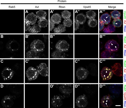
We also asked whether the results of our in vitro binding experiments reflected the protein association in vivo, by examining the subcellular localization pattern of Rabenosyn relative to each of the Rab proteins. In cultured Drosophila S2 cells, Rabenosyn is found in discrete puncta that partially overlap with Avl-positive endocytic compartments (Figure 4A). Expression of Rab5-YFP demonstrates Rbsn and Avl colocalization in Rab5-positive puncta (Figure 4B), indicating that Rbsn localizes to early endosomes in response to Rab5 activation. By contrast, in cells expressing activated forms of Rab4 or Rab11, Rbsn and Avl colocalize in puncta that are mostly discrete from those marked by Rab4 or Rab11 (Supplemental Figure S2), indicating that Rbsn is not strongly recruited to recycling endosomes, consistent with our in vitro results.
Figure 4.
In vivo colocalization between Rab5, Avl, Rbsn, and Vps45. Drosophila S2 cells were transfected with DNA constructs as indicated below and then labeled to detect Rab5, Avl, Rbsn, or Vps45 as indicated in column headings. In Vps45-V5 transfected cells (A), Vps45 (green) shows partial overlap with Avl (red) and Rbsn (blue). In cells transfected with Rab5-YFP (B), Rbsn (blue), and Avl (red) are recruited to Rab5-positive structures (green). When Rab5-YFP and Vps45-V5 are cotransfected (C and D), Vps45 (blue in C, red in D) is recruited to structures labeled with Rab5 (green) and Avl (red in C) or Rbsn (blue in D). Arrowheads indicate triple colocalization; arrows in A‴ indicate independent Vps45 localization. Bar, 5 μm.
Rbsn Is Required for Endocytosis
The above-mentioned data suggest an association between Rbsn and the endocytic pathway, and disruption of several endocytic stages has been previously shown to perturb both cell polarity and cell proliferation control (Lu and Bilder, 2005; Vaccari and Bilder, 2005). We therefore directly tested whether Rbsn was required for endocytosis. In wild-type imaginal disc cells, the apically localized transmembrane protein Notch is continuously endocytosed and lysosomally degraded; the endocytic transient population can be visualized as intracellular cytoplasmic puncta. However, in rbsn cells, Notch is present at greater than wild-type levels (Figure 2B, compared with 2A); a similar elevation is seen with the apical transmembrane protein Crumbs (Crb; Figure 2D, compare with Figure 2C). To directly analyze cargo internalization, we performed a trafficking assay in living disc tissue. This assay pulse-labels cell surface Notch by using an antibody against the Notch extracellular domain; endocytosis is then allowed to occur over varying chase periods. After 10 min of chase in WT cells, Notch is internalized and is found in early endosomes (Figure 2, E–E′), whereas after 5 h, no Notch signal remains (Figure 2, G–G′). In contrast, in rbsn mutant cells no intracellular Notch puncta are seen after 10 min (Figure 2, F–F′); instead, Notch remains in the cell periphery, and this localization persists even after 5 h (Figure 2, H–H′). This pattern strongly resembles that seen in rab5 mutants (Supplemental Figure S1), but contrasts with that seen with the late-acting ESCRT mutants (Vaccari and Bilder, 2005), in which Notch accumulates in enlarged endocytic compartments; it also contrasts with mutations in “junctional scaffold” neoplastic tumor suppressor genes such as scrib, where no effect on Notch endocytosis is seen (unpublished data). The activity of a Notch reporter is reduced in rbsn mutant discs (Figure 2, I–J) as in Rab5 and avl mutants; this is consistent with studies indicating that Notch that does not enter endosomes has reduced signaling function (Vaccari et al., 2008) and suggests that Notch accumulation is not involved in the rbsn tumor phenotype. Together, these results establish that rbsn is required for an early step in the endocytic pathway.
Figure 2.
Rbsn is required for uptake of endocytic cargo. The apical transmembrane proteins Notch (green; A and B) and Crumbs (Crb; cyan; C and D) are present at higher levels in rbsn discs (B and D) than in WT discs (A and C). (E–H′): Notch trafficking assays performed in WT (E and G) and rbsn (F and H) discs. Actin staining outlines cells (E–H; red). In WT cells, surface-labeled Notch (green) is found in internal puncta after 10 min (E′), and is gone from discs after 5 h (G′). In rbsn cells, Notch staining is elevated and retained at the cell periphery after 10 min (F′) and persists over the 5-h experiment (H′). The Notch signaling reporter, mβ-LacZ (I–J; green) is not ectopically activated compared with wild-type (I) in rbsn (J) mutant discs. Images shown are single confocal cross sections. Bars, 100 μm (A–D, I–J) and 10 μm (E′–H′).
Vps45 Binds to Rbsn and Regulates an Early Endocytic Step
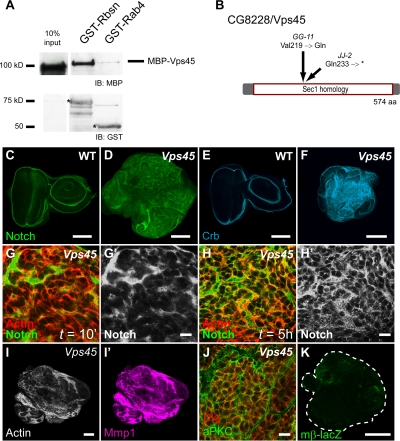
Our data indicate that rbsn has an endocytic mutant phenotype similar to Rab5 mutants, and Rbsn colocalizes with Rab5 at early endosomes and binds directly to Rab5-GTP. These results suggest that Rbsn might regulate Rab5-dependent fusion events at the early endosome. Interestingly, the Sec-1/Munc-18 (SM) protein Vps45 has been identified as a Rabenosyn-5–interacting protein (Nielsen et al., 2000). Although the function of mammalian Vps45 is unknown, the yeast homologue Vps45p has a well documented requirement in biosynthetic Golgi-to-lysosome traffic, and it interacts with a Rabenosyn-5 like protein Vac1p (Cowles et al., 1994; Peterson et al., 1999). To test whether Rbsn might associate with a Vps45-like protein, we first identified a single clear Vps45 homologue among the four SM proteins in Drosophila, which is encoded by the uncharacterized gene CG8228 (hereafter referred to as Vps45). We then expressed an MBP-Vps45 fusion protein in bacteria and we found, using in vitro binding assays, that Vps45 strongly binds to Rbsn (Figure 3A). These data suggest that Rab5 might regulate traffic through Rbsn-dependent Vps45 recruitment to the early endosome. The localization of Vps45 in animals has not been reported previously. To assess the in vivo localization of Vps45, we expressed an epitope-tagged Vps45 construct in S2 cells. In transfected cells, Vps45 shows a punctate pattern with only partial overlap to that of Rbsn and Avl (Figure 4A). Interestingly, upon overexpression of Rab5-YFP, Vps45 relocalizes to the resultant enlarged endosomes; most Vps45 in these cells colocalizes with both Rbsn and Avl (Figure 4, C and D). Together with the in vitro binding data, this result suggests that Vps45 is recruited to the early endosome in response to Rab5 activation.
Figure 3.
Loss of Drosophila Vps45 disrupts endocytic traffic and epithelial architecture. (A) Western blots demonstrate that GST-Rbsn binds to MBP-Vps45, as detected by blotting for MBP (top). GST-Rab4 is included as a negative control; anti-GST blot (bottom) is included as a loading control. Relevant bands are indicated by the asterisk (*). (B) Location of the MENE(3R)-BJJ-2 and MENE(3R)-BGG-11 mutations within the CG8228/Vps45 coding region. (C–F) Compared with WT tissues (C and E), Notch (green), and Crb (cyan) levels are elevated in Vps45 tissue (D and F). (G–H′) Notch trafficking assay performed as described in Figure 2. Vps45 mutants accumulate Notch (green) at the cell periphery, marked by actin staining (red), that persists after 5 h. (I–K) Vps45 mutant discs lose epithelial architecture (I, actin) and mislocalize polarity markers (J, aPKC in green, Dlg in red), and up-regulate Mmp1 (I′; magenta) but do not activate Notch signaling (K, mβ-LacZ reporter in green; compare with WT in Figure 2I). Bars, 100 μm (C–F, I, K) and 10 μm (G–H′, J).
Because yeast Vps45p is required for vacuolar but not endocytic traffic (Raymond et al., 1992; Bryant et al., 1998), we sought to determine whether the Rbsn–Vps45 interaction we observed in Drosophila was relevant for function at the early endosome. No mutations in Vps45 have been reported in flies. However, by testing the uncharacterized neoplastic mutants isolated in our screen, we found that MENE(3R)-B alleles fail to complement deficiencies that remove Vps45. We sequenced Vps45 and we found lesions in the coding region in each of the two MENE(3R)-B alleles (Figure 3B). MENE(3R)-BJJ2 causes premature termination of the protein at amino acid 233 of the 574-amino acid coding region, and MENE(3R)-BGG11 converts valine 219 to a glutamine. Larvae homozygous for either allele die before the third instar, and the mutant eye imaginal disc phenotypes are indistinguishable, suggesting that both represent null alleles.
We next analyzed the phenotypes of Vps45 mutant tissues. As in rbsn mutants, staining for Notch and Crb showed that these protein levels were elevated in Vps45 mutant discs (Figure 3, C–F). To test whether Vps45 mutants might cause neoplastic transformation by blocking endocytosis in a manner similar to rbsn mutants, we examined Notch trafficking. Using live trafficking assays, we found that Notch was not internalized in Vps45 mutant cells and that it accumulated near the cell surface in a manner resembling that of both rbsn and Rab5 mutant cells (Figure 3, G–H′). Moreover, the cell polarity, proliferation, Mmp1 expression and Notch signaling phenotypes were indistinguishable from those of rbsn mutant discs (Figure 3, I–K). These data suggest that in Drosophila, Vps45 function is indeed required for endocytic traffic and in particular for early endosomal stages.
Genetic and Physical Interactions between Vps45 and Avl
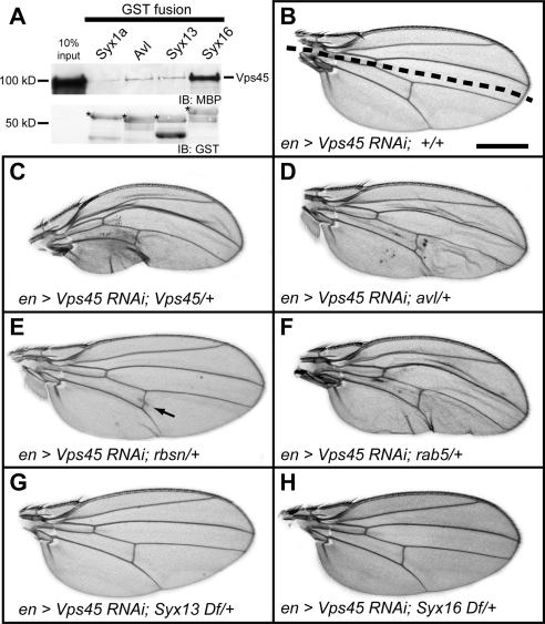
SM proteins are canonically thought to be trafficking regulators, able to bind to either SNARE proteins or complexes and govern fusion between vesicular and target membranes. In Golgi-to-vacuole traffic in yeast, the binding of Vps45p to Tlg2p is necessary for fusion into the vacuole (Nichols et al., 1998). We tested for physical interactions between the Drosophila homologues of these proteins and confirmed a strong interaction between Vps45 and Syx16 (Figure 5A). However, only a small fraction of Drosophila Syx16 localizes to endosomes, and strong expression of a Syx16 RNAi transgene did not generate defects associated with disrupted endocytosis (Supplemental Figure S3). Because similar expression of a Vps45 RNAi transgene shows strong endocytic defects resembling that seen in null mutant tissue (Supplemental Figure S3), we asked whether Vps45 might control endocytosis by associating with early endocytic SNAREs such as Avl. We found weak but consistent binding between Vps45 and Avl compared with Syx1 as a negative control (Figure 5A); this binding was comparable with that seen with the annotated Drosophila homologue of Syx13, which interacts with Vps45 in humans and Caenorhabditis elegans.
Figure 5.
Vps45 interacts with syntaxins of the endocytic pathway. (A) Western blots demonstrate that MBP-Vps45 binds to Avl, GST-Syx13, and GST-Syx16 in vitro (top). GST-Syx1a is included as a negative control. Bottom, loading control (anti-GST blot, with relevant bands labeled with an asterisk [*]). (B–H) Genetic interaction tests in a sensitized background generated by driving expression of a UAS-Vps45-RNAi transgene with the engrailed-GAL4 driver (en >Vps45 RNAi). The engrailed expression domain is below the dashed line in B. Removal of one copy of Vps45 (positive control, C), avl (D), rbsn (E) or Rab5 (F), but not Syx13 (G) or Syx16 (H) enhances the RNAi phenotype. Note posterior cross vein modification in E (arrow). Bar, 0.5 mm.
To evaluate whether Vps45 might regulate these SNAREs in vivo, we turned to genetic interaction studies. Moderately reducing the protein levels of Vps45 using an RNAi construct expressed in the posterior compartment of the adult wing produces no obvious phenotype (Figure 5B). However, removing one copy of the wild-type Vps45 gene to further reduce Vps45 protein levels results in an enhanced phenotype, including aberrant vein formation and ruffling of the posterior margin (Figure 5C). Similar defects are also seen when Avl protein levels are reduced by RNAi (Supplemental Figure S4), suggesting that this phenotype results from impaired endocytosis. We then used the Vps45 RNAi sensitized background to test whether other genes might act in the Vps45-regulated endocytic pathway. We found that removing one copy of Syx13 or Syx16 did not alter the Vps45 RNAi phenotype (Figure 5, G and H), but removing one copy of avl, as well as Rab5 and to a lesser extent Rbsn, resulted in an enhancement similar to that produced by the removal of one copy of Vps45 (Figure 5, D–F). Although weak, the interaction between Vps45 and rbsn was consistent; 64% of en>Vps45-IR; rbsn/+ wings show ectopic vein formation across the posterior cross vein, versus only 17% of en>Vps45-IR wings. Analogous results were seen when knocking down avl (Supplemental Figure S4), further validating the interactions between these genes. Along with the strong phenotypic similarity of the mutants, these results suggest that Vps45 acts together with Rab5, Rbsn, and Avl at the early endosome, and they point to Avl as a regulatory target for Vps45.
Rabenosyn, Rab5, Vps45, and avl Mutants Block Endocytosis at an Identical Early Step
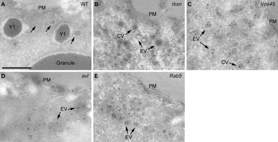
The genetic and biochemical interactions described above suggest the hypothesis that Rab5, Rbsn, Vps45, and Avl act together to promote a single stage of endocytic traffic, involving fusion of incoming endocytic vesicles into the early endosome. If this hypothesis is correct, then cells lacking any of these proteins should block endocytosis at the same step and show similar disruption of endosomal structures. Because light microscopy does not allow the resolution required to clearly distinguish these structures, we instead turned to transmission and immunoelectron microscopy to test this hypothesis. We analyzed late-stage oocytes, which are large cells with a defined endocytic pathway required for uptake of yolk proteins (Schonbaum et al., 2000). By making germline clones, we obtained oocytes mutant for Rab5, rbsn, Vps45, and avl, and we found that all four mutants are defective in the formation of yolk granules. In WT oocytes, yolk granules are late endocytic structures that have a characteristic, electron-dense appearance resulting from condensation of internalized yolk proteins (Figure 6A). These structures are strikingly absent from mutant oocytes (Figure 6, B–E; unpublished data). Because the lack of yolk granules could point to a prior block in the endocytic pathway, we analyzed endocytic intermediates by using an antibody against the yolk proteins, which are produced outside the oocyte, to trace a known endocytic cargo within oocyte vesicular compartments. In wild-type oocytes, yolk proteins are found in numerous endocytic compartments spanning a wide size range (Figure 6A; Trougakos et al., 2001). In contrast, in Rab5, avl, rbsn, and Vps45 mutant oocytes (Figure 6, B–E), yolk proteins are confined to small vesicles with a narrow size distribution; these are primarily found in dense accumulations in proximity to the plasma membrane. The diameter of the vesicles, ∼100 nm, is consistent with both the expected size of internalized clathrin-coated vesicles and the size of vesicles present in WT oocytes (Figure 6A), and both electron-dense coated and uncoated vesicles are seen. Together, these data indicate that endocytic vesicles still form in the absence of Avl, Rab5, Vps45, or Rbsn; these vesicles can uncoat but nevertheless cannot fuse to form later endoyctic structures. The strong phenotypic similarity seen among all four mutants by using immunoelectron microscopy support a model in which Vps45 and Rabenosyn act with Rab5 and Avl to promote vesicle fusion into the early endosome.
Figure 6.
rbsn, Vps45, avl and Rab5 cells accumulate endocytic vesicles but lack later endocytic structures. Images show transmission electron micrographs of oocytes, immunolabeled against yolk proteins produced in follicle cells and endocytosed by the oocyte, detected with gold particles (black puncta). In WT oocytes (A), yolk protein accumulates predominantly in large granules, but it can also be found in immature granules (Y1); ∼100-nm vesicles are also evident below the plasma membrane (PM) (arrows). Yolk granules are absent in oocytes mutant for rbsn (B), Vps45 (C), avl (D), and Rab5 (E); instead, yolk protein is found only in vesicles distributed predominantly below the PM. Some of these endocytic vesicles (EV) are uncoated, whereas others display a clearly visible coat (CV). Note the size difference between labeled endocytic structures found in WT tissue and vesicles found in mutant tissue. Bar, 500 nm.
DISCUSSION
Targeted membrane trafficking steps are vital for maintaining the proper segregation of intracellular compartments and for the sorting of their protein cargoes. Here, we have used forward genetics to identify and characterize two essential regulators of plasma membrane-to-early endosome traffic in Drosophila: Rbsn and Vps45. Rbsn and Vps45 are related to proteins implicated in endocytosis in mammalian cells, and their endocytic role in Drosophila is definitively demonstrated here by direct analysis of cargo trafficking in null mutant tissue. Loss of either protein disrupts the flow of information from the activated small GTPase Rab5 to the trans-SNARE complex and blocks the fusion of endocytic vesicles into the endosome. The endocytic defect further causes mispolarization of epithelial cells and consequent overproliferation to form “neoplastic tumors.” Although we have shown previously that Rab5 acts as a Drosophila tumor suppressor (Lu and Bilder, 2005), Rab5 has many effectors that regulate cellular processes as diverse as lipid metabolism, cytoskeletal organization, and cargo recycling (Christoforidis et al., 1999b; Lanzetti et al., 2004; Naslavsky et al., 2004). Our demonstration that Rbsn and Vps45 are effectors of the tumor suppressive-activity of Drosophila Rab5 emphasizes that growth regulation requires endosomal fusion itself. These two proteins therefore extend the list of endocytic regulators that act as tumor suppressors, confirm the critical role of endocytosis in coordinating cell polarity and cell proliferation, and they provide insight into the processes controlling entry into the early endosome.
The mechanisms linking Rab-mediated vesicle targeting and SNARE-mediated vesicle fusion are among the least well understood events in cellular trafficking. We show here that Drosophila Rbsn is a Rab5 effector, binding to Rab5-GTP and localizing to early endosomes. Like Rab5, Rbsn is required for early endocytic entry, and rbsn and Rab5 mutants are phenotypically indistinguishable. In particular, we have used, for the first time, high-resolution immunoelectron microscopy to identify the site of cargo trapping in cells completely lacking rbsn and Rab5 (as well as Vps45 and avl; see below). These mutants show a striking accumulation of endocytic cargo-containing vesicles of a size consistent with plasma membrane-derived carrier vesicles; the absence of large endosomal compartments suggests that these vesicles fail to undergo fusion to form early endosomes (Rubino et al., 2000). Together, the genetic, biochemical and in vivo phenotypic data provide strong support for a model in which Rbsn is a Rab5 effector essential for endocytic vesicles to fuse into the early endosome.
The severe endocytic block seen in rbsn tissue contrasts with the phenotype of mammalian cells depleted of the related human protein Rabenosyn-5 by RNA knockdown, which allows early endosomal entry but is defective in recycling cargo back to the plasma membrane (Naslavsky et al., 2004). The involvement of Rabenosyn-5 in the recycling pathway is supported not only by this phenotype but also by its ability to bind Rab5 and the recycling regulator Rab4 simultaneously, prompting a model in which Rabenosyn-5 acts to coordinate cargo transfer from the early to recycling endosomes (De Renzis et al., 2002). We find that Drosophila Rbsn, despite its strong association with Rab5-GTP, does not bind to Rab proteins known to regulate recycling, and note that although Rabenosyn-5 contains separate Rab4 and Rab5 binding domains (Eathiraj et al., 2005), these domains show homology to the same single domain in Drosophila Rbsn. It can be speculated that in mammalian Rabenosyn-5, duplication of the Rbsn Rab-binding domain followed by subsequent functional divergence led to its adoption into the recycling pathway, whereas the mammalian tethering protein and Rab5 effector EEA1 played a greater role in regulating early endosome entry (Christoforidis et al., 1999a). Although such an evolutionary scenario is possible, we cannot exclude the possibility that Rbsn plays a role in Drosophila recycling, particularly because the strong endocytic defects that we observed in rbsn mutants are upstream of, and thus prevent analysis of, the recycling pathway.
Our analysis of rbsn null mutant tissue demonstrates that Rbsn is required for vesicles to fuse into the early endosome. How does Rbsn promote vesicle fusion? We find that the Drosophila SM protein Vps45, which binds to Rbsn, is required for the identical step of endocytosis as Rbsn and Rab5. We further localize Vps45 for the first time in vivo, and we find localization to early endosomes, which is increased by Rab5 overexpression. Recruitment of Vps45 by Rbsn bound to active Rab5 may create a high local concentration of Vps45 (Nielsen et al., 2000); once concentrated at the endosome, Vps45 could act on SNARE proteins to enable fusion of incoming carrier vesicles. In contrast to yeast, where Vps45p is required for lysosomal delivery of biosynthetic cargo (Cowles et al., 1994), we have shown that Drosophila Vps45 is required for trafficking and degradation of surface-derived cargo, thus identifying an SM protein that acts in the endocytic pathway.
While this manuscript was in preparation, Gengyo-Ando et al. (2007) reported that C. elegans oocytes lacking homologues of Vps45 or Rbsn are defective in yolk uptake, but they did not distinguish the precise stage of endocytic traffic blocked; moreover, they could not identify any syntaxin required for endocytosis and therefore could not determine a functional target of Vps45. Here, we provide evidence that the endocytic syntaxin Avl is a key Vps45 target. We detected a clear genetic interaction specifically between Vps45 and avl, as well as a weak physical interaction between Vps45 and both Avl and Syx13. Although human and C. elegans Syx13 have been shown to bind Vps45 (Nielsen et al., 2000; Gengyo-Ando et al., 2007), orthologous relationships with Drosophila syntaxins are ambiguous: both Drosophila Avl and Syx13 are similar to human and C. elegans Syx13 as well as to human Syx7. Our data demonstrate that Avl is required for the fusion event required for cargo entry into early endosomes; although RNAi experiments do not reveal a role for Drosophila Syx13 in endocytosis (unpublished data), further experiments are needed to clarify the function of Syx13 in vesicle trafficking.
The in vitro physical interactions we observed between Vps45 and both Avl and Syx13 were notably weaker than that with Syx16. However, our data do not provide evidence for an endocytic role of Syx16. In addition, the significance of SM protein binding to an isolated SNARE remains unclear. Although in most cases it correlates with SNARE complex assembly, in some instances this interaction is not necessary for function in vivo (Carpp et al., 2006), and in others it is associated with inhibition of incorporation into a SNARE complex (Dulubova et al., 1999; Peng and Gallwitz, 2002). Considering these scenarios, our phenotypic analysis of mutant tissues completely lacking Vps45 demonstrates common phenotypes to those completely lacking Rab5, Avl, or Rbsn at the tissue, cellular, and ultrastructural levels, indicating that Vps45 acts as a positive regulator of early endocytic SNAREs; this is also consistent with the enhancing nature of the genetic interactions. Moreover, these data argue that Avl is a component of the SNARE complex whose activity in vesicle fusion requires Vps45, establishing a functional link between Rab5 and SNAREs essential for early endosomal entry.
Together, our genetic, phenotypic, and biochemical analyses provide strong support for a model in which Rbsn, by binding to Vps45 and Rab5, enables incoming cargo vesicles to fuse into the early endosome. This trafficking event is required for the proper control of surface levels of transmembrane proteins and has significant consequences for tissue development. Given that plasma membrane-to-early endosome trafficking is a process by which metazoan animals can control intercellular interactions, Rbsn may be an attractive target for cellular regulation of this event. Indeed, genetic interactions hint at a role for Rbsn in modulating several cell–cell interaction and communication pathways (Vaccari, Morrison, and Bilder, unpublished data); future work will reveal whether Rbsn activity is modulated in specific contexts to achieve different developmental outcomes.
Supplementary Material
ACKNOWLEDGMENTS
We thank Marcos Gonzalez-Gaitan, Matt Scott, Suzanne Eaton, Matt Welch, William Trimble, Ruth Palmer, Peter Gallant, and Hugo Stocker for reagents. We thank Thomas Vaccari, Han Lu, and Geena Wu for experimental support, and Youngsoo Jun and other members of the Bilder laboratory for helpful discussions. This work was supported by National Institutes of Health grant R01 GM-068675 and American Cancer Society grant RSG-07-040-01 (to D. B.); The Norwegian Research Council and Funksjonell Genomforskning, Norway (to T.E.R., H. S., and A. B.); and The Norwegian Cancer Society and the Novo Nordisk Foundation (to H. S). W.W.F., B.D.P., and S.E.C. were supported by U.S. Department of Energy contract DE-AC-02-05CH11231.
Abbreviations used:
- Avl
Avalanche
- Crb
Crumbs
- Rbsn
Rabenosyn
Footnotes
This article was published online ahead of print in MBC in Press (http://www.molbiolcell.org/cgi/doi/10.1091/mbc.E08-07-0716) on August 6, 2008.
REFERENCES
- Aalto M. K., Ronne H., Keränen S. Yeast syntaxins Sso1p and Sso2p belong to a family of related membrane proteins that function in vesicular transport. EMBO J. 1993;12:4095–4104. doi: 10.1002/j.1460-2075.1993.tb06093.x. [DOI] [PMC free article] [PubMed] [Google Scholar]
- Bänziger C., Soldini D., Schütt C., Zipperlen P., Hausmann G., Basler K. Wntless, a conserved membrane protein dedicated to the secretion of Wnt proteins from signaling cells. Cell. 2006;125:509–522. doi: 10.1016/j.cell.2006.02.049. [DOI] [PubMed] [Google Scholar]
- Barbieri M. A., Hoffenberg S., Roberts R., Mukhopadhyay A., Pomrehn A., Dickey B. F., Stahl P. D. Evidence for a symmetrical requirement for Rab5-GTP in in vitro endosome-endosome fusion. J. Biol. Chem. 1998;273:25850–25855. doi: 10.1074/jbc.273.40.25850. [DOI] [PubMed] [Google Scholar]
- Bilder D., Li M., Perrimon N. Cooperative regulation of cell polarity and growth by Drosophila tumor suppressors. Science. 2000;289:113–116. doi: 10.1126/science.289.5476.113. [DOI] [PubMed] [Google Scholar]
- Bilder D., Perrimon N. Localization of apical epithelial determinants by the basolateral PDZ protein Scribble. Nature. 2000;403:676–680. doi: 10.1038/35001108. [DOI] [PubMed] [Google Scholar]
- Bock J. B., Matern H. T., Peden A. A., Scheller R. H. A genomic perspective on membrane compartment organization. Nature. 2001;409:839–841. doi: 10.1038/35057024. [DOI] [PubMed] [Google Scholar]
- Bryant N. J., Piper R. C., Gerrard S. R., Stevens T. H. Traffic into the prevacuolar/endosomal compartment of Saccharomyces cerevisiae: a VPS45-dependent intracellular route and a VPS45-independent, endocytic route. Eur. J. Cell Biol. 1998;76:43–52. doi: 10.1016/S0171-9335(98)80016-2. [DOI] [PubMed] [Google Scholar]
- Bucci C., Parton R. G., Mather I. H., Stunnenberg H., Simons K., Hoflack B., Zerial M. The small GTPase rab5 functions as a regulatory factor in the early endocytic pathway. Cell. 1992;70:715–728. doi: 10.1016/0092-8674(92)90306-w. [DOI] [PubMed] [Google Scholar]
- Carpp L. N., Ciufo L. F., Shanks S. G., Boyd A., Bryant N. J. The Sec1p/Munc18 protein Vps45p binds its cognate SNARE proteins via two distinct modes. J. Cell Biol. 2006;173:927–936. doi: 10.1083/jcb.200512024. [DOI] [PMC free article] [PubMed] [Google Scholar]
- Carr C. M., Grote E., Munson M., Hughson F. M., Novick P. J. Sec1p binds to SNARE complexes and concentrates at sites of secretion. J. Cell Biol. 1999;146:333–344. doi: 10.1083/jcb.146.2.333. [DOI] [PMC free article] [PubMed] [Google Scholar]
- Chen Y. A., Scheller R. H. SNARE-mediated membrane fusion. Nat. Rev. Mol. Cell Biol. 2001;2:98–106. doi: 10.1038/35052017. [DOI] [PubMed] [Google Scholar]
- Chou T. B., Perrimon N. The autosomal FLP-DFS technique for generating germline mosaics in Drosophila melanogaster. Genetics. 1996;144:1673–1679. doi: 10.1093/genetics/144.4.1673. [DOI] [PMC free article] [PubMed] [Google Scholar]
- Christoforidis S., McBride H. M., Burgoyne R. D., Zerial M. The Rab5 effector EEA1 is a core component of endosome docking. Nature. 1999a;397:621–625. doi: 10.1038/17618. [DOI] [PubMed] [Google Scholar]
- Christoforidis S., Miaczynska M., Ashman K., Wilm M., Zhao L., Yip S.-C., Waterfield M. D., Backer J. M., Zerial M. Phosphatidylinositol-3-OH kinases are Rab5 effectors. Nat. Cell Biol. 1999b;1:249–252. doi: 10.1038/12075. [DOI] [PubMed] [Google Scholar]
- Cowles C. R., Emr S. D., Horazdovsky B. F. Mutations in the VPS45 gene, a SEC1 homologue, result in vacuolar protein sorting defects and accumulation of membrane vesicles. J. Cell Sci. 1994;107:3449–3459. doi: 10.1242/jcs.107.12.3449. [DOI] [PubMed] [Google Scholar]
- De Renzis S., Sönnichsen B., Zerial M. Divalent Rab effectors regulate the sub-compartmental organization and sorting of early endosomes. Nat. Cell Biol. 2002;4:124–133. doi: 10.1038/ncb744. [DOI] [PubMed] [Google Scholar]
- Dollar G., Struckhoff E., Michaud J., Cohen R. S. Rab11 polarization of the Drosophila oocyte: a novel link between membrane trafficking, microtubule organization, and oskar mRNA localization and translation. Development. 2002;129:517–526. doi: 10.1242/dev.129.2.517. [DOI] [PubMed] [Google Scholar]
- Dubreuil R., Byers T. J., Branton D., Goldstein L. S., Kiehart D. P. Drosophilia spectrin. I. Characterization of the purified protein. J. Cell Biol. 1987;105:2095–2102. doi: 10.1083/jcb.105.5.2095. [DOI] [PMC free article] [PubMed] [Google Scholar]
- Dulubova I., Sugita S., Hill S., Hosaka M., Fernandez I., Sudhof T. C., Rizo J. A conformational switch in syntaxin during exocytosis: role of munc18. EMBO J. 1999;18:4372–4382. doi: 10.1093/emboj/18.16.4372. [DOI] [PMC free article] [PubMed] [Google Scholar]
- Eathiraj S., Pan X., Ritacco C., Lambright D. G. Structural basis of family-wide Rab GTPase recognition by rabenosyn-5. Nature. 2005;436:415–419. doi: 10.1038/nature03798. [DOI] [PMC free article] [PubMed] [Google Scholar]
- Fasshauer D., Sutton R. B., Brunger A. T., Jahn R. Conserved structural features of the synaptic fusion complex: SNARE proteins reclassified as Q- and R-SNAREs. Proc. Natl. Acad. Sci. USA. 1998;95:15781–15786. doi: 10.1073/pnas.95.26.15781. [DOI] [PMC free article] [PubMed] [Google Scholar]
- Gengyo-Ando K., Kuroyanagi H., Kobayashi T., Murate M., Fujimoto K., Okabe S., Mitani S. The SM protein VPS-45 is required for RAB-5-dependent endocytic transport in Caenorhabditis elegans. EMBO Rep. 2007;8:152–157. doi: 10.1038/sj.embor.7400882. [DOI] [PMC free article] [PubMed] [Google Scholar]
- Gorvel J. P., Chavrier P., Zerial M., Gruenberg J. rab5 controls early endosome fusion in vitro. Cell. 1991;64:915–925. doi: 10.1016/0092-8674(91)90316-q. [DOI] [PubMed] [Google Scholar]
- Grosshans B. L., Ortiz D., Novick P. Rabs and their effectors: achieving specificity in membrane traffic. Proc. Natl. Acad. Sci. USA. 2006;103:11821–11827. doi: 10.1073/pnas.0601617103. [DOI] [PMC free article] [PubMed] [Google Scholar]
- Lanzetti L., Palamidessi A., Areces L., Scita G., Di Fiore P. P. Rab5 is a signalling GTPase involved in actin remodelling by receptor tyrosine kinases. Nature. 2004;429:309–314. doi: 10.1038/nature02542. [DOI] [PubMed] [Google Scholar]
- Lawrence P. A., Johnston P., Morata G. Methods of marking cells. In: Roberts D. B., editor. Drosophila: A Practical Approach. Oxford, United Kingdom: IRL Press Limited; 1986. pp. 229–242. [Google Scholar]
- Lu H., Bilder D. Endocytic control of epithelial polarity and proliferation in Drosophila. Nat. Cell Biol. 2005;7:1232–1239. doi: 10.1038/ncb1324. [DOI] [PubMed] [Google Scholar]
- Lu Y., Settleman J. The Drosophila Pkn protein kinase is a Rho/Rac effector target required for dorsal closure during embryogenesis. Genes Dev. 1999;13:1168–1180. doi: 10.1101/gad.13.9.1168. [DOI] [PMC free article] [PubMed] [Google Scholar]
- McBride H. M., Rybin V., Murphy C., Giner A., Teasdale R., Zerial M. Oligomeric complexes link Rab5 effectors with NSF and drive membrane fusion via interactions between EEA1 and Syntaxin 13. Cell. 1999;98:377–386. doi: 10.1016/s0092-8674(00)81966-2. [DOI] [PubMed] [Google Scholar]
- McNew J. A., Parlati F., Fukuda R., Johnston R. J., Paz K., Paumet F., Sollner T. H., Rothman J. E. Compartmental specificity of cellular membrane fusion encoded in SNARE proteins. Nature. 2000;407:153–159. doi: 10.1038/35025000. [DOI] [PubMed] [Google Scholar]
- Menut L., Vaccari T., Dionne H., Hill J., Wu G., Bilder D. A mosaic genetic screen for Drosophila neoplastic tumor suppressor genes based on defective pupation. Genetics. 2007;177:1667–1677. doi: 10.1534/genetics.107.078360. [DOI] [PMC free article] [PubMed] [Google Scholar]
- Naslavsky N., Boehm M., Backlund P. S., Jr, Caplan S. Rabenosyn-5 and EHD1 Interact and sequentially regulate protein recycling to the plasma membrane. Mol. Biol. Cell. 2004;15:2410–2422. doi: 10.1091/mbc.E03-10-0733. [DOI] [PMC free article] [PubMed] [Google Scholar]
- Nichols B. J., Holthuis J. C., Pelham H. R. The Sec1p homologue Vps45p binds to the syntaxin Tlg2p. Eur. J. Cell Biol. 1998;77:263–268. doi: 10.1016/s0171-9335(98)80084-8. [DOI] [PubMed] [Google Scholar]
- Nielsen E., Christoforidis S., Uttenweiler-Joseph S., Miaczynska M., Dewitte F., Wilm M., Hoflack B., Zerial M. Rabenosyn-5, a novel Rab5 effector, is complexed with hVPS45 and recruited to endosomes through a FYVE finger domain. J. Cell Biol. 2000;151:601–612. doi: 10.1083/jcb.151.3.601. [DOI] [PMC free article] [PubMed] [Google Scholar]
- Parks A. L., et al. Systematic generation of high-resolution deletion coverage of the Drosophila melanogaster genome. Nat. Genet. 2004;36:288–292. doi: 10.1038/ng1312. [DOI] [PubMed] [Google Scholar]
- Peng R., Gallwitz D. Sly1 protein bound to Golgi syntaxin Sed5p allows assembly and contributes to specificity of SNARE fusion complexes. J. Cell Biol. 2002;157:645–655. doi: 10.1083/jcb.200202006. [DOI] [PMC free article] [PubMed] [Google Scholar]
- Peterson M. R., Burd C. G., Emr S. D. Vac1p coordinates Rab and phosphatidylinositol 3-kinase signaling in Vps45p-dependent vesicle docking/fusion at the endosome. Curr. Biol. 1999;9:159–162. doi: 10.1016/s0960-9822(99)80071-2. [DOI] [PubMed] [Google Scholar]
- Raymond C. K., Howald-Stevenson I., Vater C. A., Stevens T. H. Morphological classification of the yeast vacuolar protein sorting mutants: evidence for a prevacuolar compartment in class E vps mutants. Mol. Biol. Cell. 1992;3:1389–1402. doi: 10.1091/mbc.3.12.1389. [DOI] [PMC free article] [PubMed] [Google Scholar]
- Rubino M., Miaczynska M., Lippe R., Zerial M. Selective membrane recruitment of EEA1 suggests a role in directional transport of clathrin-coated vesicles to early endosomes. J. Biol. Chem. 2000;275:3745–3748. doi: 10.1074/jbc.275.6.3745. [DOI] [PubMed] [Google Scholar]
- Schonbaum C. P., Perrino J. J., Mahowald A. P. Regulation of the vitellogenin receptor during Drosophila melanogaster oogenesis. Mol. Biol. Cell. 2000;11:511–521. doi: 10.1091/mbc.11.2.511. [DOI] [PMC free article] [PubMed] [Google Scholar]
- Seals D. F., Eitzen G., Margolis N., Wickner W. T., Price A. A Ypt/Rab effector complex containing the Sec1 homolog Vps33p is required for homotypic vacuole fusion. Proc. Natl. Acad. Sci. USA. 2000;97:9402–9407. doi: 10.1073/pnas.97.17.9402. [DOI] [PMC free article] [PubMed] [Google Scholar]
- Srivastava A., Pastor-Pareja J. C., Igaki T., Pagliarini R., Xu T. Basement membrane remodeling is essential for Drosophila disc eversion and tumor invasion. Proc. Natl. Acad. Sci. USA. 2007;104:2721–2726. doi: 10.1073/pnas.0611666104. [DOI] [PMC free article] [PubMed] [Google Scholar]
- Stenmark H., Olkkonen V. M. The Rab GTPase family. Genome Biol. 2001;2:REVIEWS3007. doi: 10.1186/gb-2001-2-5-reviews3007. [DOI] [PMC free article] [PubMed] [Google Scholar]
- Stenmark H., Parton R. G., Steele-Mortimer O., Lütcke A., Gruenberg J., Zerial M. Inhibition of rab5 GTPase activity stimulates membrane fusion in endocytosis. EMBO J. 1994;13:1287–1296. doi: 10.1002/j.1460-2075.1994.tb06381.x. [DOI] [PMC free article] [PubMed] [Google Scholar]
- Tepass U., Knust E. Crumbs and Stardust act in a genetic pathway that controls the organization of epithelia in Drosophila melanogaster. Dev. Biol. 1993;159:311–326. doi: 10.1006/dbio.1993.1243. [DOI] [PubMed] [Google Scholar]
- Trougakos I. P., Papassideri I. S., Waring G. L., Margaritis L. H. Differential sorting of constitutively co-secreted proteins in the ovarian follicle cells of Drosophila. Eur. J. Cell Biol. 2001;80:271–284. doi: 10.1078/0171-9335-00163. [DOI] [PubMed] [Google Scholar]
- Tsukada M., Will E., Gallwitz D. Structural and functional analysis of a novel coiled-coil protein involved in Ypt6 GTPase-regulated protein transport in yeast. Mol. Biol. Cell. 1999;10:63–75. doi: 10.1091/mbc.10.1.63. [DOI] [PMC free article] [PubMed] [Google Scholar]
- Uhlirova M., Bohmann D. JNK- and Fos-regulated Mmp1 expression cooperates with Ras to induce invasive tumors in Drosophila. EMBO J. 2006;25:5294–5304. doi: 10.1038/sj.emboj.7601401. [DOI] [PMC free article] [PubMed] [Google Scholar]
- Ullrich O., Reinsch S., Urbe S., Zerial M., Parton R. G. Rab11 regulates recycling through the pericentriolar recycling endosome. J. Cell Biol. 1996;135:913–924. doi: 10.1083/jcb.135.4.913. [DOI] [PMC free article] [PubMed] [Google Scholar]
- Ungermann C., Langosch D. Functions of SNAREs in intracellular membrane fusion and lipid bilayer mixing. J. Cell Sci. 2005;118:3819–3828. doi: 10.1242/jcs.02561. [DOI] [PubMed] [Google Scholar]
- Vaccari T., Bilder D. The Drosophila tumor suppressor vps25 prevents nonautonomous overproliferation by regulating notch trafficking. Dev. Cell. 2005;9:687–698. doi: 10.1016/j.devcel.2005.09.019. [DOI] [PubMed] [Google Scholar]
- Vaccari T., Lu H., Kanwar R., Fortini M. E., Bilder D. Endosomal entry regulates Notch receptor activation in Drosophila melanogaster. J. Cell Biol. 2008;180:755–762. doi: 10.1083/jcb.200708127. [DOI] [PMC free article] [PubMed] [Google Scholar]
- Waters M. G., Hughson F. M. Membrane tethering and fusion in the secretory and endocytic pathways. Traffic. 2000;1:588–597. doi: 10.1034/j.1600-0854.2000.010802.x. [DOI] [PubMed] [Google Scholar]
- Whyte J.R.C., Munro S. Vesicle tethering complexes in membrane traffic. J. Cell Sci. 2002;115:2627–2637. doi: 10.1242/jcs.115.13.2627. [DOI] [PubMed] [Google Scholar]
- Wucherpfennig T., Wilsch-Brauninger M., Gonzalez-Gaitan M. Role of Drosophila Rab5 during endosomal trafficking at the synapse and evoked neurotransmitter release. J. Cell Biol. 2003;161:609–624. doi: 10.1083/jcb.200211087. [DOI] [PMC free article] [PubMed] [Google Scholar]
- Xu H., Boulianne G. L., Trimble W. S. Drosophila syntaxin 16 is a Q-SNARE implicated in Golgi dynamics. J. Cell Sci. 2002;115:4447–4455. doi: 10.1242/jcs.00139. [DOI] [PubMed] [Google Scholar]
- Zerial M., McBride H. M. Rab proteins as membrane organizers. Nat. Rev. Mol. Cell Biol. 2001;2:107–117. doi: 10.1038/35052055. [DOI] [PubMed] [Google Scholar]
Associated Data
This section collects any data citations, data availability statements, or supplementary materials included in this article.


